Copyright
©2005 Baishideng Publishing Group Inc.
World J Gastroenterol. May 28, 2005; 11(20): 3080-3084
Published online May 28, 2005. doi: 10.3748/wjg.v11.i20.3080
Published online May 28, 2005. doi: 10.3748/wjg.v11.i20.3080
Figure 1 Determining NF-κB activity in ECV304WT and ECV304MT cells.
A: IκBα level were detected after treated with PMA (50 nmol/L) for 12 h; B: EMSA was performed to determine the activity of NF-κB after treated with PMA (50 nmol/L) for 12 h. Band of NF-κB was marked.
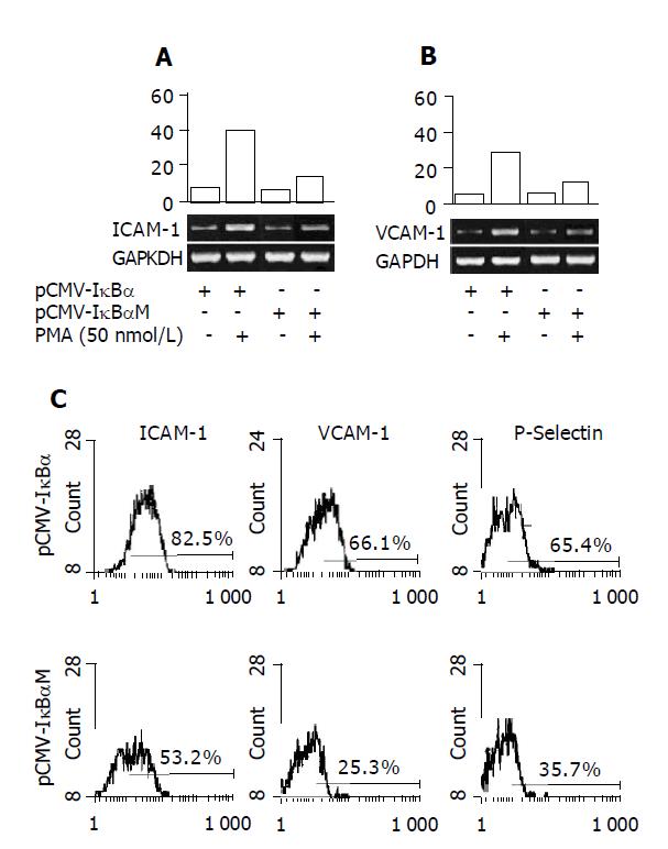
Figure 2 Down-regulation of PMA-induced adhesion molecules expression in ECV cells transfected with pCMV-IκBαM compared with pCMV-IκBα.
RT-PCR analysis was performed with primers specific for ICAM-1 (A) and VCAM-1 (B). C: The expression of ICAM-1, VCAM-1 and P-selectin were detected using specific antibodies by flow cytometry. Results are representative of at least three independent experiments.
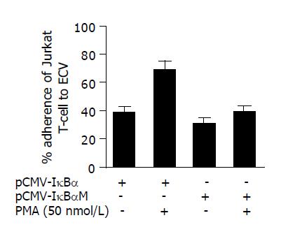
Figure 3 Adhesion of human Jurkat T-cells to PMA-activated endothelial (ECV) cells is inhibited by transfection with pCMV-IκBαM compared with pCMV-IκBα.
ECV cells were activated with PMA (50 nmol/L) for 12 h. Cells were washed thrice with PBS and then co-cultured with Jurkat T-cells for 2 h. Then the cells were washed with PBS thrice. Jurkat cell adhesion was determined by visual counting under a phase-contrast microscope. P<0.05 when compared ECVMT with ECVWT.
- Citation: Wei JF, Sun K, Xu SG, Xie HY, Zheng SS. Inhibition of PMA-induced endothelial cell activation and adhesion by over-expression of domain negative IκBα protein. World J Gastroenterol 2005; 11(20): 3080-3084
- URL: https://www.wjgnet.com/1007-9327/full/v11/i20/3080.htm
- DOI: https://dx.doi.org/10.3748/wjg.v11.i20.3080


