©2005 Baishideng Publishing Group Inc.
World J Gastroenterol. Apr 28, 2005; 11(16): 2431-2437
Published online Apr 28, 2005. doi: 10.3748/wjg.v11.i16.2431
Published online Apr 28, 2005. doi: 10.3748/wjg.v11.i16.2431
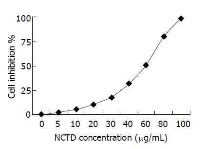
Figure 1 The dose-response curves of effect of NCTD on GBC-SD cells for 72 h.
Inhibition of the growth of human gallbladder carcinoma GBC-SD cells by various concentrations of NCTD. Cell number was counted by the MTT method.
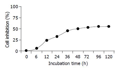
Figure 2 The inhibitory effect curves of IC50 NCTD on GBC-SD cells at different times.
Cell number was counted by the MTT method.
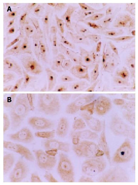
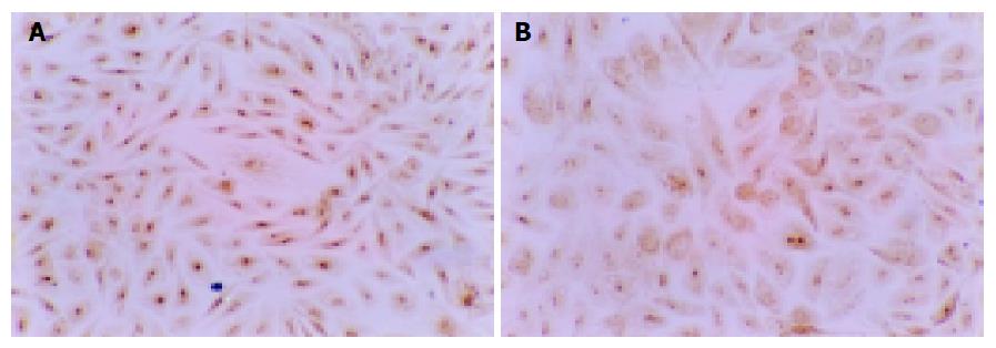
Figure 3 Positive expression occurred in cell nucleoli, with brown or yellow dye, of PCNA protein of GBC-SD cells (immunohistochemistry SABC method, ×200).
A: The brown dye of PCNA was shown positively in most cells of the control group. B: In the experiment group with treatment of IC50 NCTD for 48 h, the positive cells of PCNA expression decreased significantly and the dye in cell nucleoli became light and shallow.
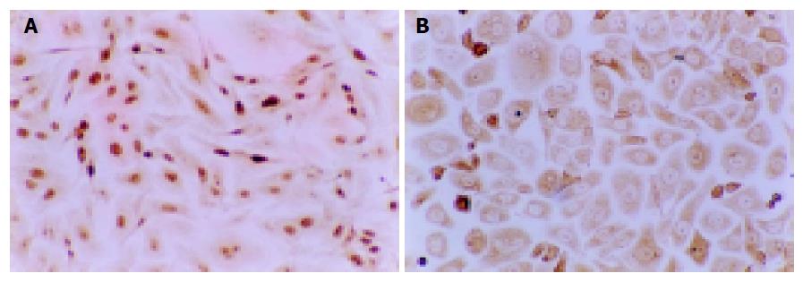
Figure 4 The positive expression occurred in cell nucleoli, with brown or yellow dye, of Ki-67 protein of GBC-SD cells (immunohistochemistry SABC method, ×100).
A: The brown dye of Ki-67 was shown positively in most cells of the control group. B: In the experiment group with treatment of IC50 NCTD for 48 h, the positive cells of Ki-67 expression decreased significantly and the dye in cell nucleoli became light and shallow.
Figure 5 Positive expression with brown dye occurred in cytoplast of MMP2 protein of GBC-SD cells (immunohistochemistry SABC method, ×100).
A: The brown dye of MMP2 was shown positively in most cells of the control group. B: In the experiment group with treatment of NCTD (5 μg/mL) for 48 h, the positive cells of MMP2 expression decreased significantly and the dye in the cytoplast became light.
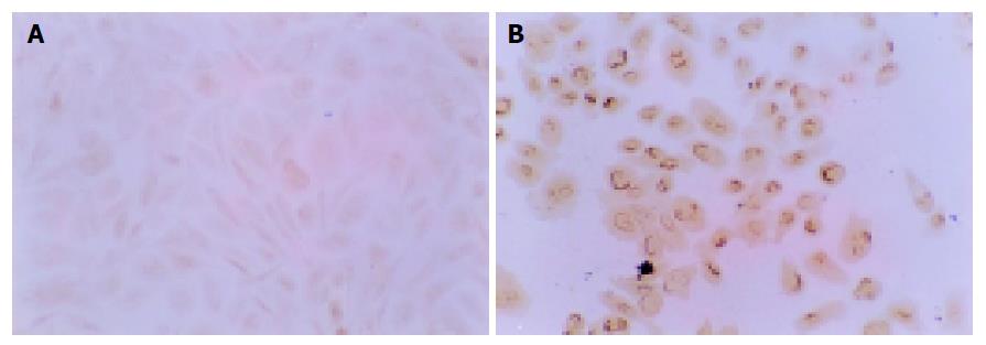
Figure 6 Positive expression with brown dye occurred on nucleoli membrane of TIMP2 protein of GBC-SD cells (immunohistochemistry SABC method, ×100).
A: The negative expression of TIMP2 was observed in most GBC-SD cells of the control group. B: In the experiment group with treatment of NCTD (5 μg/mL) for 48 h, the positive cells of TIMP2 expression were increased significantly and the dye on nucleoli membrane represent brown.
- Citation: Fan YZ, Fu JY, Zhao ZM, Chen CQ. Effect of norcantharidin on proliferation and invasion of human gallbladder carcinoma GBC-SD cells. World J Gastroenterol 2005; 11(16): 2431-2437
- URL: https://www.wjgnet.com/1007-9327/full/v11/i16/2431.htm
- DOI: https://dx.doi.org/10.3748/wjg.v11.i16.2431


