©2005 Baishideng Publishing Group Inc.
World J Gastroenterol. Apr 28, 2005; 11(16): 2398-2401
Published online Apr 28, 2005. doi: 10.3748/wjg.v11.i16.2398
Published online Apr 28, 2005. doi: 10.3748/wjg.v11.i16.2398
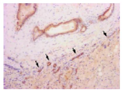
Figure 1 Constitutive expression of β-catenin in normal bile ducts.
Membranous expression is conspicuous in normal bile ducts and newly formed bile ducts (arrow heads), but no expression is observed in normal hepatocytes.
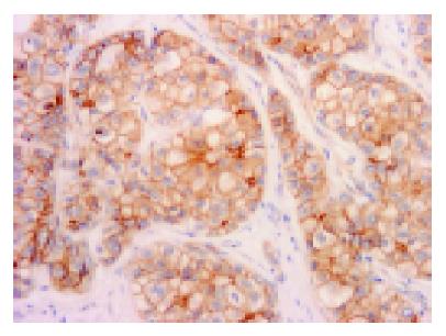
Figure 2 Membranous expression.
Membranous expression is conspicuous in tumor cell membranes. This case is moderately-differentiated HCC.
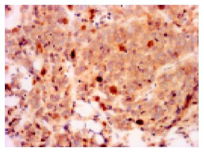
Figure 3 Nuclear expression.
Nuclear expression is encountered in the poorly-differentiated HCC (arrows). Most cells co-expressed β-catenin in the cytoplasm.
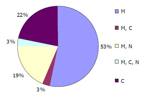
Figure 4 Localization of β-catenin expression in cell level.
M: membranous, M+C: membranous and cytoplasmic, M+N: membranous and nuclear, M+N+C: membranous, cytoplasmic, and nuclear, C: cytoplasmic.
- Citation: Tien LT, Ito M, Nakao M, Niino D, Serik M, Nakashima M, Wen CY, Yatsuhashi H, Ishibashi H. Expression of β-catenin in hepatocellular carcinoma. World J Gastroenterol 2005; 11(16): 2398-2401
- URL: https://www.wjgnet.com/1007-9327/full/v11/i16/2398.htm
- DOI: https://dx.doi.org/10.3748/wjg.v11.i16.2398


