©2005 Baishideng Publishing Group Inc.
World J Gastroenterol. Jan 7, 2005; 11(1): 41-45
Published online Jan 7, 2005. doi: 10.3748/wjg.v11.i1.41
Published online Jan 7, 2005. doi: 10.3748/wjg.v11.i1.41
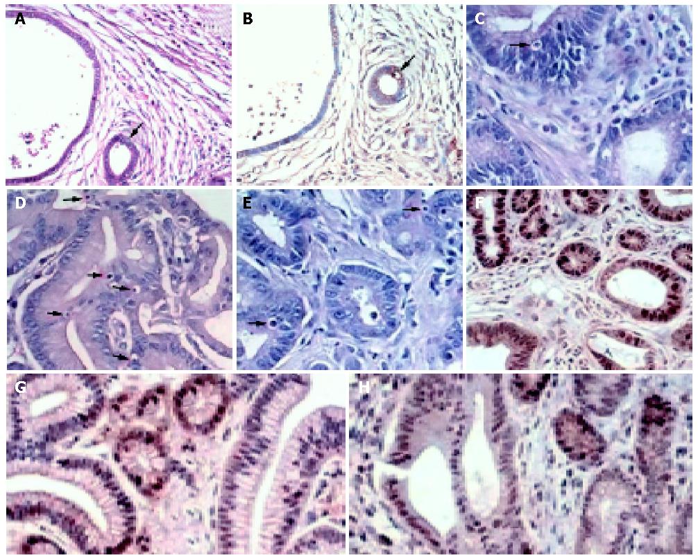
Figure 1 Histological examination of apoptosis and proliferation.
Apoptosis was examined by apoptotic nuclei counting (A) and verified by TUNEL (B). A representative apoptotic nucleus is illustrated by the black arrow. Representative H&E stained sections showing apoptotic bodies (red arrow) in (C) MNNG-treated tumors, (D) celecoxib-treated tumors and (E) indomethacin-treated tumors. (F-H) Ki-67 immunostaining was used in the assessment of proliferation. Representative proliferating cells in (F) MNNG treated tumors, (G) celecoxib-treated tumors and (H) indomethacin-treated tumors indicated by positive immunoreactivity against Ki-67.
Figure 2 Effects of celecoxib/indomethacin treatment on gastric cell apoptosis and proliferation.
A: Effects of celecoxib/indomethacin treatment on gastric cell apoptosis. The mean apoptotic index with standard error was shown. The apoptotic indexes were significantly higher in MNNG-induced tumor than in untreated control (P = 0.001). Moreover, the levels of apoptosis were significantly different among tumors (T), their adjacent non-tumor tissues (NT) and normal tissues from non-tumor rats (N) in all treatment groups (aP<0.005, ANOVA). Treatment with celecoxib was associated with a higher apoptotic index in tumors (P<0.05, ANOVA) and their adjacent non-tumor tissues (P = 0.003, ANOVA). There appeared to be a dose-dependent increase in apoptotic index in celecoxib-treated tumors when compared to tumors treated with MNNG alone, but there was no significant increase in apoptotic index in indomethacin-treated tumors; B: The mean proliferation indexes with standard error. There were significant differences in the proliferation indexes among tumors (P≤0.001, ANOVA) and their adjacent normal gastric tissues (P = 0.01, ANOVA). Specifically, tumors in MNNG group had the highest proliferation index than other treatment groups (group B vs all other groups, P<0.003).
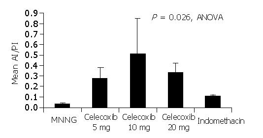
Figure 3 Effects of celecoxib or indomethacin on the apoptosis index to proliferation ratio (AI/PI) of gastric tumors.
The mean AI/PI ratio with standard error was shown. There was a significant difference in the AI/PI ratio among different treatment groups (P = 0.026, ANOVA). The highest ratio was seen in gastric tumors treated with celecoxib at 10 mg/(kg·d) whereas the lowest ratio was seen in tumors from MNNG group. The AI/PI ratio appeared to inversely correlate with the tumor incidence reported in different treatment groups (Table 1).
- Citation: Yu J, Tang BD, Leung WK, To KF, Bai AH, Zeng ZR, Ma PK, Go MY, Hu PJ, Sung JJ. Different cell kinetic changes in rat stomach cancer after treatment with celecoxib or indomethacin: Implications on chemoprevention. World J Gastroenterol 2005; 11(1): 41-45
- URL: https://www.wjgnet.com/1007-9327/full/v11/i1/41.htm
- DOI: https://dx.doi.org/10.3748/wjg.v11.i1.41


