©2005 Baishideng Publishing Group Inc.
World J Gastroenterol. Jan 7, 2005; 11(1): 118-121
Published online Jan 7, 2005. doi: 10.3748/wjg.v11.i1.118
Published online Jan 7, 2005. doi: 10.3748/wjg.v11.i1.118
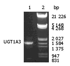
Figure 1 Agarose gel electrophoresis of UGT1A3 PCR products.
Lane 1: UGT1A3 PCR products; lane 2: DNA molecular markers.
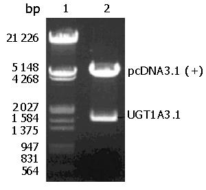
Figure 2 Restriction enzyme analysis of pcDNA3.
1 (+)-UGT1A3 recombinant plasmids. Lane 1: DNA molecular markers; lane 2: pcDNA3.1 (+)-UGT1A3 plasmids after digestion with Xho I and Hind III.
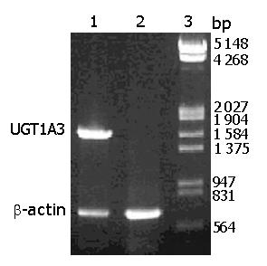
Figure 3 Agarose gel electrophoresis of the result of RT-PCR.
Lane 1: CHL-UGT1A3; lane 2: untransfected CHL cells; lane 3: DNA molecular markers.
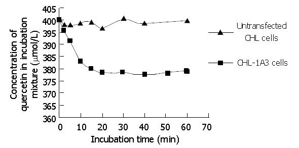
Figure 4 Time course of quercetin incubated with S9 prepared from CHL-UGT1A3 cells or untransfected CHL cells.
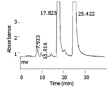
Figure 5 Typical chromatograms of quercetin after incubation with S9 prepared from CHL-UGT1A3 Cells.
For quercetin-glucuronide, tR = 7.933 min; for morin, tR = 17.825 min; and for quercetin, tR = 25.422 min.
- Citation: Chen YK, Li X, Chen SQ, Zeng S. Heterologous expression of active human uridine diphosphate glucuronosyltransferase 1A3 in Chinese hamster lung cells. World J Gastroenterol 2005; 11(1): 118-121
- URL: https://www.wjgnet.com/1007-9327/full/v11/i1/118.htm
- DOI: https://dx.doi.org/10.3748/wjg.v11.i1.118


