Copyright
©The Author(s) 2004.
World J Gastroenterol. Nov 1, 2004; 10(21): 3107-3111
Published online Nov 1, 2004. doi: 10.3748/wjg.v10.i21.3107
Published online Nov 1, 2004. doi: 10.3748/wjg.v10.i21.3107
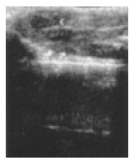
Figure 1 Ultrasonic image of implanted tumors in nude mice liver.
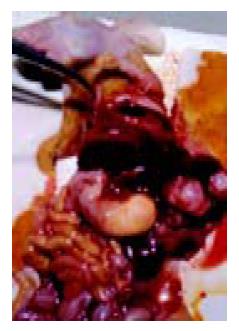
Figure 2 Implanted tumors in nude mice liver by autopsy.
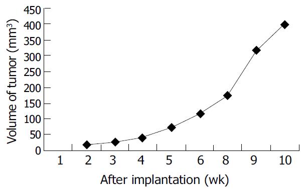
Figure 3 Relation between volume of implanted tumor and time after implantation.
Figure 4 Implanted tumor in nude mice liver metastases to lungs.
The first passage just formed a few metastatic lesions A; The 4th passage formed multiple metastatic lesions B; and metastatic lesions in lungs were found under microscope C.
Figure 5 Implanted tumor in nude mice liver spontaneously metastasized to bone.
The first passage metastasized to vertebra thoracica just as the donor A, and the 4th passage showed multiple bone metastases B, C. Metastatic lesions were shown in bones under microscope D.
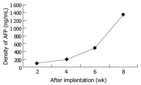
Figure 6 Relation between density of AFP in serum and time of tumor after implantation.
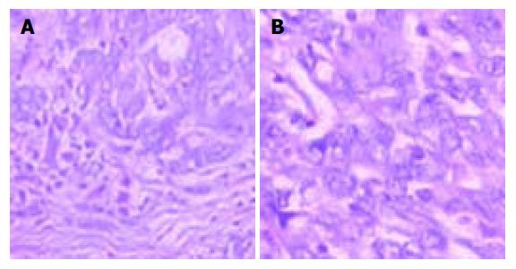
Figure 7 Edmonson grade II-III HCC specimen from patients A and in tumor tissue from nude mice model B.
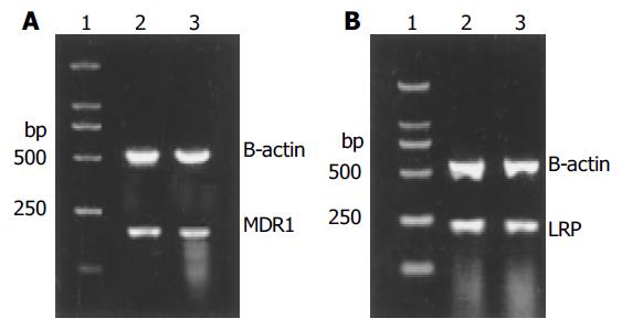
Figure 8 MDR1 and LRP expressed in tumors of the donor (2) and the mude mice model (3), and the expression had no sig-nificant difference between both.
A: MDR1 expressed in tumors. B: LRP expressed in tumors.
-
Citation: Gao YS, Chen XP, Li KY, Wu ZD. Nude mice model of human hepatocellular carcinoma
via orthotopic implantation of histologically intact tissue. World J Gastroenterol 2004; 10(21): 3107-3111 - URL: https://www.wjgnet.com/1007-9327/full/v10/i21/3107.htm
- DOI: https://dx.doi.org/10.3748/wjg.v10.i21.3107


