©The Author(s) 2004.
World J Gastroenterol. Nov 1, 2004; 10(21): 3094-3098
Published online Nov 1, 2004. doi: 10.3748/wjg.v10.i21.3094
Published online Nov 1, 2004. doi: 10.3748/wjg.v10.i21.3094
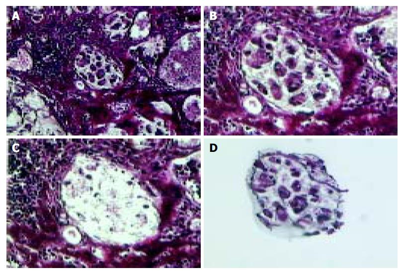
Figure 1 Laser capture microdissection of cells from gastric carcinoma.
A: Morphology map view for pathological diagnosis (× 100). B: Group of gastric carcinoma cells were selected for LCM (× 200). C: The same section revealed where carcinoma cells were lifted from the section (× 200). D: The carcinoma cells were removed on the plastic film to provide a template for PCR.
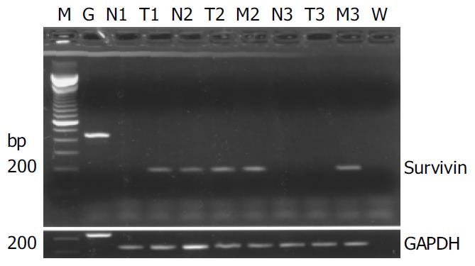
Figure 2 Representative RT-PCR results of survivin expres-sion in normal (N), tumor (T) and metastatic carcinoma (M) cells obtained by LCM from 3 patients suffering gastric cancer.
Case 1 was a patient without lymphatic metastasis, cases 2 and 3 were patients with lymphatic metastasis. G: Genomic DNA from human peripheral blood. W: Water as negative control.
- Citation: Wang ZN, Xu HM, Jiang L, Zhou X, Lu C, Zhang X. Expression of survivin in primary and metastatic gastric cancer cells obtained by laser capture microdissection. World J Gastroenterol 2004; 10(21): 3094-3098
- URL: https://www.wjgnet.com/1007-9327/full/v10/i21/3094.htm
- DOI: https://dx.doi.org/10.3748/wjg.v10.i21.3094


