Copyright
©The Author(s) 2004.
World J Gastroenterol. Sep 15, 2004; 10(18): 2628-2631
Published online Sep 15, 2004. doi: 10.3748/wjg.v10.i18.2628
Published online Sep 15, 2004. doi: 10.3748/wjg.v10.i18.2628
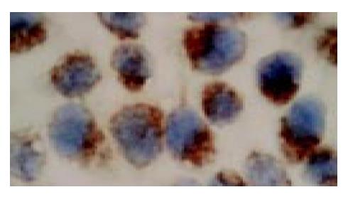
Figure 1 In situ hybridization (ISH) analysis of progesterone receptor (PR) expression in human gastric adenocarcinoma cell line SGC7901 (ISH, × 1000).
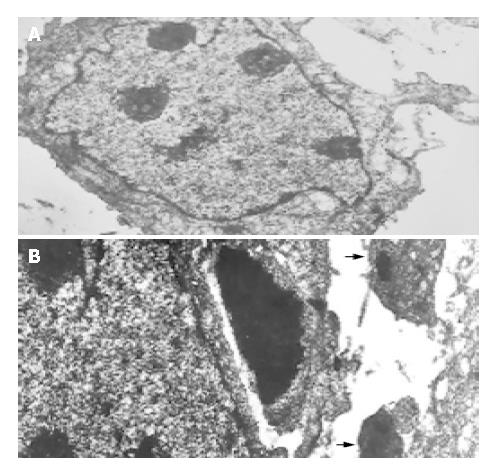
Figure 2 Transmission electron microscopic photographs of the SGC7901 cells cultured for 96 h in the absence (A) or the presence of 20 μmol/L mifepristone (B) in vitro (TEM, × 2000).
Arrows indicate apoptotic bodies which were formed in the cells of mifepristone-treated group.
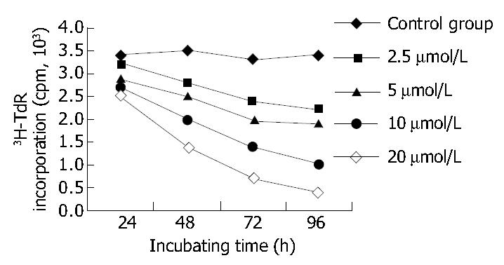
Figure 3 Effect of various concentrations of mifepristone on the 3H-TdR incorporation of SGC7901 cells at various time intervals in vitro.
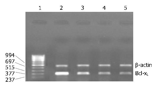
Figure 4 RT-PCR analysis of Bcl-XL mRNA expression in the SGC7901 cells cultured for 96 h in the absence or the presence of various concentrations of mifepristone in vitro.
Lanes 1-5: Marker (bp), contro l, 5, 10, 20 μmol/L mifepristone, respectively.
-
Citation: Li DQ, Wang ZB, Bai J, Zhao J, Wang Y, Hu K, Du YH. Effects of mifepristone on proliferation of human gastric adenocarcinoma cell line SGC-7901
in vitro . World J Gastroenterol 2004; 10(18): 2628-2631 - URL: https://www.wjgnet.com/1007-9327/full/v10/i18/2628.htm
- DOI: https://dx.doi.org/10.3748/wjg.v10.i18.2628


