Copyright
©The Author(s) 2004.
World J Gastroenterol. Jul 1, 2004; 10(13): 1939-1942
Published online Jul 1, 2004. doi: 10.3748/wjg.v10.i13.1939
Published online Jul 1, 2004. doi: 10.3748/wjg.v10.i13.1939
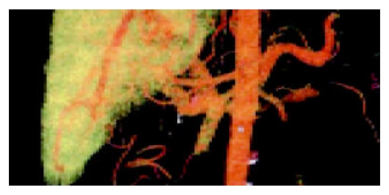
Figure 1 Portal vein in artery phase and in portal vein phase.
A: Portal vein in artery phase. VR technique could display artery-portal vein fistula clearly in artery phase, the portal vein was visualized early, even portal vein blood up the stream. The arrow points the fistula aperture between the right hepatic artery and right portal vein. B: Portal vein in portal vein phase. The portal vein was very slightly developed and portal vein-vena cava shunt emerged in the portal vein phase.
Figure 2 Enlarged portal vein and a non-flow region in the trunk (arrow).
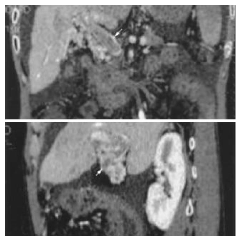
Figure 3 Image of portal vein reconstructed by MPVR.
We could see the emboli’s scopes and sizes at different planes (arrow).
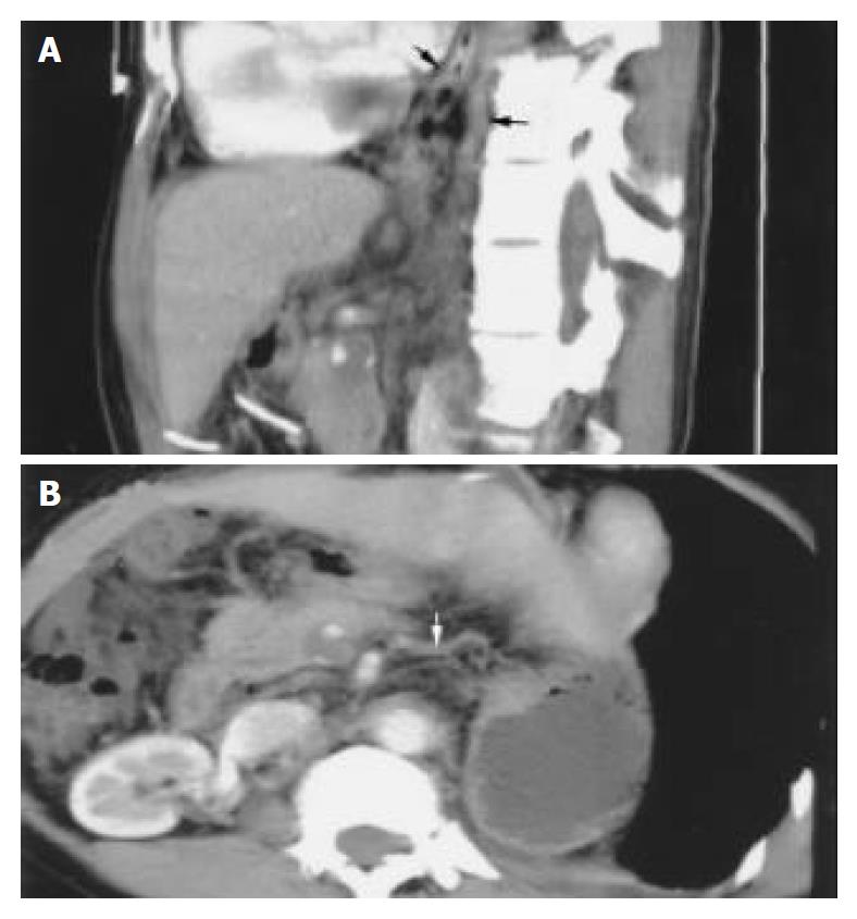
Figure 4 Portal vein-vena cave shunt, portal vein emboli and fistula of hepatic artery-portal vein on MPVR images.
A: Paraesophageal varices and dilated azygos vein on MPVR image (arrow). B: Paragastric varices on MPVR image (arrow).
- Citation: Chu Q, Li Z, Zhang SM, Hu DY, Xiao M. Relationship between encephalopathy and portal vein-vena cava shunt: Value of computed tomography during arterial portography. World J Gastroenterol 2004; 10(13): 1939-1942
- URL: https://www.wjgnet.com/1007-9327/full/v10/i13/1939.htm
- DOI: https://dx.doi.org/10.3748/wjg.v10.i13.1939


