Published online Jul 1, 2004. doi: 10.3748/wjg.v10.i13.1943
Revised: January 5, 2004
Accepted: January 12, 2004
Published online: July 1, 2004
AIM: To investigate the role of curved planar reformations using multidetector spiral CT (MSCT) in diagnosis of pancreatic and peripancreatic diseases.
METHODS: From October 2001 to September 2003, 47 consecutive patients with pancreatic or peripancreatic diseases, which were confirmed by operation, endoscopic retrograde cholangiopancreatography and clinical follow-up, were enrolled in this study. CT scanning was performed at a MSCT with four rows of detector. A set of images with an effective thickness of 1.0-2.0 mm and a gap of 0.5-1.0 mm (50% overlap) were acquired in all patients for post-processing. Curved planar reformations were carried out by drawing a curved line on transverse source images, coronal or sagittal multiplanar reformations according to certain anatomic structures (such as cholangiopancreatic ducts or peripancreatic vessels) and the position of lesion.
RESULTS: With thin collimation, MSCT could acquire high-quality curved planar reformations to display the profile of the whole pancreas, to trace the cholangiopancreatic ducts and peripancreatic vessels, and to show the relationship of lesions with pancreas and peripancreatic anatomic structures in one curved plane, which facilitates diagnosis and rapid communication of diagnostic information with referring physicians.
CONCLUSION: MSCT with thin collimation could be used to create high-quality curved planar reformations in evaluating pancreatic and peripancreatic diseases with pertinent anatomic information and relative pathologic signs to facilitate the diagnosis and enhance communication with the referring physician. Curved planar reformations can serve as supplements for transverse images in diagnosis and management of pancreatic and peripancreatic diseases.
- Citation: Gong JS, Xu JM. Role of curved planar reformations using multidetector spiral CT in diagnosis of pancreatic and peripancreatic diseases. World J Gastroenterol 2004; 10(13): 1943-1947
- URL: https://www.wjgnet.com/1007-9327/full/v10/i13/1943.htm
- DOI: https://dx.doi.org/10.3748/wjg.v10.i13.1943
Pancreas is located in the retroperitoneum with a tortuous course, which is difficult for CT and MR to display the whole pancreas in one slice. There are abundant vessels around the pancreas. It is important to visualize these vessels and the cholangiopancreatic ducts for diagnosing and evaluating the local involvement of pancreatic and peripancreatic lesions. Curved planar reformations can delineate a curved path and display the whole course of pancreas in a single cross-section image according to a curved line drawn on the transverse source images, coronal or sagittal multiplanar reformations (MPR) from a three dimension (3-D) volumetric data set acquired by spiral CT[1,2]. They have been widely used to display vessels and evaluate vascular abnormalities in conjunction with CT angiography[1-7]. Recently the new techniques have been applied to evaluate the pancreas and bile duct system pathologically[8-11]. This article focuses on the role of curved planar reformations using MSCT in the diagnosis of pancreatic and peripancreatic diseases.
From October 2001 to September 2003, 47 consecutive patients (21 female and 26 male, age range: 13-72 years, mean age: 53 years) with pancreatic or peripancreatic diseases, which were confirmed by operation (n = 18), endoscopic retrograde cholangiopancreatography (ERCP) (n = 6) and clinical follow-up (n = 23), were enrolled in this study. The diseases included p ancreatic carcinoma (n = 9), cy stadenoma (n = 3), neuroendocrine tumor (n = 4), chronic pancreatitis with pseudocyst (n = 16), real cyst of pancreas (n = 1), choledochal cyst (n = 2), ampullary carcinoma (n = 3), choledocholithiasis (n = 5), duodenal diverticulum (n = 1), peripancreatic adenopathy (n = 3) and splenic arterial aneurysm (n = 1).
CT scans were performed with a MSCT scanner (Toshiba Aquilion, Tokyo, Japan). Seven patients underwent plain scanning, 38 patients underwent both plain and enhanced scanning, and the other two patients a drip infusion CT cholangiography. A scanning with parameters of 120 kVp, 300 mA, 4 × 1-2 mm collimation, rotation time of 0.5 s, and a pitch of 5.5 was included in all patients during a breathhold of 10-15 s. In patients who underwent both plain and enhanced scanning, this protocol was obtained at the pancreatic parenchyma phase (a delay of 40 s after the beginning of intravenous injection of nonionic contrast media with an iodine content of 300 mg/mL [Omnipaque, Nycomed-Amersham, Shanghai] at 3 mL/s using a power injector). The CT cholangiographic image was acquired 60 min after intravenous drip infusion of iodipamide methyglucamine. A set of images with an effective thickness of 1-2 mm and a gap of 0.5-1.0 mm (50% overlap) was reconstructed and transmitted to workstation for post-processing.
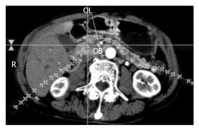
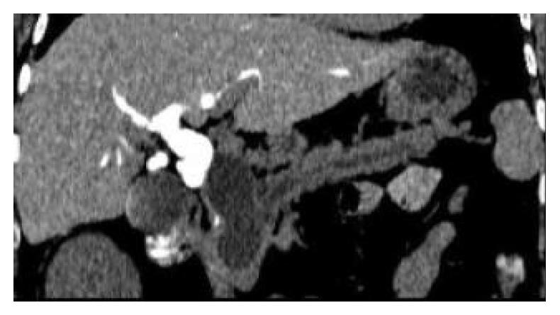
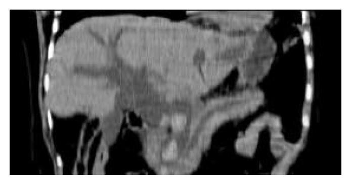
Curved planar reformations were obtained using a cursor to draw a curved line along a special anatomic structure on a stack of axial, sagittal, coronal section at a SGI workstation (Alatoview 1.21) by an experienced radiologist who was familiar with the pancreatic and peripancreatic anatomic structures and had carefully reviewed all transverse source images. The operator was asked to adjust the trace of the cursor interactively to create several curved planes to display essential anatomic structures, such as cholangiopancreatic ducts and peripancreatic vessels, and the relationship of the lesion with those structures according to diagnostic and clinical need (Figure 1). The section thickness of curved plane will never be larger than the effective section thickness or smaller than the transverse pixel dimensions.
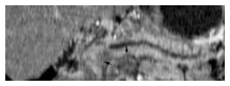
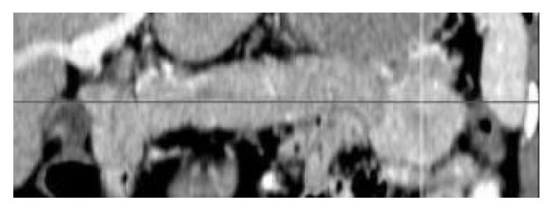
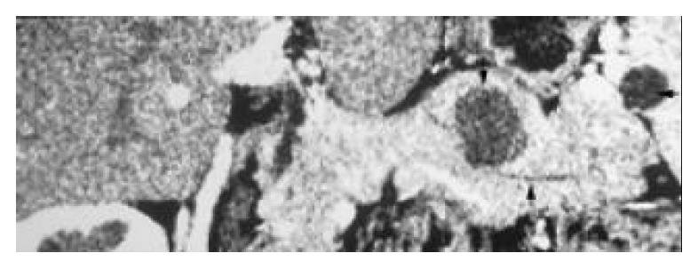
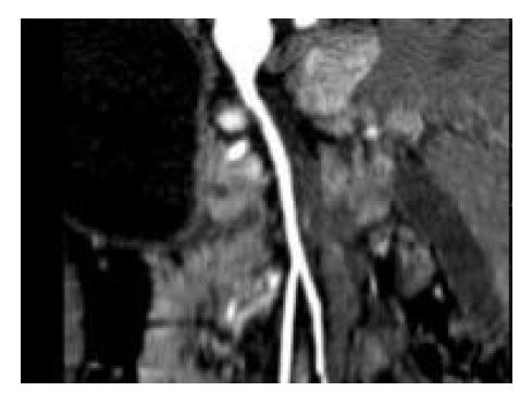
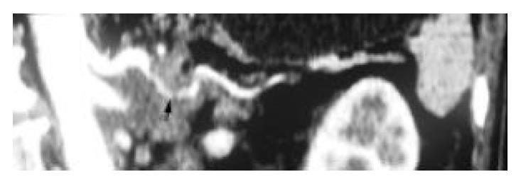
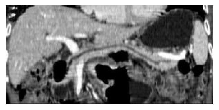
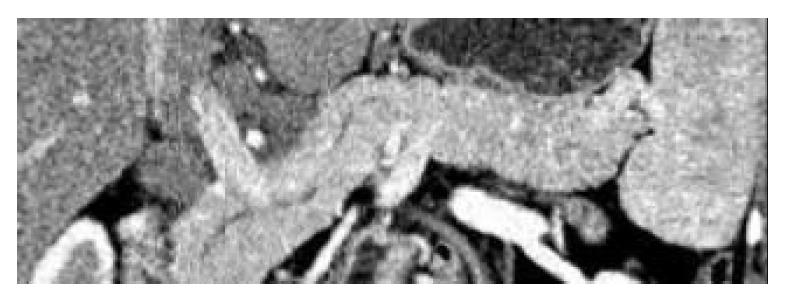
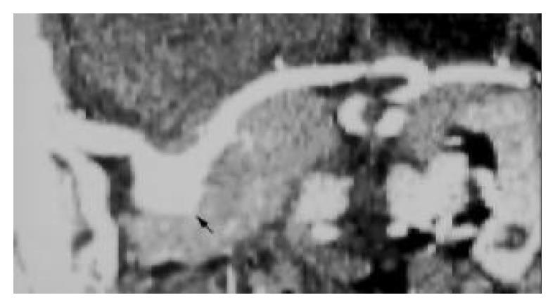
With thinner collimation and overlap image data sets, high quality curved planar reformations were obtained in all the 47 patients, displaying the whole pancreas and tracing cholangiopancreatic ducts or peripancreatic vessels in a single cross-section image without any misregistration and steps artifacts (Figure 2, Figure 3, Figure 4, Figure 5, Figure 6, Figure 7, Figure 8, Figure 9, Figure 10, Figure 11, Figure 12). For the 20 pancreatic local lesions (9 cases of pancreatic adenocarcinoma, 3 cystic adenomas, 4 neuroendocrine tumors, 3 intrapancreatic pseudocysts and 1 real cyst of pancreas), curved planar reformations demonstrated that the whole or major portions of the lesion was located in the pancreatic parenchyma clearly, which made it easier to determine lesion’s location (Figure 2, Figure 3, Figure 4, Figure 5). Curved planar reformations, by tracing a certain vessel, such as the celiac artery, the superior mesenteric artery, the portal vein, could demonstrate changes in the caliber of the vessel and wall invasion, which was helpful for determining whether these vessels were involved and the length of involvement, as in the 9 patients with pancreatic adenocarcinoma and the 3 patients with ampullary carcinoma (Figure 6, Figure 7). Combining with transverse images, curved planar reformations demonstrated 2 positivities and 1 false positivity in predicting respectability, 6 positivities in assessment of unresectability in 9 patients with pancreatic adenocarcinoma. The dilatation of cholangiopancreatic ducts in patients with chronic pancreatitis, pancreatic or ampullary carcinoma and peripancreas lymph nodes enlargement were well depicted on curved planar reformations (Figure 8, Figure 9). In 1 patient with pancreatic real cyst, the curved planar reformations after CT cholangiography revealed the beaded dilatation of pancreatic duct and the dilatation and stenosis of the common bile duct. The cyst lay beside the dilated common bile duct (Figure 4). In the 13 patients with pseudocyst beyond the pancreas and the 2 patients with choledochal cyst, curved planar reformations displayed the cystic lesion and the relationship of cysts with pancreas well. Stones’ location and cholangiopancreatic ducts morphologic changes distal and proximal to obstruction were depicted on curved planar reformations in all patients with choledocholithiasis (Figure 10). The location and relationship with pancreas of peripancreatic enlarged lymph nodes were easily identified on curved planar reformations, which made differential diagnosis from pancreatic mass more simple (Figure 11). In 1 case of splenic arterial aneurysm, the dilatation of the artery was detected on curved planar reformations but missed on transverse source images due to its small diameter and dilatation along the z axis of CT imaging (Figure 12).
Computed tomography (CT) is the most frequently used imaging modality for the evaluation of patients with suspected pancreatic and peripancreatic diseases, especially for diagnosing and staging pancreatic adenocarcinoma[12-17]. In detection and staging of pancreatic ductal adenocarcinoma, CT had a sensitivity of more than 90% and a positive predictive value in the range of 96% to 100% for determining surgical unresectability[9,12-14]. In determining resectability, the accuracy of CT remained low with positive predictive values ranging from 53% to 79% due to underestimation of vascular invasion and failure to detect small peritoneal and liver metastases[9,12,14]. The introduction of MSCT and the development of three-dimensional (3D) imaging software have significantly improved the ability of CT to image the pancreas and evaluate a wide range of pancreatic and peripancreatic pathology[8-11,17,18].
One major advantage of MSCT over single-detector spiral CT is substantial improvement in the speed of scan acquisition, which permits routine use of very thin collimation covering a large region during breath-hold to acquire volumetric data sets with high resolution along all three axes, especially along z axis, even with isotropic image[9,10,18,19]. The improvement of resolution along the z axis is very helpful for generating high quality multiplanar reformations (MPR) and 3D images. Curved planar reformations are a special form of MPR; which depict the cross-section profile along any curved line the operator draws from the volumetric data sets to trace the course of an anatomic structure. The plane presents results in a 2D image that displays the entire course of the anatomic structure[9]. Thus the techniques are very useful in displaying a complex anatomic structure with a tortuous course. They can delineate the curved vascular structure and display its whole course in one cross-section image. Therefore, curved planar reformations were used to display vessels and evaluate their abnormality[2-7]. This provided a convenient and fast review method for radiologists and an effective format for communicating the pertinent findings to referring physicians. These techniques were especially useful in evaluating those vessels, such as the carotid siphon, which has a tortuous course and is surrounded by complex bony structures and thus the maximal intensity projection (MIP) and 3D images were difficult to depict them[5]. Recently, the application of curved planar reformations in the pancreatic and bile duct system showed that these techniques could provide helpful unique anatomic information, highlight critical anatomic and pathologic relationships, which were useful for surgical planning[8-11]. The other advantage of curved planar reformations was that the referring physicians could review a few curved planes to acquire critical information instead of reviewing a large number of images in great detail, which enhanced rapid communication[8].
In the present series, curved planar reformations using MSCT were performed in 47 patients with pancreatic and peripancreatic diseases. With thin collimation and overlap reconstruction, high quality curved planar reformations were obtained in all the patients displaying the whole tortuous pancreas in one cross-section image, tracing critical surrounding anatomic structures, such as cholangiopancreatic ducts and peripancreatic vessels, or highlighting relationships of the lesion with essential anatomic structures. All these were helpful for determining the lesion’s location and evaluating its invasion to surrounding anatomic structures. The latter was important to determining the resectability of pancreatic ductal adenocarcinoma. In 9 patients with pancreatic carcinoma, curved planar reformations tracing critical peripancreatic vessels could not only demonstrate the vascular involvement of tumor, but also depict the length of the affected segment. Although one study showed that curved planar reformations were equivalent to transverse images in the detection of pancreatic tumors and determination of surgical resectability[10], we did not assess the resectability according to curved planar reformations only due to their operator dependence, which wound be discussed later. In this series, by combination of curved planar reformations with transverse images, 2 positivities and 1 false positivity in predicting resectability and 6 positivities in determining unresectability in 9 patients with pancreatic adenocarcinoma were obtained. Whether it would improve the accuracy of staging of pancreatic adenocarcinoma by combining curved planar reformation with transverse images was not carried out in this study due to the small sample size of pancreatic carcinoma, which was one limitation of this study. In one case of pancreatic real cyst, curved planar reformations after CT cholangiography demonstrated the beaded dilatation of pancreatic duct and the dilatation and obstruction of the bile duct system. The relationship of the cystic lesion with the pancreatic and bile duct system was depicted clearly. All these could provide guides for operation, although a correct diagnosis was not acquired before operation. For the peripancreatic lesions, such as peripancreatic adenopathy, ampullary carcinoma, duodenal diverticulum and pseudocysts, curved planar reformations depicted the changes of the bile duct system and pancreatic duct, and relationships of lesions with surrounding important anatomic structures, which were helpful for making diagnostic and treatment strategies. Direct display of morphologic changes of bile duct system and stones location in curved planar reformations would be very useful for surgical planning, especially for endoscopic management of choledocholithiasis. With their advantage in demonstrating vascular abnormality, curved planar reformations displayed the small splenic arterial aneurysm easily in this series although it was missed in transverse images. On the one hand, it needs the operator interactively to adjust the path of cursor and work intensively to generate proper curved planar reformations with highlight important diagnostic information. On the other hand, several curved planes may transfer the important information to referring physicians. This reduces the referring physicians’ burden to review all the transverse images for useful information.
One limitation of curved planar reformations is that they are exceedingly operator-dependent. Improper path of the cursor may generate spurious stenoses of cholangiopancreatic ducts and vessels, distort the anatomic structures or cause important lesion missed. Therefore, the operator should be familiar with the anatomy of pancreas and carefully review all transverse source images before beginning to draw the line. Although careful view of all transverse source images may be a time-consuming and tedious job, it is helpful to create proper curved planar reformations to highlight critical anatomic structures and pathologic signs. Due to this limitation, in this series a comparison with standard transverse images was not carried out. Curved planar reformations can serve as supplements in evaluating pancreatic and peripancreatic diseases while not a substitute. Recently, Raman et al[20] developed an automated method to generate curved planar reformations with reduced time and effort. With the development of post-processing software, a well-trained radiologic technologists may generate curved planar reformations routinely.
In summary, MSCT with thin collimation can be used to generate high quality curved planar reformations for evaluating pancreatic and peripancreatic diseases. Our preliminary study showed that curved planar reformations could clearly depict essential anatomic structures and highlight critical anatomic information and pathologic signs in several cross-section images, which enhanced the rapid communication with referring physicians. Curved planar reformations can serve as supplements for transverse images in diagnosis and management of pancreatic and peripancreatic diseases.
We are grateful to Mr. Xiang-Peng Zheng, Ph.D candidate for radiological biology at University of Texas Health Science Center, for language review.
Edited by Zhu LH and Chen WW Proofread by Xu FM
| 1. | Rubin GD, Dake MD, Semba CP. Current status of three-dimensional spiral CT scanning for imaging the vasculature. Radiol Clin North Am. 1995;33:51-70. [PubMed] |
| 2. | Achenbach S, Moshage W, Ropers D, Bachmann K. Curved multiplanar reconstructions for the evaluation of contrast-enhanced electron beam CT of the coronary arteries. AJR Am J Roentgenol. 1998;170:895-899. [RCA] [PubMed] [DOI] [Full Text] [Cited by in Crossref: 81] [Cited by in RCA: 58] [Article Influence: 2.1] [Reference Citation Analysis (0)] |
| 3. | Portugaller HR, Schoellnast H, Tauss J, Tiesenhausen K, Hausegger KA. Semitransparent volume-rendering CT angiography for lesion display in aortoiliac arteriosclerotic disease. J Vasc Interv Radiol. 2003;14:1023-1030. [RCA] [PubMed] [DOI] [Full Text] [Cited by in Crossref: 14] [Cited by in RCA: 10] [Article Influence: 0.4] [Reference Citation Analysis (0)] |
| 4. | Takase K, Sawamura Y, Igarashi K, Chiba Y, Haga K, Saito H, Takahashi S. Demonstration of the artery of Adamkiewicz at multi- detector row helical CT. Radiology. 2002;223:39-45. [RCA] [PubMed] [DOI] [Full Text] [Cited by in Crossref: 131] [Cited by in RCA: 116] [Article Influence: 4.8] [Reference Citation Analysis (0)] |
| 5. | Teasdale E. Curved planar reformatted CT angiography: utility for the evaluation of aneurysms at the carotid siphon. AJNR Am J Neuroradiol. 2000;21:985. [PubMed] |
| 6. | Ochi T, Shimizu K, Yasuhara Y, Shigesawa T, Mochizuki T, Ikezoe J. Curved planar reformatted CT angiography: usefulness for the evaluation of aneurysms at the carotid siphon. AJNR Am J Neuroradiol. 1999;20:1025-1030. [PubMed] |
| 7. | Prokesch RW, Coulam CH, Chow LC, Bammer R, Rubin GD. CT angiography of the subclavian artery: utility of curved planar reformations. J Comput Assist Tomogr. 2002;26:199-201. [RCA] [PubMed] [DOI] [Full Text] [Cited by in Crossref: 19] [Cited by in RCA: 18] [Article Influence: 0.8] [Reference Citation Analysis (0)] |
| 8. | Nino-Murcia M, Jeffrey RB, Beaulieu CF, Li KC, Rubin GD. Multidetector CT of the pancreas and bile duct system: value of curved planar reformations. AJR Am J Roentgenol. 2001;176:689-693. [RCA] [PubMed] [DOI] [Full Text] [Cited by in Crossref: 89] [Cited by in RCA: 67] [Article Influence: 2.7] [Reference Citation Analysis (0)] |
| 9. | Nino-Murcia M, Jeffrey RB. Multidetector-row CT and volumetric imaging of pancreatic neoplasms. Gastroenterol Clin North Am. 2002;31:881-896. [RCA] [PubMed] [DOI] [Full Text] [Cited by in Crossref: 12] [Cited by in RCA: 12] [Article Influence: 0.5] [Reference Citation Analysis (0)] |
| 10. | Prokesch RW, Chow LC, Beaulieu CF, Nino-Murcia M, Mindelzun RE, Bammer R, Huang J, Jeffrey RB. Local staging of pancreatic carcinoma with multi-detector row CT: use of curved planar reformations initial experience. Radiology. 2002;225:759-765. [RCA] [PubMed] [DOI] [Full Text] [Cited by in Crossref: 107] [Cited by in RCA: 82] [Article Influence: 3.4] [Reference Citation Analysis (0)] |
| 11. | Nino-Murcia M, Tamm EP, Charnsangavej C, Jeffrey RB. Multidetector-row helical CT and advanced postprocessing techniques for the evaluation of pancreatic neoplasms. Abdom Imaging. 2003;28:366-377. [RCA] [PubMed] [DOI] [Full Text] [Cited by in Crossref: 50] [Cited by in RCA: 42] [Article Influence: 1.8] [Reference Citation Analysis (0)] |
| 12. | Tabuchi T, Itoh K, Ohshio G, Kojima N, Maetani Y, Shibata T, Konishi J. Tumor staging of pancreatic adenocarcinoma using early- and late-phase helical CT. AJR Am J Roentgenol. 1999;173:375-380. [RCA] [PubMed] [DOI] [Full Text] [Cited by in Crossref: 69] [Cited by in RCA: 50] [Article Influence: 1.9] [Reference Citation Analysis (0)] |
| 13. | Boland GW, O'Malley ME, Saez M, Fernandez-del-Castillo C, Warshaw AL, Mueller PR. Pancreatic-phase versus portal vein-phase helical CT of the pancreas: optimal temporal window for evaluation of pancreatic adenocarcinoma. AJR Am J Roentgenol. 1999;172:605-608. [RCA] [PubMed] [DOI] [Full Text] [Cited by in Crossref: 99] [Cited by in RCA: 85] [Article Influence: 3.1] [Reference Citation Analysis (0)] |
| 14. | O'Malley ME, Boland GW, Wood BJ, Fernandez-del Castillo C, Warshaw AL, Mueller PR. Adenocarcinoma of the head of the pancreas: determination of surgical unresectability with thin-section pancreatic-phase helical CT. AJR Am J Roentgenol. 1999;173:1513-1518. [RCA] [PubMed] [DOI] [Full Text] [Cited by in Crossref: 114] [Cited by in RCA: 101] [Article Influence: 3.7] [Reference Citation Analysis (0)] |
| 15. | Sheth S, Hruban RK, Fishman EK. Helical CT of islet cell tumors of the pancreas: typical and atypical manifestations. AJR Am J Roentgenol. 2002;179:725-730. [RCA] [PubMed] [DOI] [Full Text] [Cited by in Crossref: 74] [Cited by in RCA: 53] [Article Influence: 2.2] [Reference Citation Analysis (0)] |
| 16. | Demos TC, Posniak HV, Harmath C, Olson MC, Aranha G. Cystic lesions of the pancreas. AJR Am J Roentgenol. 2002;179:1375-1388. [RCA] [PubMed] [DOI] [Full Text] [Cited by in Crossref: 83] [Cited by in RCA: 53] [Article Influence: 2.2] [Reference Citation Analysis (0)] |
| 17. | McNulty NJ, Francis IR, Platt JF, Cohan RH, Korobkin M, Gebremariam A. Multi--detector row helical CT of the pancreas: effect of contrast-enhanced multiphasic imaging on enhancement of the pancreas, peripancreatic vasculature, and pancreatic adenocarcinoma. Radiology. 2001;220:97-102. [RCA] [PubMed] [DOI] [Full Text] [Cited by in Crossref: 237] [Cited by in RCA: 192] [Article Influence: 7.7] [Reference Citation Analysis (0)] |
| 18. | Rydberg J, Liang Y, Teague SD. Fundamentals of multichannel CT. Radiol Clin North Am. 2003;41:465-474. [RCA] [PubMed] [DOI] [Full Text] [Cited by in Crossref: 41] [Cited by in RCA: 35] [Article Influence: 1.5] [Reference Citation Analysis (0)] |
| 19. | Hu H, He HD, Foley WD, Fox SH. Four multidetector-row helical CT: image quality and volume coverage speed. Radiology. 2000;215:55-62. [RCA] [PubMed] [DOI] [Full Text] [Cited by in Crossref: 387] [Cited by in RCA: 295] [Article Influence: 11.3] [Reference Citation Analysis (0)] |
| 20. | Raman R, Napel S, Beaulieu CF, Bain ES, Jeffrey RB, Rubin GD. Automated generation of curved planar reformations from volume data: method and evaluation. Radiology. 2002;223:275-280. [RCA] [PubMed] [DOI] [Full Text] [Cited by in Crossref: 43] [Cited by in RCA: 34] [Article Influence: 1.4] [Reference Citation Analysis (0)] |


