©The Author(s) 2021.
Artif Intell Gastroenterol. Aug 28, 2021; 2(4): 94-104
Published online Aug 28, 2021. doi: 10.35712/aig.v2.i4.94
Published online Aug 28, 2021. doi: 10.35712/aig.v2.i4.94
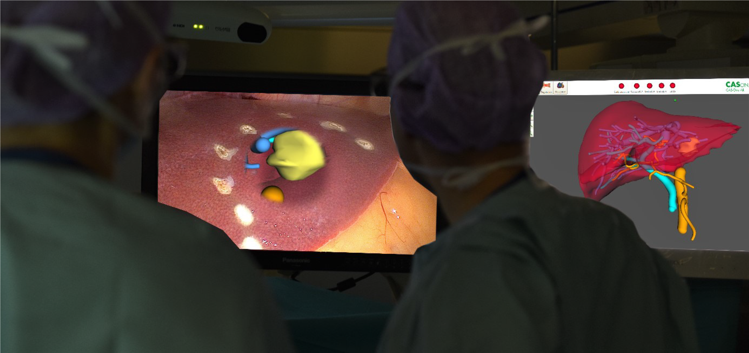
Figure 1 The use of augmented reality during laparoscopic liver resection using a 3D passive polarizing display technique.
The complete virtual three-dimensional model of the liver is visible on a second screen (right picture). On the main screen augmented reality is created (left picture). Citation: Prevost GA, Eigl B, Paolucci I, Rudolph T, Peterhans M, Weber S, Beldi G, Candinas D, Lachenmayer A, Efficiency, Accuracy and Clinical Applicability of a New Image-Guided Surgery System in 3D Laparoscopic Liver Surgery. J Gastrointest Surg 2020; 24(10): 2251-2258, Copyright © The Author(s) 2020, Published by Springer Nature[26].
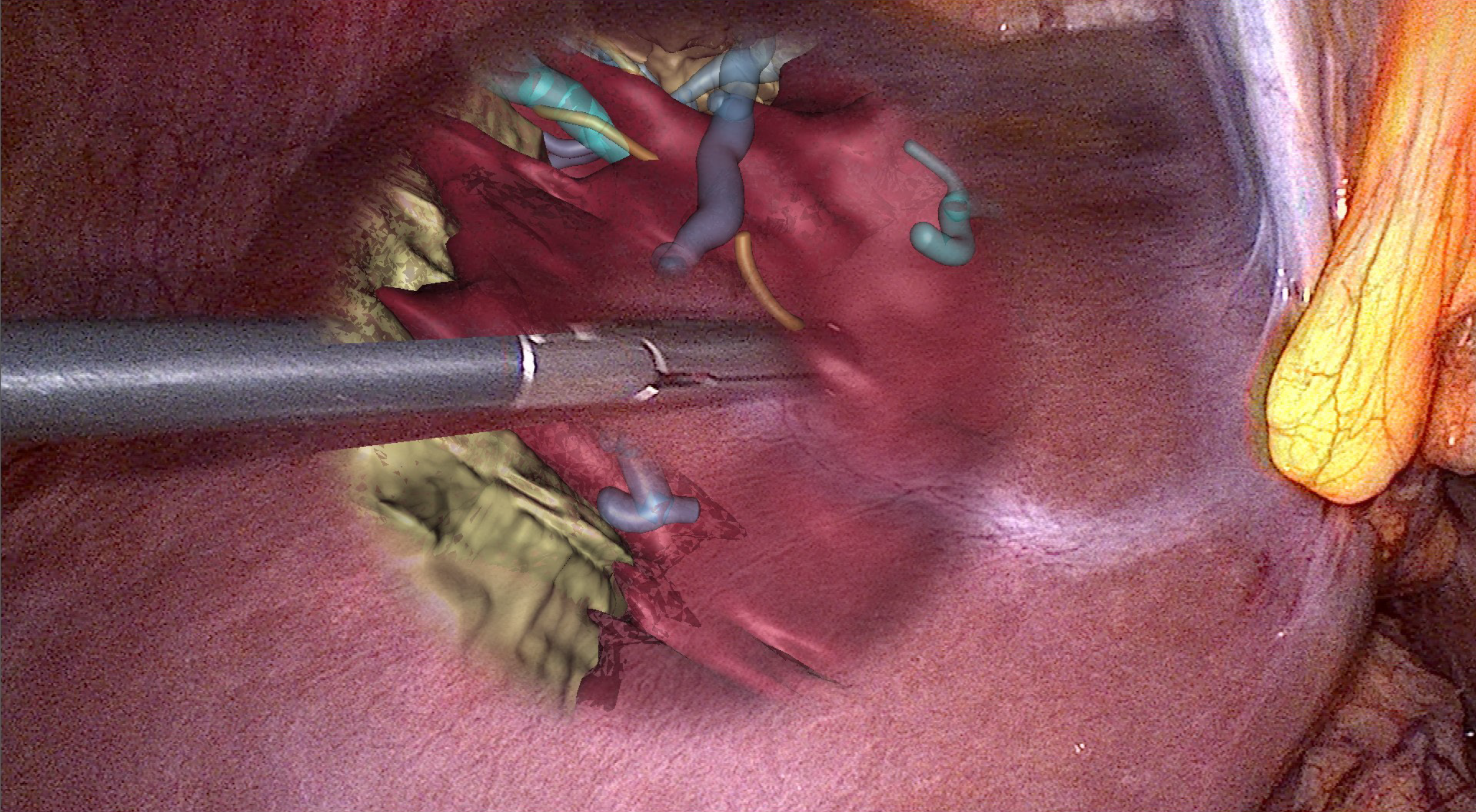
Figure 2 Directly on the laparoscopic three-dimensional image of the liver there is only that part of the virtual three-dimensional model superimposed on an area, which is relevant for the parenchyma dissection during that phase of the operation.
At the area, where the lesion is located and the parenchyma dissection will be performed, the virtual three-dimensional model is matched around the tracked/navigated dissection tool. Citation: Prevost GA, Eigl B, Paolucci I, Rudolph T, Peterhans M, Weber S, Beldi G, Candinas D, Lachenmayer A, Efficiency, Accuracy and Clinical Applicability of a New Image-Guided Surgery System in 3D Laparoscopic Liver Surgery. J Gastrointest Surg 2020, 24(10), 2251-2258, Copyright © The Author(s) 2020, Published by Springer Nature[26].
- Citation: Wahba R, Thomas MN, Bunck AC, Bruns CJ, Stippel DL. Clinical use of augmented reality, mixed reality, three-dimensional-navigation and artificial intelligence in liver surgery. Artif Intell Gastroenterol 2021; 2(4): 94-104
- URL: https://www.wjgnet.com/2644-3236/full/v2/i4/94.htm
- DOI: https://dx.doi.org/10.35712/aig.v2.i4.94


