©The Author(s) 2021.
World J Meta-Anal. Apr 28, 2021; 9(2): 176-192
Published online Apr 28, 2021. doi: 10.13105/wjma.v9.i2.176
Published online Apr 28, 2021. doi: 10.13105/wjma.v9.i2.176
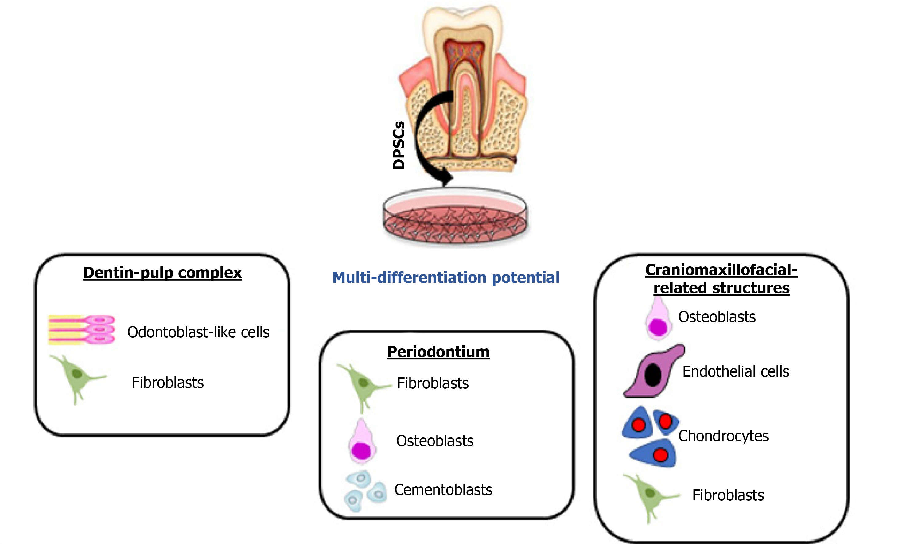
Figure 1 Different types of regeneration possible from dental pulp stem cells.
Schematic diagram showing multi-differentiation capability of dental pulp stem cells (DPSCs) for regenerating dentin-pulp complex, periodontal tissues, stromal tissues of salivary glands and craniomaxillofacial bone defects.
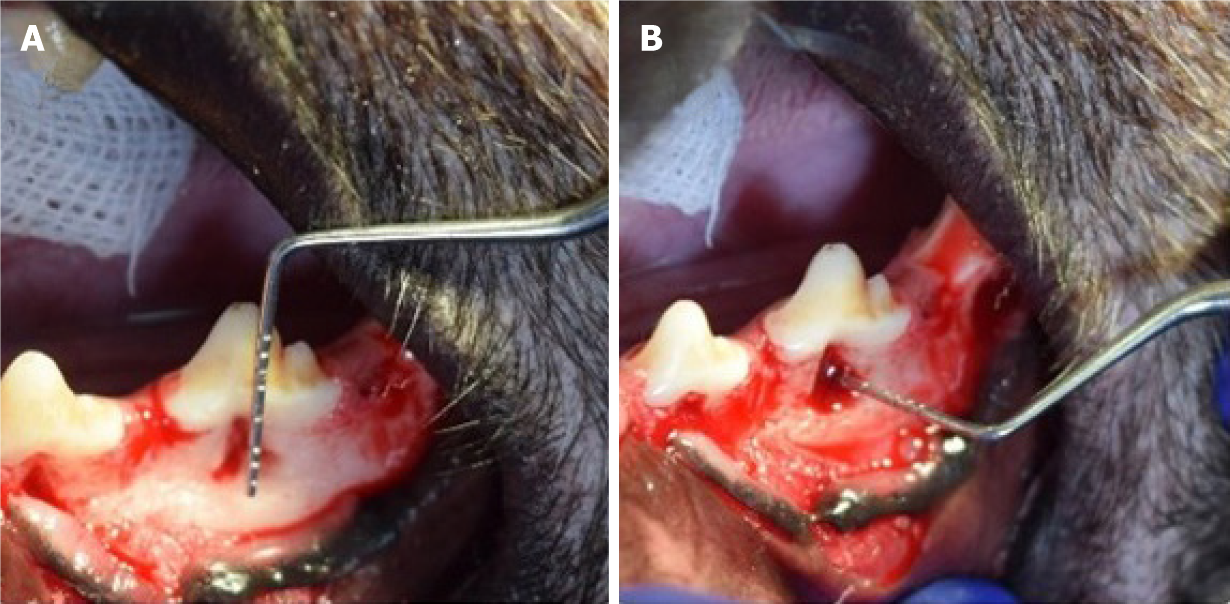
Figure 2 Periodontal bone defect.
A and B: Photographs showing the periodontal defect of vertical height and horizontal width and depth of a dog’s interradicular bone of the first molar. Courtesy provided by the staff members of Oral Biology, Faculty of Dentistry, Mansoura University, Mansoura, Egypt.
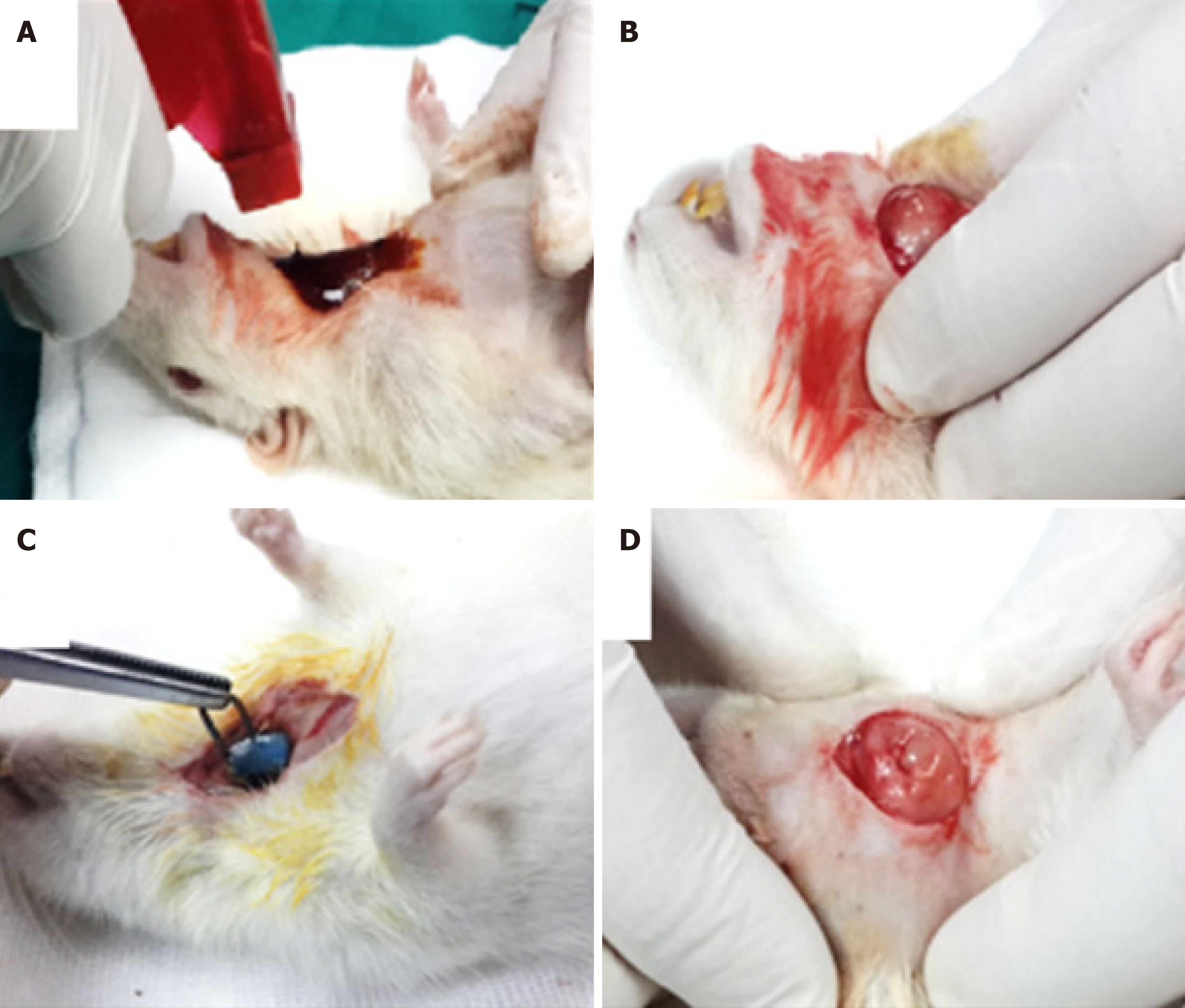
Figure 3 Punch biopsy of submandibular gland.
A: Photographs showing a tissue punch of submandibular (SMG) as a model of mechanical injury through disinfecting the skin of operation site by betadine; B: Exposing the right SMG; C: Applying the tissue punch on the center of right SMG; D: The arrow points to a rectangular cut area of 5 mm × 8 mm × 1.5 mm dimensions. Courtesy provided by the staff members of Oral Biology, Faculty of Dentistry, Mansoura University, Mansoura, Egypt.
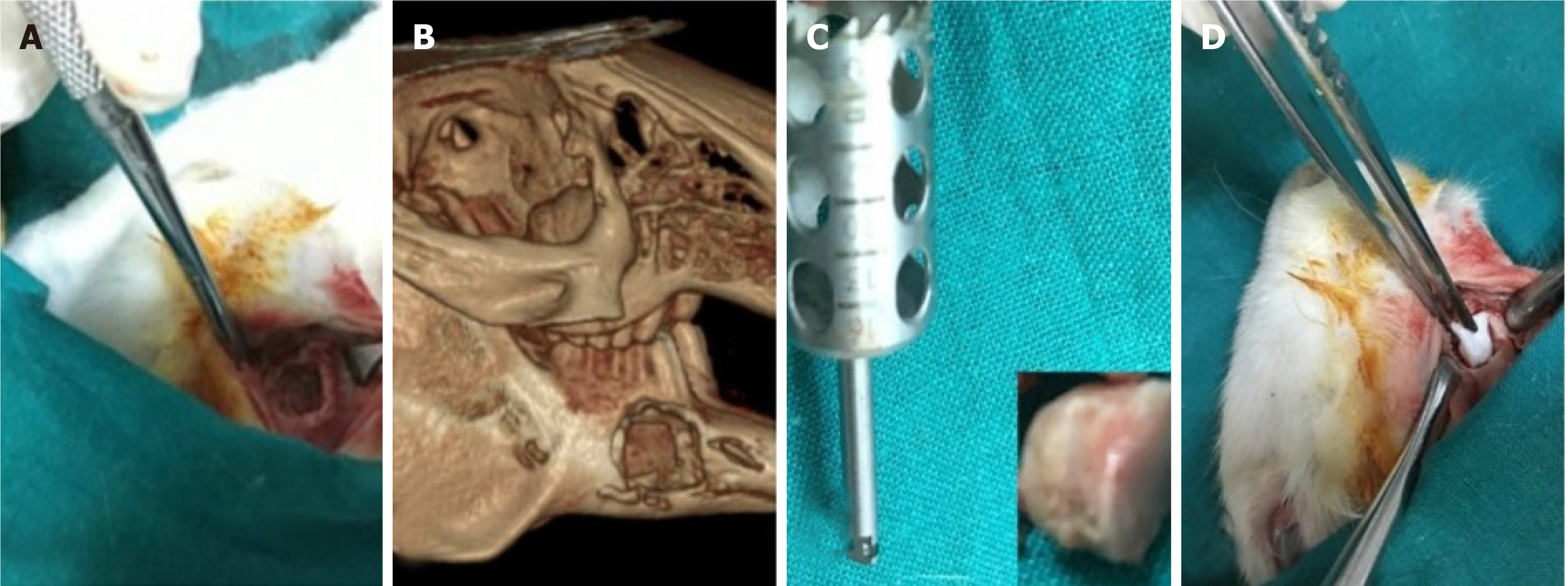
Figure 4 Mandibular bone defect.
A: Photograph showing a 10-mm diameter, full-thickness mandibular defect with the removal of the mandibular first premolar and both cortical plates; B: Cone-beam computed tomography systems reconstruction of the critical-size bone defect; C: The 10-mm trephine bur with autogenous tibial bone graft; D: Gelfoam placed on the defect. Courtesy provided by the staff members of Oral Biology, Faculty of Dentistry, Mansoura University, Mansoura, Egypt.
- Citation: Grawish ME, Saeed MA, Sultan N, Scheven BA. Therapeutic applications of dental pulp stem cells in regenerating dental, periodontal and oral-related structures. World J Meta-Anal 2021; 9(2): 176-192
- URL: https://www.wjgnet.com/2308-3840/full/v9/i2/176.htm
- DOI: https://dx.doi.org/10.13105/wjma.v9.i2.176


