Published online Dec 20, 2025. doi: 10.5662/wjm.v15.i4.106708
Revised: May 4, 2025
Accepted: May 29, 2025
Published online: December 20, 2025
Processing time: 152 Days and 21.3 Hours
We report a unique case of bilateral femoral stem fractures in a patient with Dorr A femoral morphology, underscoring the need for a critical reassessment of implant selection strategies. The initial failure involved a cemented revision stem placed using the cement-within-cement technique combined with an extended trochanteric osteotomy (ETO). A second revision was subsequently performed using a cortical window osteotomy and a distally fixed uncemented stem, which resulted in successful recovery. A similar approach was used to treat a subsequent contralateral stem fracture, also with favorable outcomes. This case emphasizes three key considerations: First, that standard-length cemented stems may lead to oversizing and increased stress concentration in Dorr A femurs with narrow canals; second, that ETO may compromise femoral integrity and contribute to implant failure; and third, that cortical window osteotomy enables safer implant removal and reimplantation. Based on these findings, we advocate for an indi
Core Tip: This case highlights bilateral femoral stem fractures in a patient with Dorr A femoral morphology, raising concerns about the mechanical reliability of cemented fixation in such cases. Through successful management with cortical window osteotomy and uncemented distal fixation, we emphasize the importance of tailoring revision strategies to femoral anatomy. Our experience reinforces the need to reconsider implant selection in Dorr A femurs to prevent recurrent failure.
- Citation: Lucero CM, Luco JB, Albani Forneris A, Buttaro MA. Recurrent femoral stem fractures in Dorr A femurs: Lessons learned and a call for alternative strategies. World J Methodol 2025; 15(4): 106708
- URL: https://www.wjgnet.com/2222-0682/full/v15/i4/106708.htm
- DOI: https://dx.doi.org/10.5662/wjm.v15.i4.106708
Following our previously published case report on femoral stem refracture in a Dorr A femur[1], we find ourselves revisiting our own conclusions in light of an unexpected recurrence in the same patient. This reinforces key considerations regarding implant selection and revision strategies in this particular femoral morphology.
In our original publication, we described a refracture of a cemented femoral stem in a Dorr A femur after revision surgery involving extended trochanteric osteotomy (ETO) and a cement-within-cement (CWC) technique[1]. The failure of the revised stem led us to perform a second revision using a cortical window osteotomy, allowing safe extraction of the distal stem fragment and cement, followed by implantation of a distally fixed uncemented stem. The patient had an uneventful recovery, reinforcing the efficacy of this approach[1,2].
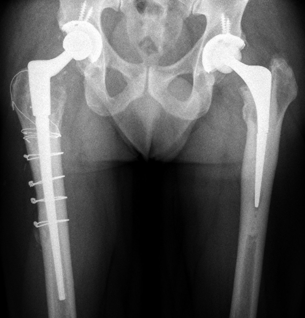
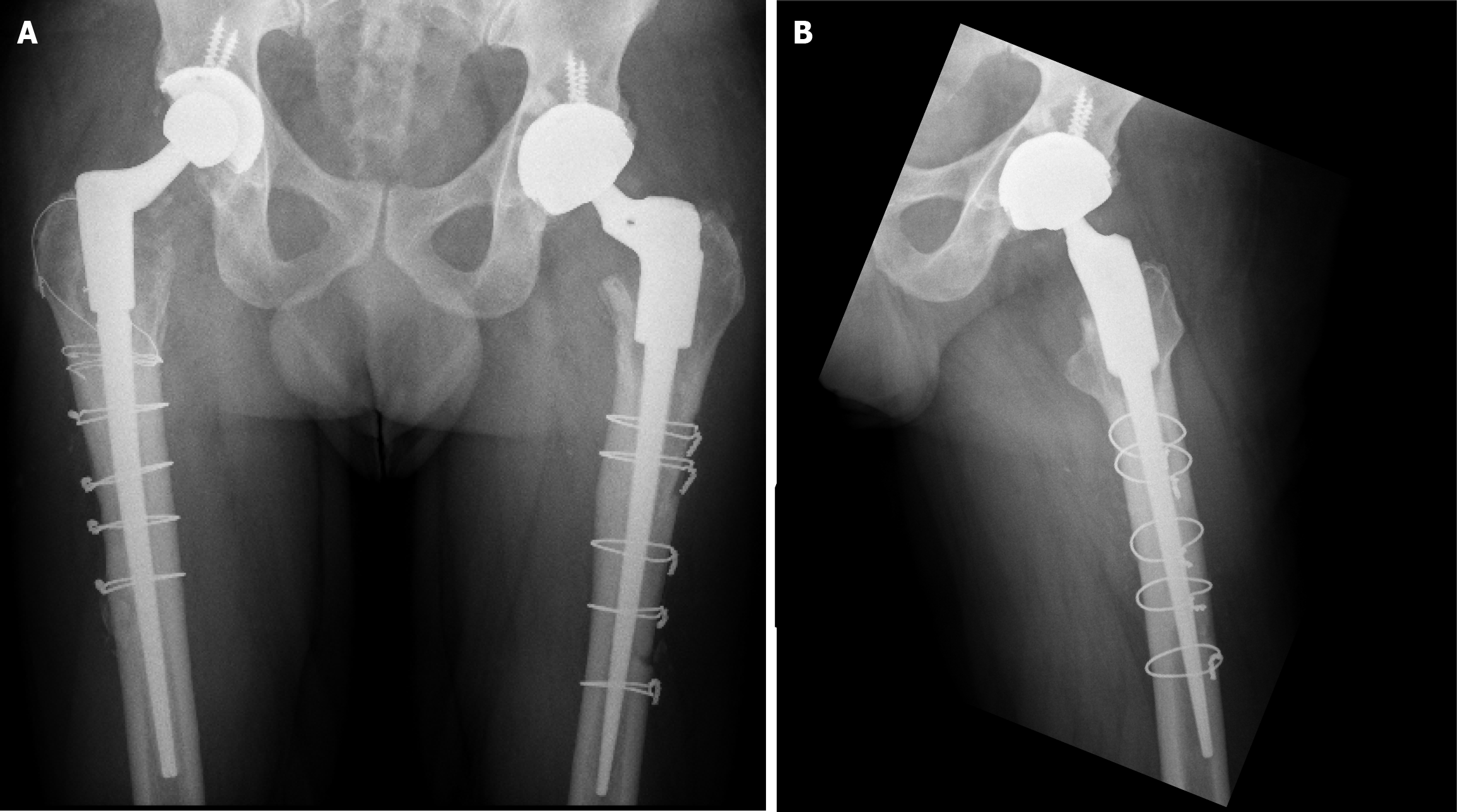
Recently, however, the same patient presented with a femoral stem fracture in the contralateral hip, further challenging the reliability of cemented stems in Dorr A femurs (Figure 1) With the knowledge gained from our prior experience, we opted for the same approach: A cortical window osteotomy for safe removal of the fractured stem and cement, followed by the implantation of a distally fixed uncemented stem. This method once again allowed direct visualization, ensured a controlled cement removal process, and minimized damage to the femoral cortex (Figure 2)[3]. The patient’s posto
Cemented stems in Dorr A femurs require careful consideration: The high cortical thickness and narrow diaphyseal canal characteristic of Dorr A femurs may lead to mechanical mismatch with standard-length cemented stems, increasing the risk of cement mantle thinning and stress concentration at the stem-body junction[1-4]. However, this does not preclude cemented fixation altogether. When appropriately sized, such as with short or specially designed cemented stems, successful outcomes have been reported even in narrow canals. The key lies in tailoring the implant choice to the patient’s specific femoral morphology.
ETO may increase mechanical risk in Dorr A femurs: Although widely used in revision procedures, ETO can compromise the femoral structure—particularly in patients with a dense cortex and limited canal diameter—potentially predisposing to refracture[5]. Our case reinforces the need for alternative extraction techniques in such femoral types.
Cortical window osteotomy is a viable revision strategy: This technique allowed for direct visualization and safe extraction of the fractured stem and cement while minimizing additional cortical damage. It also facilitated accurate implant placement and may be preferred over ETO in select patients, especially those with stiff, narrow femurs[2,3].
Our experience suggests that fixation strategies in Dorr A femurs should not adopt a one-size-fits-all approach. Rather than categorically avoiding cemented stems, surgeons should carefully evaluate canal geometry, patient age, bone quality, and mechanical demands. Cemented short stems or stems specifically designed for narrow femoral canals may be appropriate in older or low-demand patients. Conversely, distally fixed uncemented stems or short cementless stems may offer better outcomes in younger or more active individuals, provided they achieve stable metaphyseal fixation. This case underscores the importance of individualized implant selection and highlights the need for further research into fixation principles specifically tailored to the unique characteristics of Dorr A femoral anatomy.
| 1. | Lucero CM, Luco JB, Garcia-Mansilla A, Slullitel PA, Zanotti G, Comba F, Buttaro MA. Successful hip revision surgery following refracture of a modern femoral stem using a cortical window osteotomy technique: A case report and review of literature. World J Methodol. 2023;13:502-509. [RCA] [PubMed] [DOI] [Full Text] [Full Text (PDF)] [Cited by in RCA: 1] [Reference Citation Analysis (3)] |
| 2. | Singh A, Mukherjee S, Patel K, Herlekar D, Gandavaram S, Charalambous N. Medium term outcome of Lancaster cortical window technique for extraction of femoral stem in revision hip arthroplasty. J Orthop Surg Res. 2021;16:314. [RCA] [PubMed] [DOI] [Full Text] [Full Text (PDF)] [Cited by in Crossref: 1] [Cited by in RCA: 3] [Article Influence: 0.6] [Reference Citation Analysis (0)] |
| 3. | Park CH, Yeom J, Park JW, Won SH, Lee YK, Koo KH. Anterior Cortical Window Technique Instead of Extended Trochanteric Osteotomy in Revision Total Hip Arthroplasty: A Minimum 10-Year Follow-up. Clin Orthop Surg. 2019;11:396-402. [RCA] [PubMed] [DOI] [Full Text] [Full Text (PDF)] [Cited by in Crossref: 10] [Cited by in RCA: 11] [Article Influence: 1.6] [Reference Citation Analysis (0)] |
| 4. | Wilson LJ, Richards CJ, Irvine D, Tillie A, Crawford RW. Risk of periprosthetic femur fracture after anterior cortical bone windowing: a mechanical analysis of short versus long cemented stems in pigs. Acta Orthop. 2011;82:674-678. [RCA] [PubMed] [DOI] [Full Text] [Full Text (PDF)] [Cited by in Crossref: 1] [Cited by in RCA: 3] [Article Influence: 0.2] [Reference Citation Analysis (0)] |
| 5. | Malahias MA, Gkiatas I, Selemon NA, De Filippis R, Gu A, Greenberg A, Sculco PK. Outcomes and Risk Factors of Extended Trochanteric Osteotomy in Aseptic Revision Total Hip Arthroplasty: A Systematic Review. J Arthroplasty. 2020;35:3410-3416. [RCA] [PubMed] [DOI] [Full Text] [Cited by in Crossref: 10] [Cited by in RCA: 20] [Article Influence: 3.3] [Reference Citation Analysis (0)] |


