©The Author(s) 2016.
World J Methodol. Mar 26, 2016; 6(1): 20-24
Published online Mar 26, 2016. doi: 10.5662/wjm.v6.i1.20
Published online Mar 26, 2016. doi: 10.5662/wjm.v6.i1.20
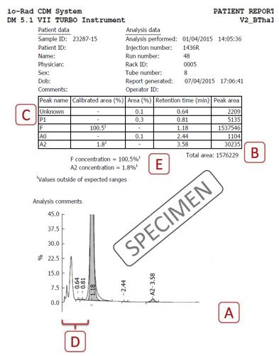
Figure 1 Specimen cation-exchange high-performance liquid chromatography output from the Bio-Rad Variant II Turbo instrument (Bio-Rad Laboratories, Hercules, United States) using the β-Thal short programme.
Label A indicates the total time of analysis (X-axis) is approximately 5 to 6 min; Label B indicates that the total area of analysis should lie between 1 and 3 million; Label C shows the unknown peaks that may occur, especially in the P2 and P3 regions; Label D depicts the preintegration phase (< 1 min) is not reflected in the table above and should be analyzed on the chromatogram; Label E shows where the problem of HbF concentration being calculated as > 100% is present in this case. Please see text for details of resolution of this problem.
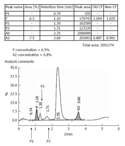
Figure 2 A specimen chromatogram to show the various peaks that may occur in health and disease.
The patient, with elevated HbA2 and mildly elevated HbF, most likely had β-thalassemia trait with increased HbF (HbF levels between 1%-5% are found in approximately 30% of β-thalassemia trait cases, and occasional ones may have even higher levels).Calibrator data had shown above.
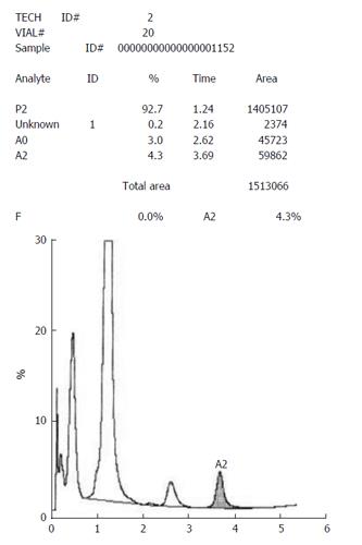
Figure 3 A case of thalassemia major with a very prominent and broad-based HbF being reported by the instrument (an older Bio-Rad Variant) as a P2-peak.
- Citation: Sharma P, Das R. Cation-exchange high-performance liquid chromatography for variant hemoglobins and HbF/A2: What must hematopathologists know about methodology? World J Methodol 2016; 6(1): 20-24
- URL: https://www.wjgnet.com/2222-0682/full/v6/i1/20.htm
- DOI: https://dx.doi.org/10.5662/wjm.v6.i1.20


