Copyright
©The Author(s) 2025.
World J Methodol. Dec 20, 2025; 15(4): 106216
Published online Dec 20, 2025. doi: 10.5662/wjm.v15.i4.106216
Published online Dec 20, 2025. doi: 10.5662/wjm.v15.i4.106216
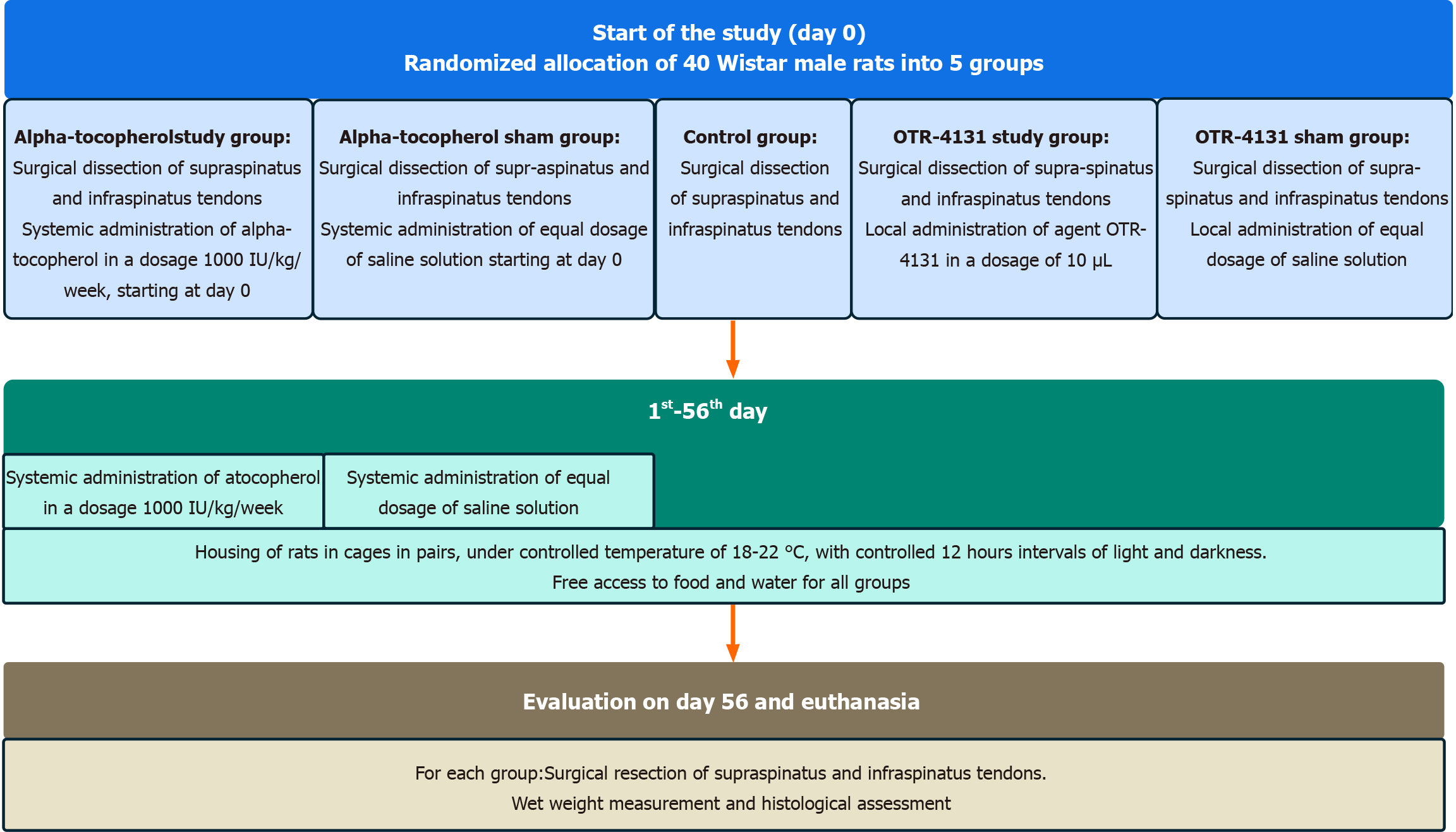
Figure 1 Study process flowchart.
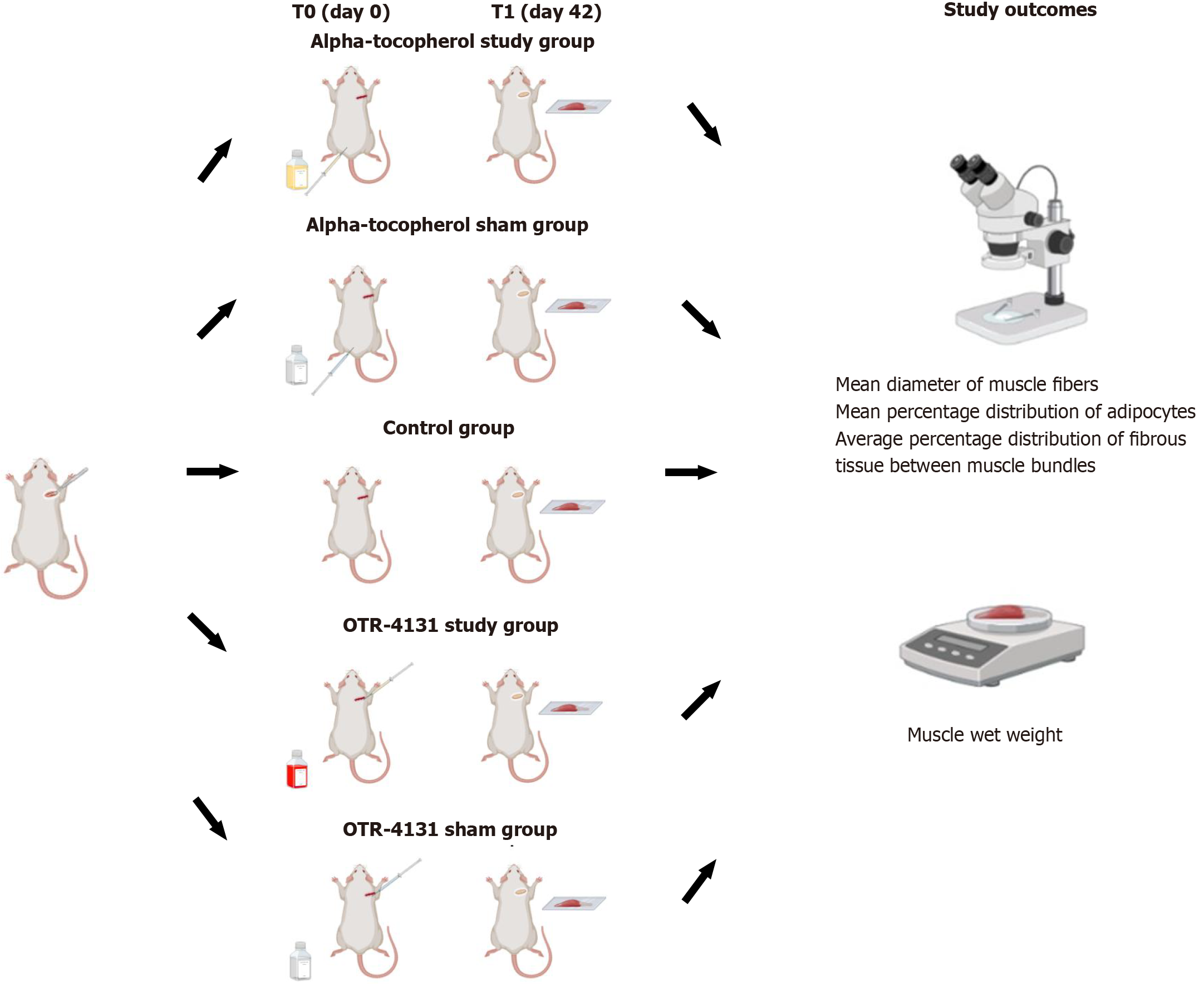
Figure 2 An overview of the experimental design.
This figure was created with BioRender.com.
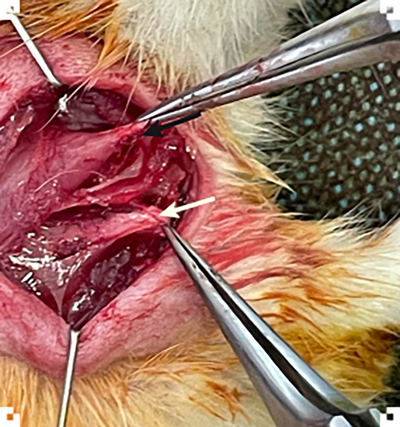
Figure 3 Image from the pilot surgery showing the identification of the supraspinatus (black arrow) and infraspinatus tendons (white arrow).
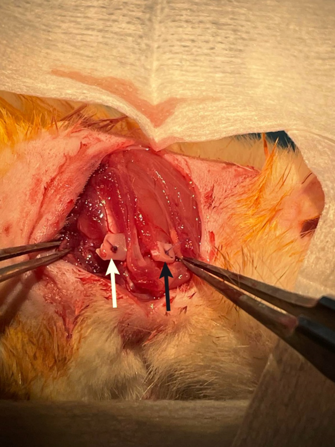
Figure 4 Image from the pilot surgery during the second procedure, illustrating the complete visualization of supraspinatus (black arrow) and infraspinatus (white arrow) muscles prior to harvesting.
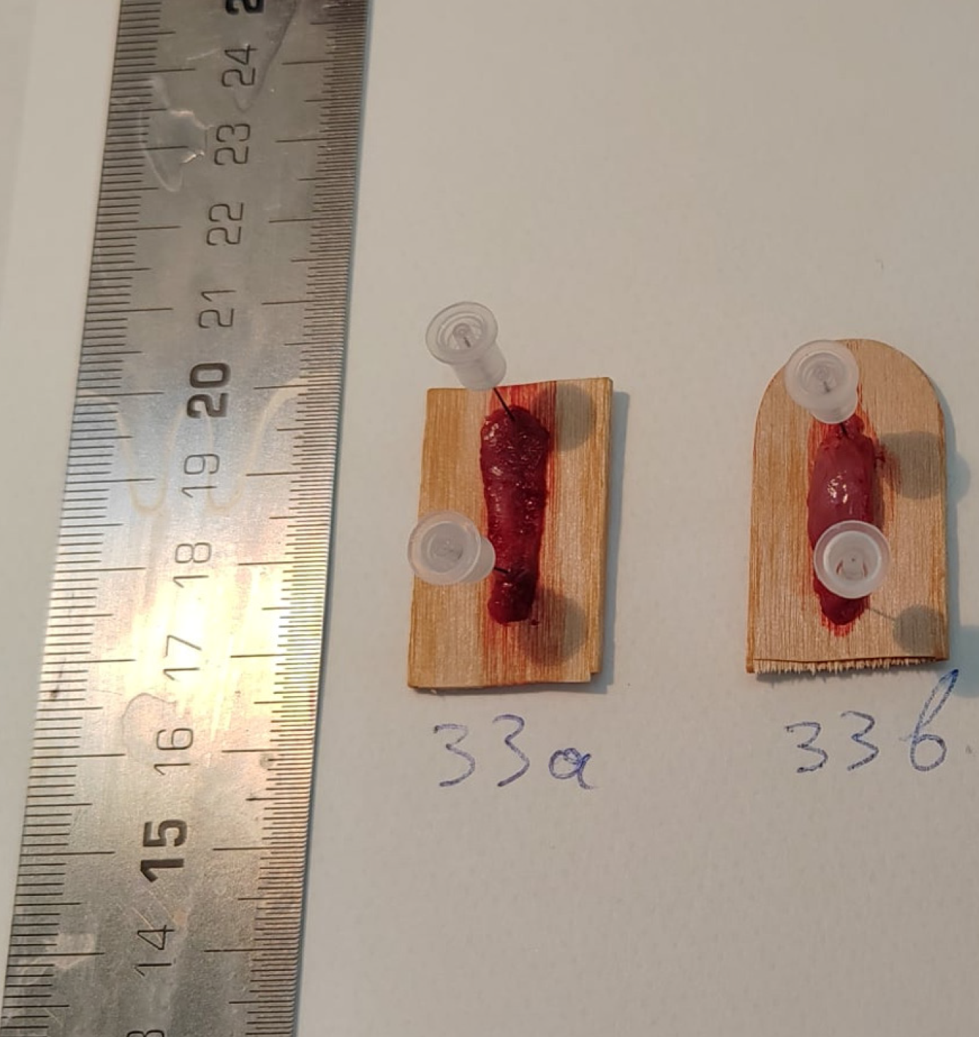
Figure 5 Specimens of infraspinatus (left) and supraspinatus (right) muscle units obtained during the pilot surgery.
- Citation: Stamiris S, Cheva A, Potoupnis M, Anestiadou E, Stamiris D, Bekiari C, Loukousia A, Kyriakos P, Tsiridis E, Sarris I. Effect of alpha-tocopherol and OTR-4131 on muscle degeneration after rotator cuff tear in rats: An experimental protocol. World J Methodol 2025; 15(4): 106216
- URL: https://www.wjgnet.com/2222-0682/full/v15/i4/106216.htm
- DOI: https://dx.doi.org/10.5662/wjm.v15.i4.106216


