Copyright
©The Author(s) 2025.
World J Methodol. Dec 20, 2025; 15(4): 104529
Published online Dec 20, 2025. doi: 10.5662/wjm.v15.i4.104529
Published online Dec 20, 2025. doi: 10.5662/wjm.v15.i4.104529
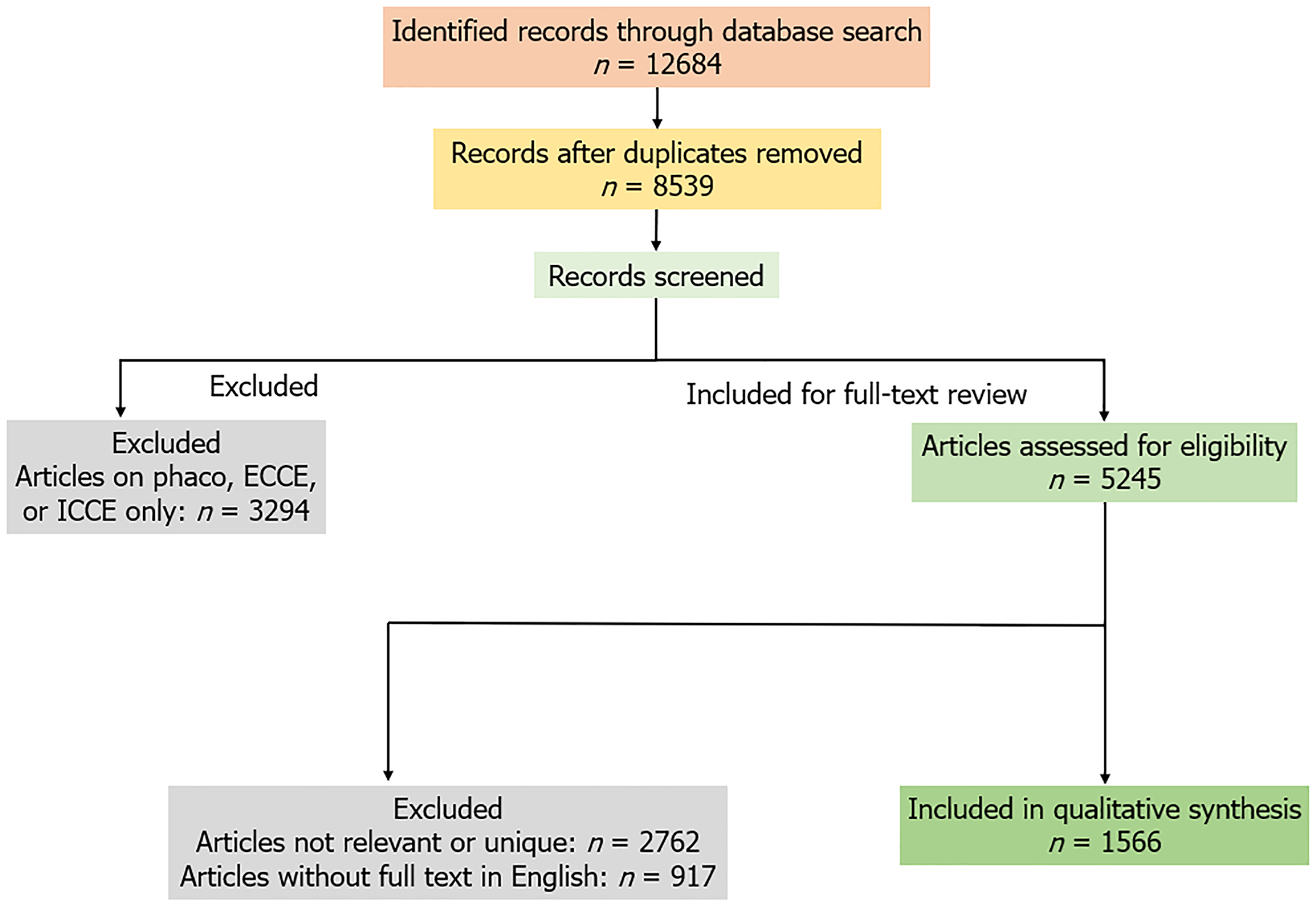
Figure 1 PRISMA of the methodology.
ECCE: Extracapsular cataract extraction; ICCE: Intracapsular cataract extraction.
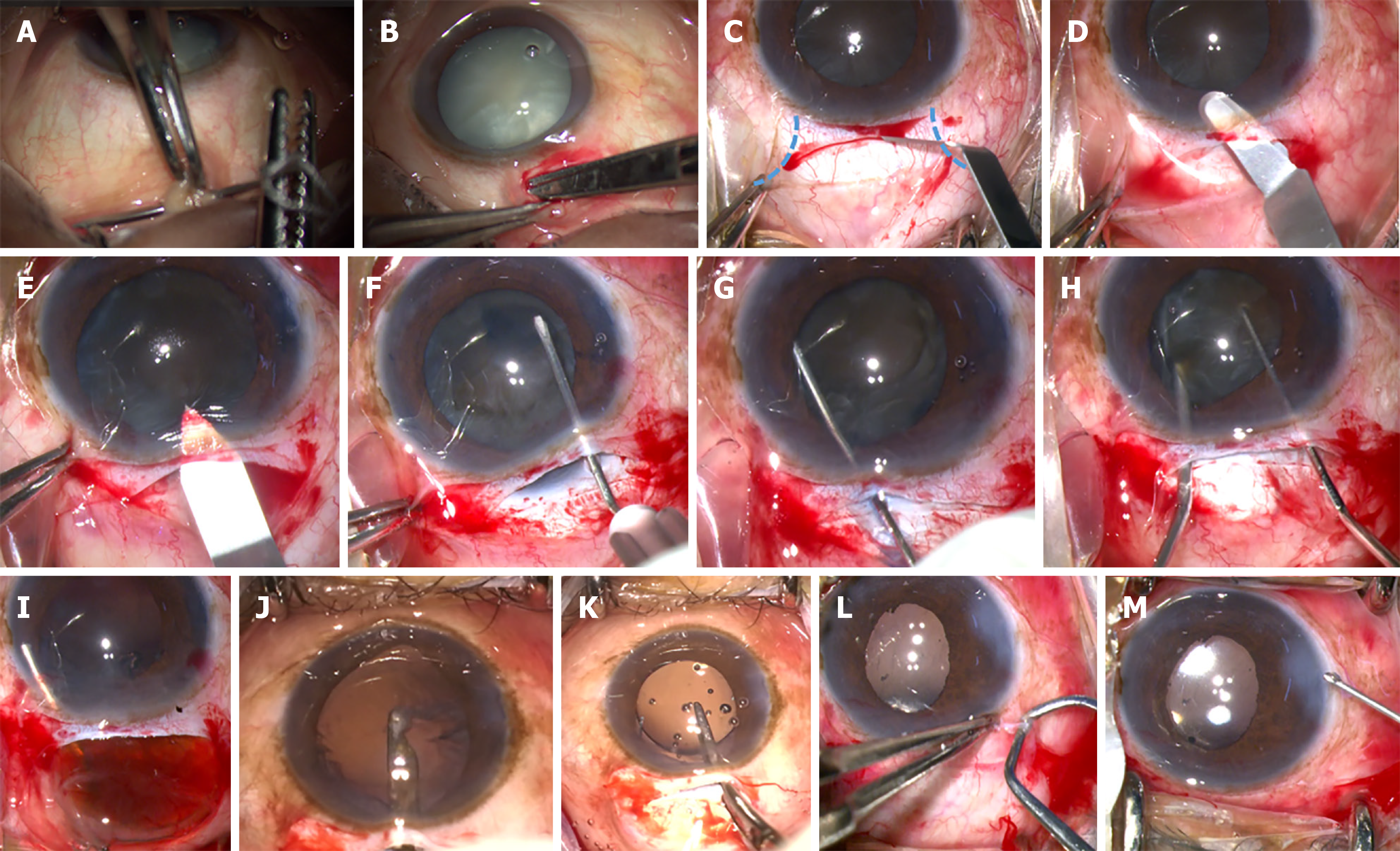
Figure 2 Steps of manual small incision cataract surgery.
A: Superior rectus bridle suture; B: Superior conjunctival peritomy; C: Scleral groove (dashed lines estimate the extent of Koch’s Funnel); D: Sclerocorneal tunnel dissection; E: Keratome entry; F: Continuous curvilinear capsulorhexis; G: Hydrodissection; H: Nuclear rotation and prolapse; I: Nuclear delivery by viscoexpression; J: Irrigation and aspiration by simcoe cannula; K: Intraocular lens implantation; L: Cautery to repose the conjunctiva; M: Sideport hydration.
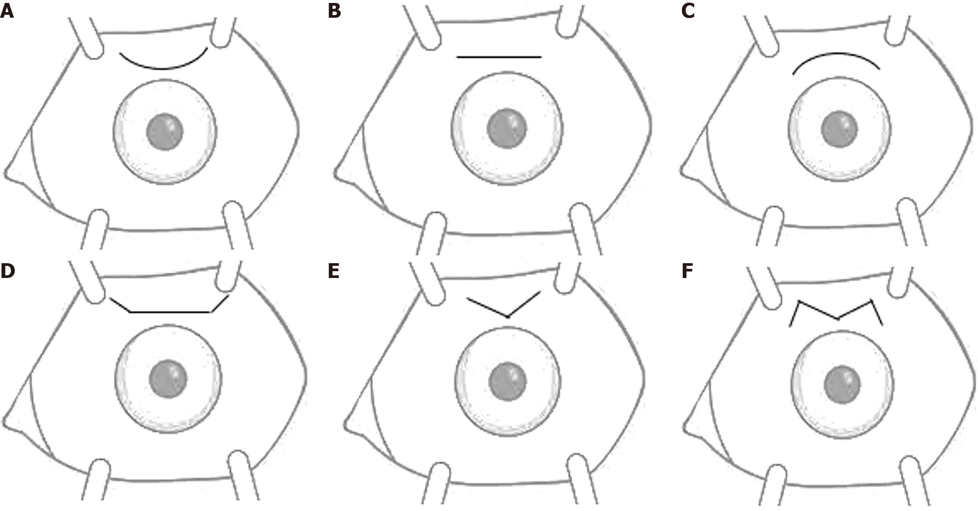
Figure 3 Various incision designs in manual small incision cataract surgery.
A: Frown; B: Straight; C: Smile; D: Straight with Blumenthal back cuts; E: Chevron; F: W-shaped or M-shaped incision for manual small incision cataract surgery with trabeculectomy.
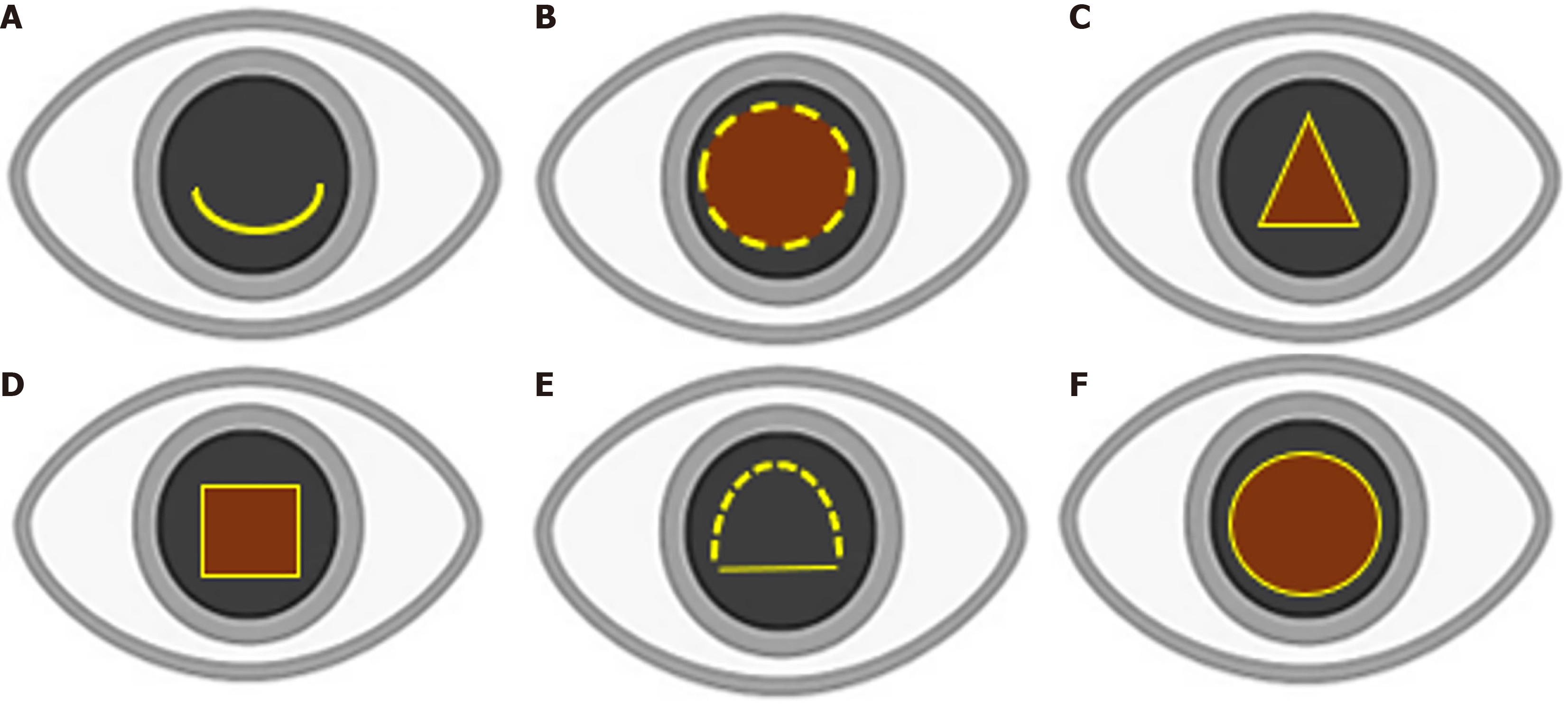
Figure 4 Various techniques and shapes of anterior capsulotomy in manual small incision cataract surgery.
A: Envelop, B: Can opener; C: Christmas tree; D: Square; E: D-shaped; F: Continuous curvilinear capsulorhexis (preferred).
- Citation: Nishant P, Singh A, Morya AK, Alam MA, Sinha S. Manual small incision cataract surgery: An ergonomic solution to tackle cataract backlog and challenging situations. World J Methodol 2025; 15(4): 104529
- URL: https://www.wjgnet.com/2222-0682/full/v15/i4/104529.htm
- DOI: https://dx.doi.org/10.5662/wjm.v15.i4.104529


