Copyright
©The Author(s) 2025.
World J Methodol. Dec 20, 2025; 15(4): 104497
Published online Dec 20, 2025. doi: 10.5662/wjm.v15.i4.104497
Published online Dec 20, 2025. doi: 10.5662/wjm.v15.i4.104497
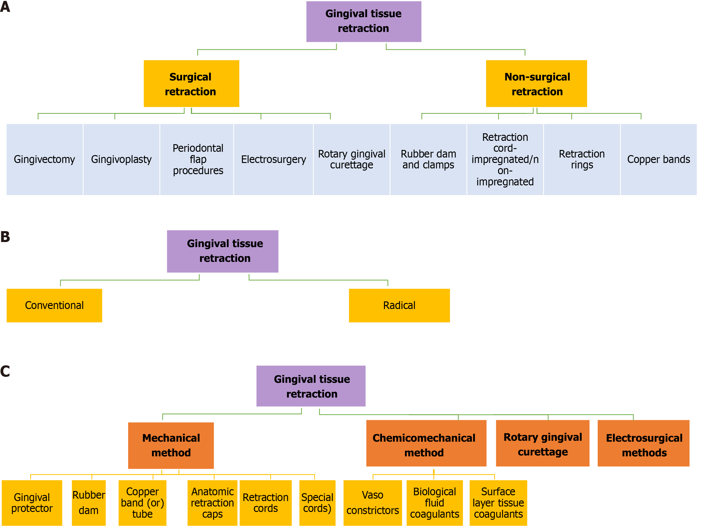
Figure 1 Classification of gingival tissue retraction.
A: The classification of surgical and non-surgical gingival tissue retraction; B: The classification of conventional and radical gingival tissue retraction; C: The classification of gingival tissue retraction methods.
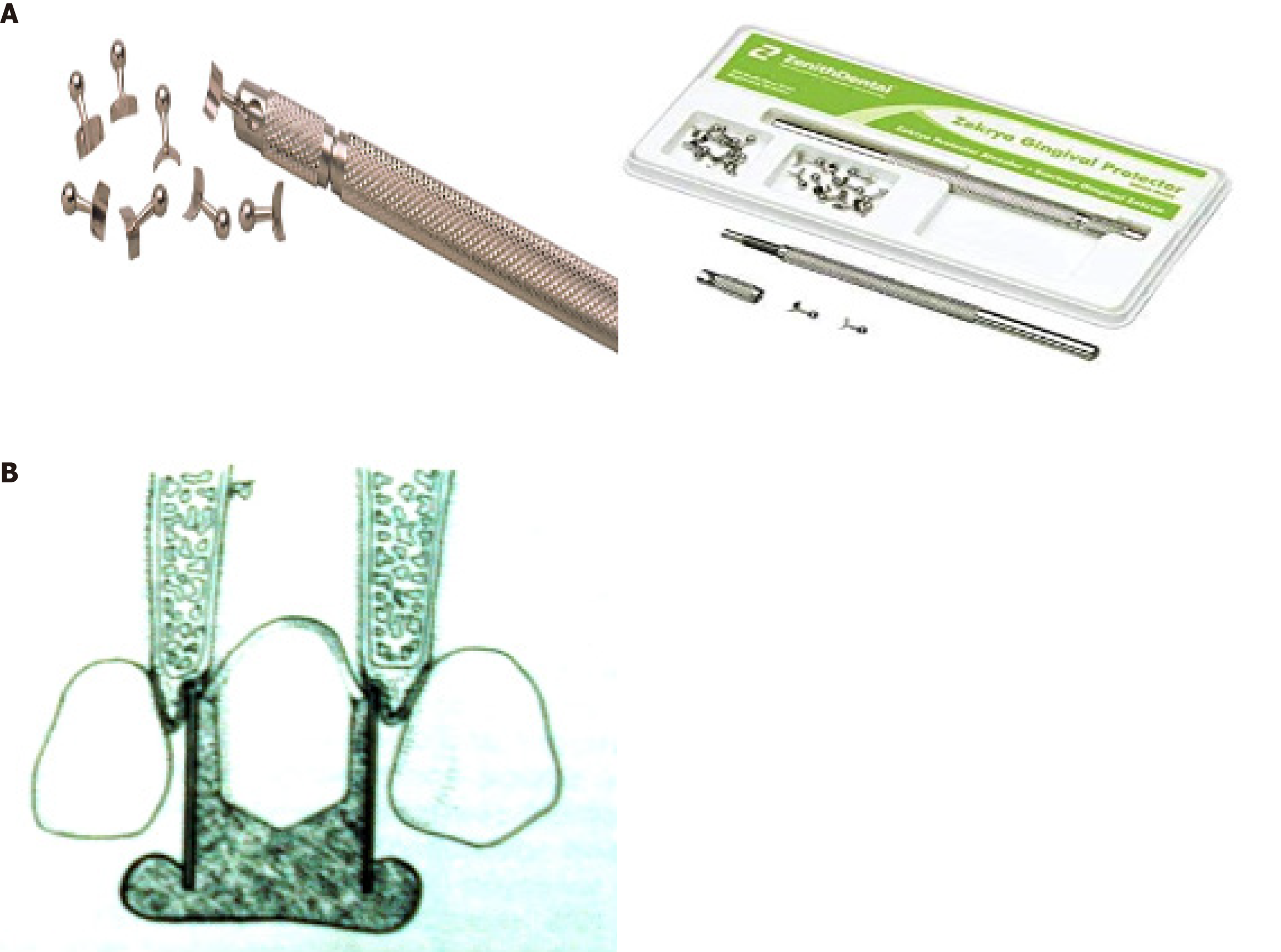
Figure 2 Gum protector and copper band.
A: The gingival protector; B: The copper band.
Figure 3 Rubber dam and anatomic retraction cap.
A: The role of the rubber dam in fixed bridge isolation; B: The anatomic retraction cap.
Figure 4
The types of retraction cord.
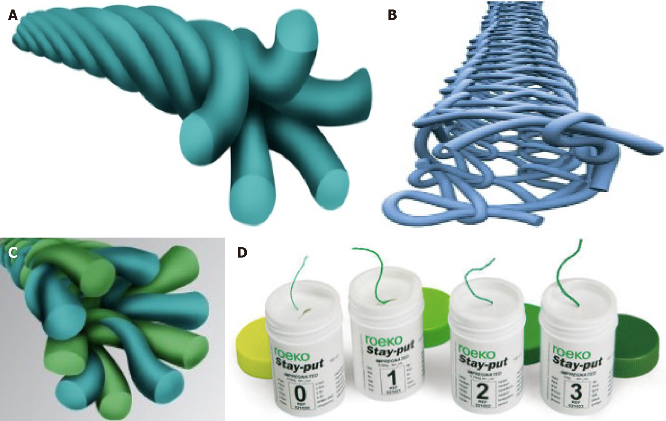
Figure 5 Characteristics of different retraction cords.
A: The braided cord; B: The knitted cord; C: The twisted cord; D: The stay-put retraction cord.
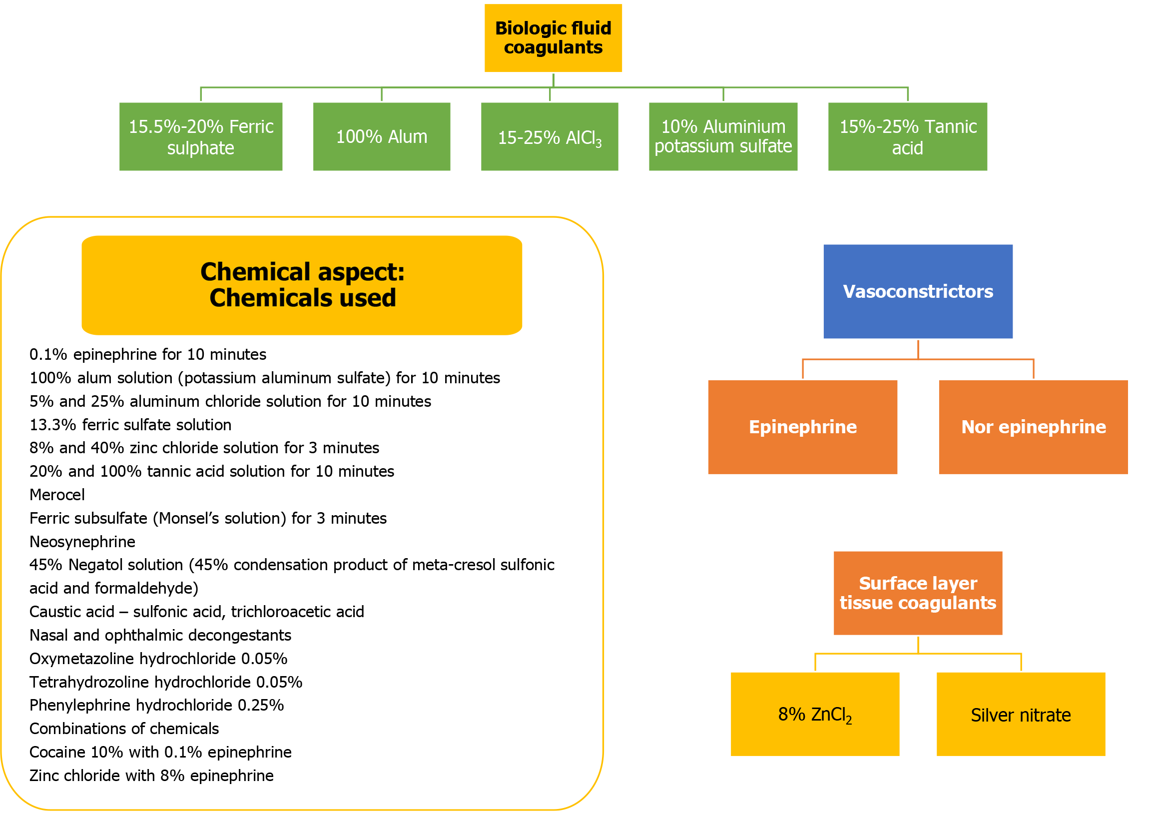
Figure 6
The chemicals used for tissue retraction.
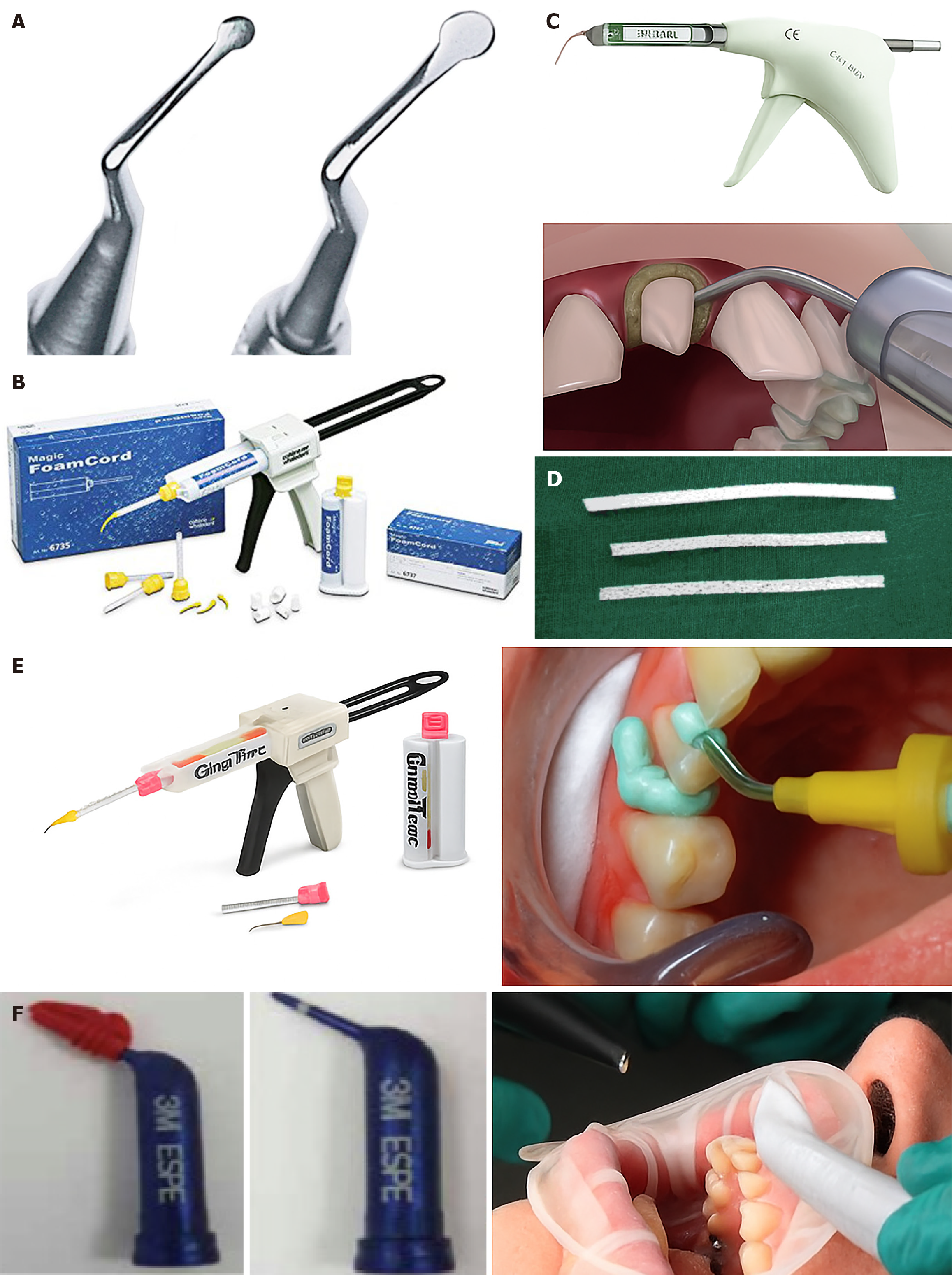
Figure 7 Instruments.
A: The cord-packing instruments; B: The magic foam cord; C: The Expasyl; D: The Merocel; E: The GingiTrac; F: The retraction capsule.
- Citation: Chauhan R, Chauhan S, Padiyar N, Kaurani P, Gupta A, Khan FN. Present status and future directions: Soft tissue management in prosthodontics. World J Methodol 2025; 15(4): 104497
- URL: https://www.wjgnet.com/2222-0682/full/v15/i4/104497.htm
- DOI: https://dx.doi.org/10.5662/wjm.v15.i4.104497


