©The Author(s) 2025.
World J Nephrol. Sep 25, 2025; 14(3): 109470
Published online Sep 25, 2025. doi: 10.5527/wjn.v14.i3.109470
Published online Sep 25, 2025. doi: 10.5527/wjn.v14.i3.109470
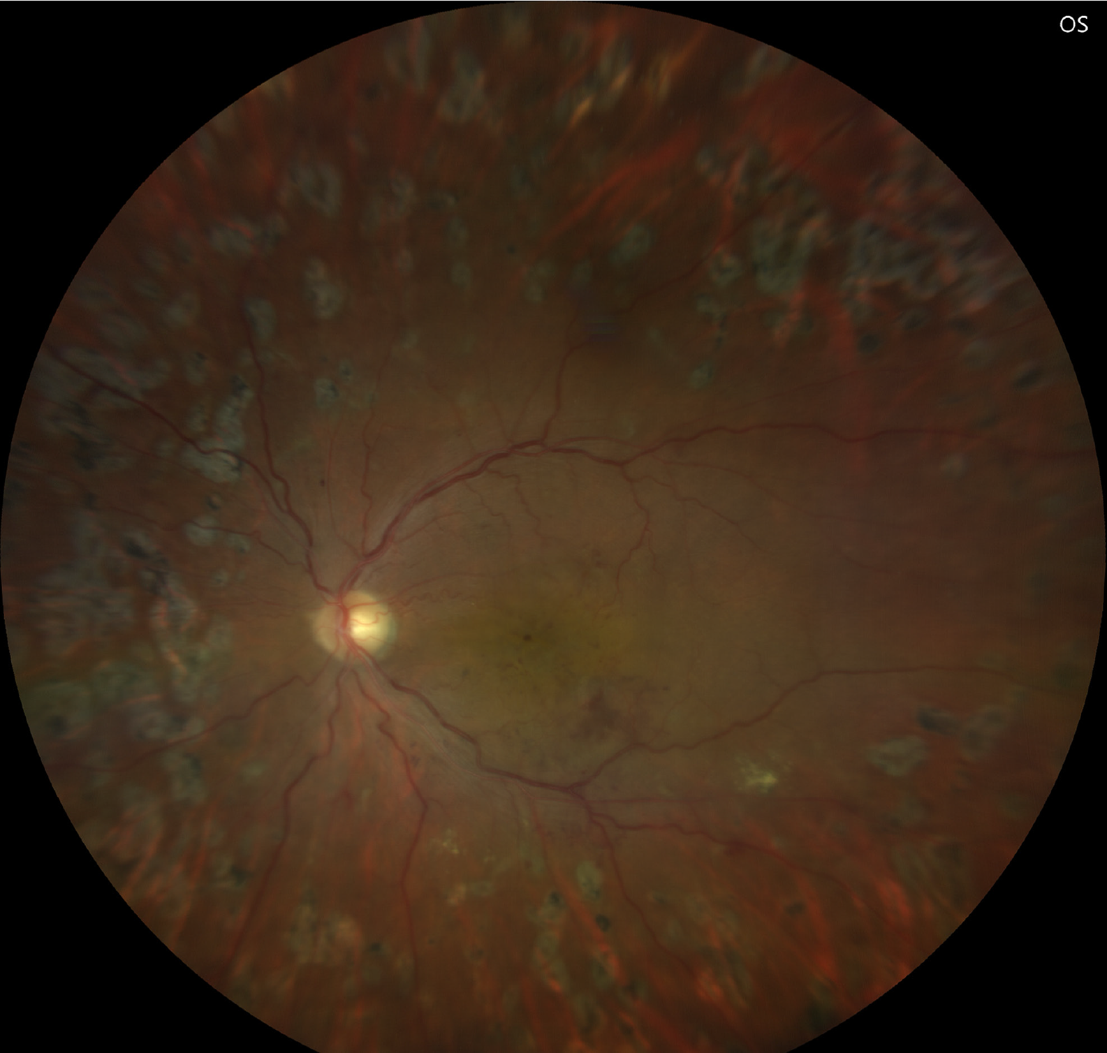
Figure 1
Fundus Image of a patient with laser-treated proliferative diabetic retinopathy with clinically significant macular edema and diabetic nephropathy.
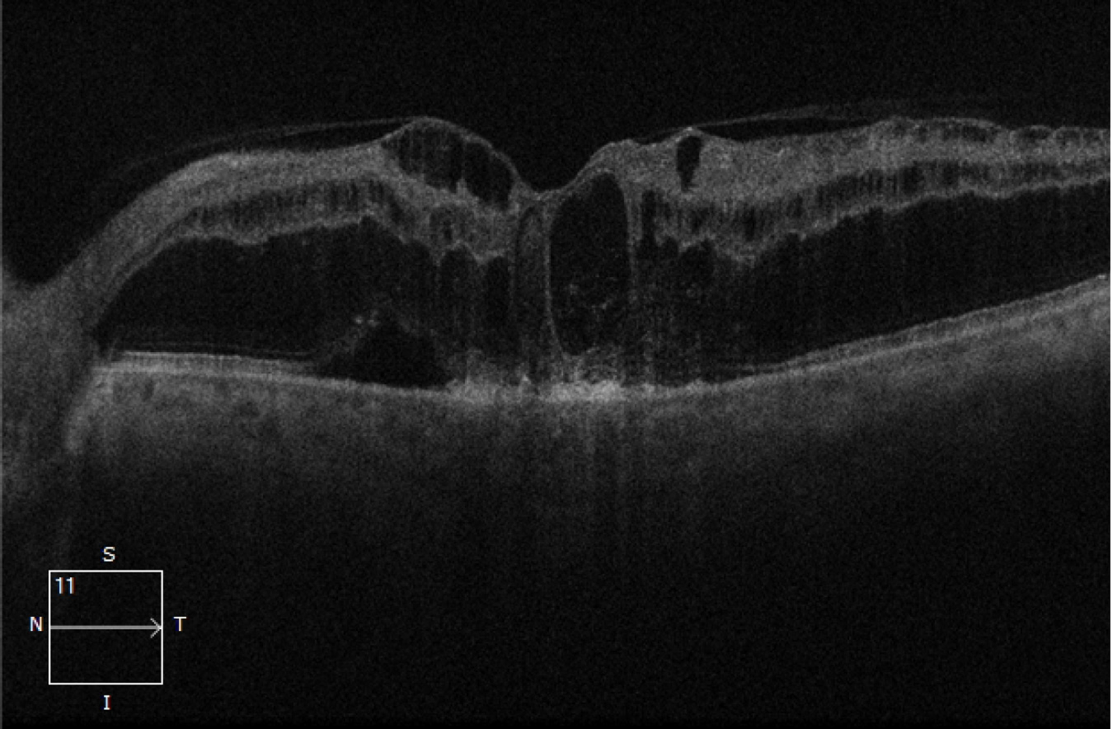
Figure 2
Optical coherence tomography image showing cystoid spaces with hyper-reflective dots and disorganization of outer and inner retinal layers.
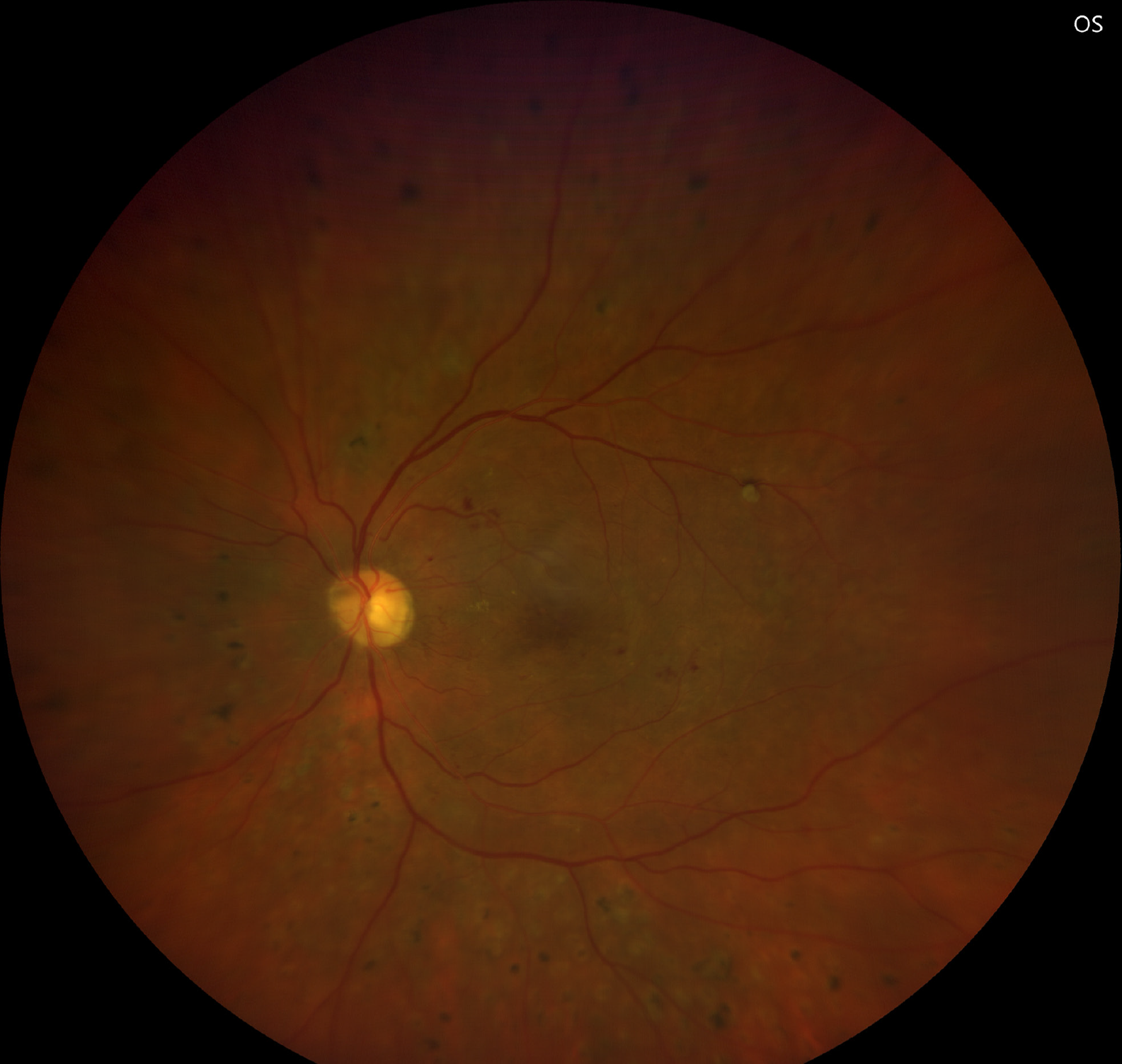
Figure 3 Left eye treated for proliferative diabetic retinopathy with dull foveal reflex in a patient with diabetic nephropathy.
OS: Outer segment.
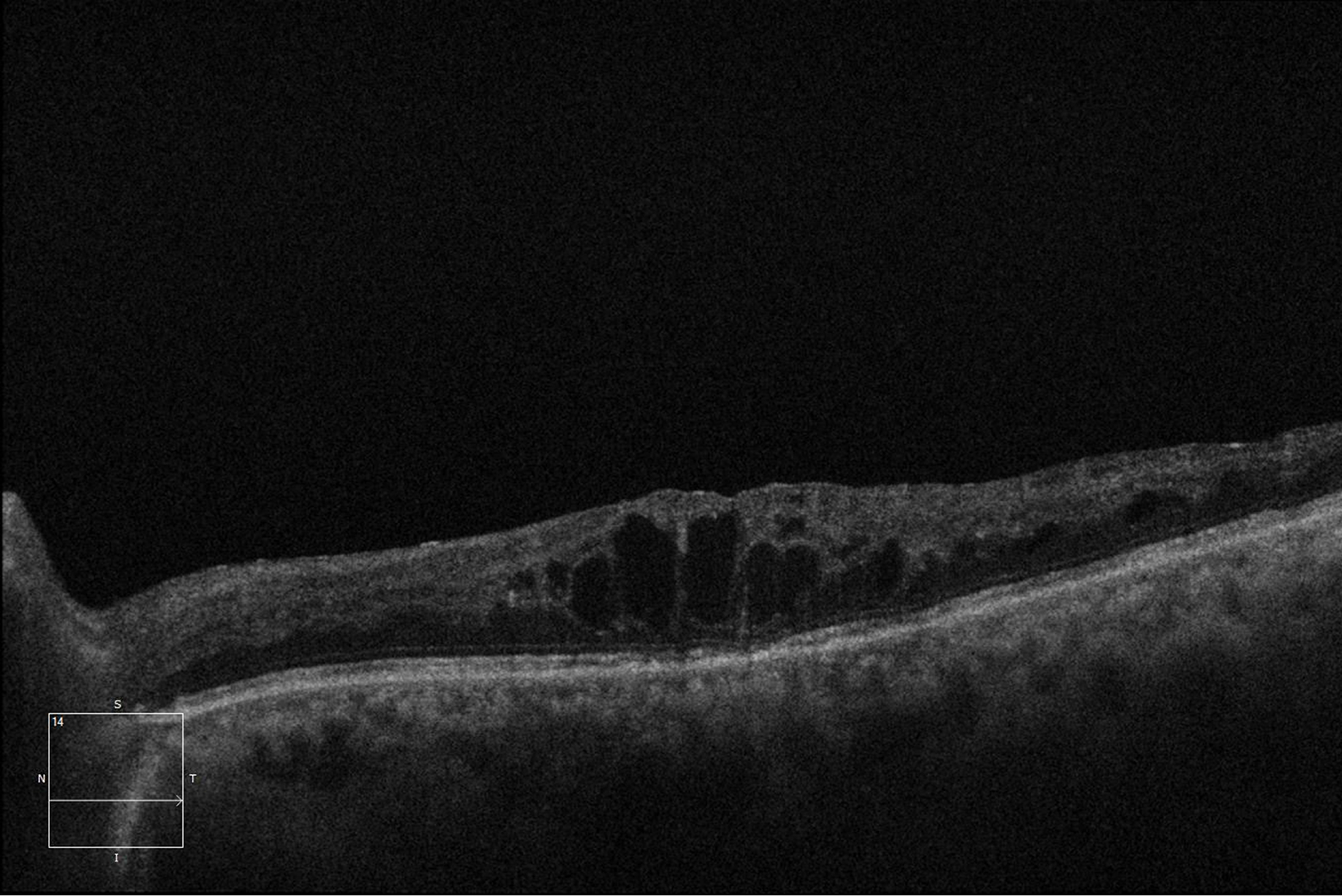
Figure 4
Optical coherence tomography photograph of the left eye showing massive cystoid spaces and intraretinal fluid leading to diffuse retinal thickness and disorganization of inner retinal layers.
- Citation: Menia NK, Morya AK, Gupta PC, Ramachandran R. Ocular biomarkers in diabetes mellitus with diabetic kidney disease: A minireview. World J Nephrol 2025; 14(3): 109470
- URL: https://www.wjgnet.com/2220-6124/full/v14/i3/109470.htm
- DOI: https://dx.doi.org/10.5527/wjn.v14.i3.109470


