©2014 Baishideng Publishing Group Inc.
World J Virology. Nov 12, 2014; 3(4): 22-29
Published online Nov 12, 2014. doi: 10.5501/wjv.v3.i4.22
Published online Nov 12, 2014. doi: 10.5501/wjv.v3.i4.22
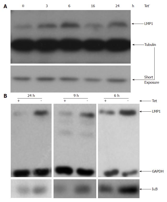
Figure 1 Blockage of nuclear factor-κB increases the expression of latent membrane protein 1 in Epstein-Barr virus-transformed cells.
Inducible IκB-expression IB4 line were washed three times with fresh RPMI1640 medium, and re-suspended in tetracycline (Tet) plus or minus medium. Cells were isolated at indicated time, and used for Western blot analysis with indicated antibodies. The expression of latent membrane protein 1 (LMP1) was shown in (A) and the expression of IκB was shown in (B). GAPDH: Glyceraldehyde-3-phosphate dehydrogenase.
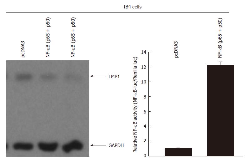
Figure 2 Overexpression of nuclear factor-κB decreases the expression of latent membrane protein 1 in Epstein-Barr virus-transformed cells.
IB4 cells were transfected with CD4 expression plasmid along with pcDNA3 (vector) or nuclear factor κB (NF-κB) expression plasmids (p50 and p65 expression plasmids at 1:1 ratio). One day after the transfection, the transfected cells were enriched using the CD4 magnetic beads, and the cell lysates were used for Western blot analysis. The identity of the proteins is as shown. The right panel: IB4 cells were transfected with the indicated plasmid as shown at the top along with NF-κB specific reporter construct and Renilla luciferase reporter plasmid. One day later, luciferase and Renilla luciferase activities were measured. Relative promoter reporter activities are shown. GAPDH: Glyceraldehyde-3-phosphate dehydrogenase; LMP1: Latent membrane protein-1.
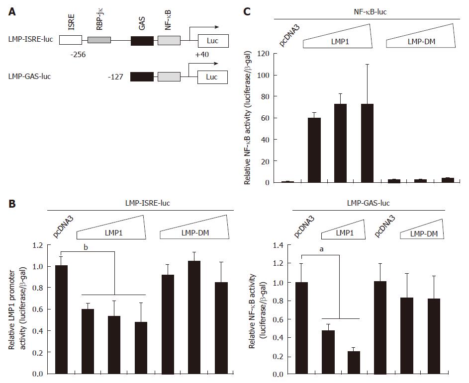
Figure 3 Latent membrane protein 1 negatively regulates its own promoter activity.
A: Schematic diagram of Epstein-Barr virus (EBV) latent membrane protein-1 (LMP1) promoter reporter constructs. RNA start site is shown. The drawing is not to scale; B: 293T cells were transfected with LMP1 promoter reporter construct and expression plasmid (0.01, 0.05, and 0.1 μg) as shown at the top. The LMP-DM has mutations in the critical domains in LMP1 for signaling. Cell lysates were used for the luciferase and β-galactosidase assays. Relative promoter reporter activities (luciferase/β-galactosidase) are shown. The results represented an average of triplicate transfections; Standard error bars are shown. Results from one representative experiment are of shown. The statistically significant difference between the indicated samples is denoted as aP < 0.05; bP < 0.01; C: 293T cells were transfected with the indicated plasmid as shown at the top. Nuclear factor κB (NF-κB) specific reporter construct was used. One day later, luciferase and β-galactosidase activities were measured. Relative promoter reporter activities are shown.
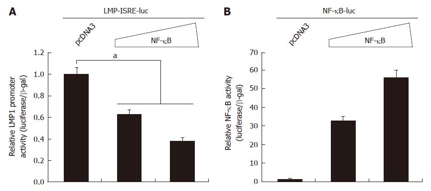
Figure 4 Nuclear factor κB represses latent membrane protein 1 promoter activity.
A: 293T cells were transfected with latent membrane protein-1 (LMP1) promoter reporter construct and nuclear factor κB (NF-κB) expression plasmid (0.05 and 0.1 μg; p65 and p50 at 1:1 ratio) as shown at the top. Cell lysates were used for the luciferase and β-galactosidase assays one day later. Relative promoter reporter activities (luciferase/β-galactosidase) are shown. Standard error bars are shown. The statistically significant difference between the indicated samples is denoted as aP < 0.05; B: 293T cells were transfected with the indicated plasmid as shown at the top. NF-κB specific reporter construct was used. One day later, luciferase and β-galactosidase activities were measured. Relative promoter reporter activities are shown.
- Citation: Cao M, Wang Q, Lingel A, Zhang L. Nuclear factor κB represses the expression of latent membrane protein 1 in Epstein-Barr virus transformed cells. World J Virology 2014; 3(4): 22-29
- URL: https://www.wjgnet.com/2220-3249/full/v3/i4/22.htm
- DOI: https://dx.doi.org/10.5501/wjv.v3.i4.22


