©The Author(s) 2017.
World J Transplant. Jun 24, 2017; 7(3): 193-202
Published online Jun 24, 2017. doi: 10.5500/wjt.v7.i3.193
Published online Jun 24, 2017. doi: 10.5500/wjt.v7.i3.193
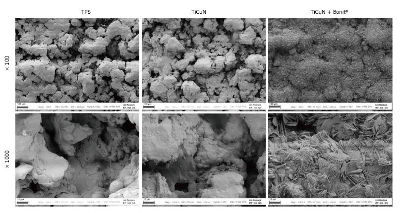
Figure 1 Surface topography of the coated materials vs titanium plasma spray control (field emission scanning electron microscopy, magnification × 100, × 1000, bars = 100 μm, 10 μm, respectively).
TPS: Titanium plasma spray; TiCuN: Titanium-copper-nitride.
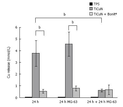
Figure 2 Copper release in Dulbecco’s modified Eagle medium.
A high amount of copper is released from the TiCuN layer after incubation in DMEM for 24 h. The copper release is reduced on TiCuN + BONIT® due to the BONIT® layer. A complete exchange of the medium and seeding with MG-63 cells for another 24 h reveals significantly reduced copper release from TiCuN. The amount is equalized to the level on TiCuN + BONIT® (n = 3, mean value ± SD, t-test, bP < 0.01). TPS: Titanium plasma spray; TiCuN: Titanium-copper-nitride; DMEM: Dulbecco’s modified Eagle medium.
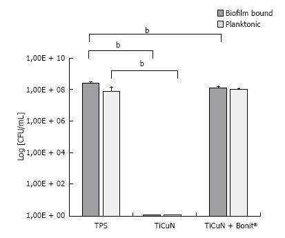
Figure 3 Antibacterial effect of the Titanium-copper-nitride coating on Staphylococcus epidermidis bacteria for planktonic and biofilm state after 2 d.
On TiCuN, planktonic and biofilm bound bacteria were killed completely. On TiCuN + BONIT®, no antibacterial effect could be observed (n = 6, mean value ± SD, U-test, bP < 0.01). TPS: Titanium plasma spray; TiCuN: Titanium-copper-nitride.
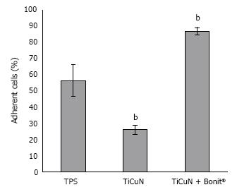
Figure 4 Initial cell adhesion of MG-63 osteoblasts on the titanium-copper-nitride.
Surfaces compared to the titanium plasma spray control after 10 min. The MG-63 cells were directly seeded onto the samples and cultivated for 10 min. Cell adhesion was significantly reduced on TiCuN, but TiCuN + BONIT® enhanced cell adherence significantly (n = 3, mean value ± SD, t-test, bP < 0.01). TPS: Titanium plasma spray; TiCuN: Titanium-copper-nitride.
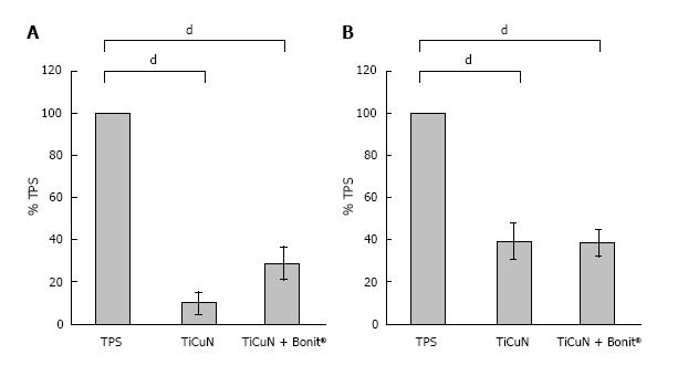
Figure 5 Viability of MG-63 osteoblasts on the titanium-copper-nitride surfaces.
Two different experimental arrangements were used: (A) the MG-63 cells were directly cultivated on the TiCuN surfaces for 24 h and (B) the samples were pre-incubated in DMEM for 24 h and after this the cells were seeded onto the surfaces for another 24 h. Cell viability was significantly reduced after direct seeding on TiCuN. Cell viability was higher on TiCuN + BONIT® compared to TiCuN. Pre-incubation of the samples in DMEM for 24 h before seeding elevated cell viability on both samples (n = 3, mean value ± SD, t-test, dP < 0.001). TPS: Titanium plasma spray; TiCuN: Titanium-copper-nitride; DMEM: Dulbecco’s modified Eagle medium.
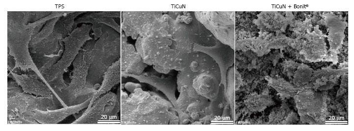
Figure 6 Scanning electron microscopy images of MG-63 osteoblasts on the pre-incubated surfaces.
Samples were pre-incubated in DMEM for 24 h. After a complete exchange of medium, cells were seeded onto the surface and cultivated for another 24 h. Cells spread well on TPS and TiCuN surfaces but seem to be smaller on TiCuN + BONIT® (magnification × 1000). TPS: Titanium plasma spray; TiCuN: Titanium-copper-nitride; DMEM: Dulbecco’s modified Eagle medium.
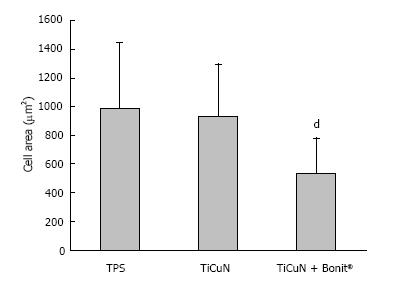
Figure 7 Spreading of MG-63 osteoblasts on pre-incubated samples after 24 h.
Cell area is unchanged on TiCuN compared to TPS but significantly reduced on TiCuN + BONIT® due to the additional BONIT® layer (n = 60, mean value ± SD, t-test, dP < 0.001). Titanium plasma spray; TiCuN: Titanium-copper-nitride.
- Citation: Bergemann C, Zaatreh S, Wegner K, Arndt K, Podbielski A, Bader R, Prinz C, Lembke U, Nebe JB. Copper as an alternative antimicrobial coating for implants - An in vitro study. World J Transplant 2017; 7(3): 193-202
- URL: https://www.wjgnet.com/2220-3230/full/v7/i3/193.htm
- DOI: https://dx.doi.org/10.5500/wjt.v7.i3.193


