©2014 Baishideng Publishing Group Inc.
World J Transplant. Sep 24, 2014; 4(3): 196-205
Published online Sep 24, 2014. doi: 10.5500/wjt.v4.i3.196
Published online Sep 24, 2014. doi: 10.5500/wjt.v4.i3.196
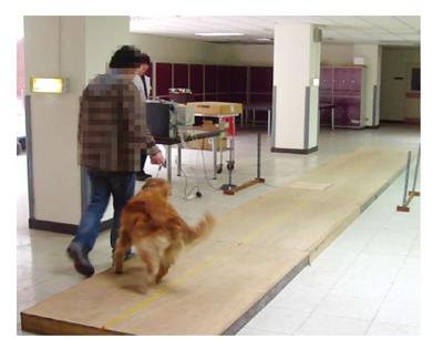
Figure 1 Gait analysis platform.
The platform was a 10 m × 1.22 m × 0.19 m wooden walkway and in the middle of which embedded a pressure sensor. During the analysis the dog was leash-walked by its owner from one end toward the other end of the walkway. The walking speed was recorded by two sets of photoelectric cells. The walk was repeated until three valid speeds were recorded for each leg.
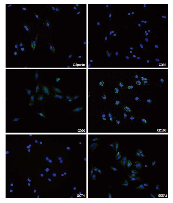
Figure 2 Stem cell marker expression.
Porcine adipose-derived stem cells were stained for the indicated cell markers (green) and nuclei (blue).
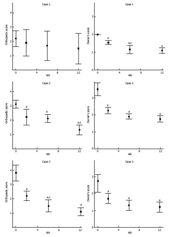
Figure 3 Orthopedic and owner’s evaluation.
Dogs (cases 1 to 3) were evaluated by veterinarians and owners for stifle joint function and pain according to the scoring criteria listed in Tables 1 and 2. Data presented are the average and range of these scores. aP < 0.05, orthopedic score after stem cells injection vs pre-treatment (week 0); cP < 0.05, orthopedic score after stem cells injection vs the previous time point. Lower scores indicate better joint function or less pain.
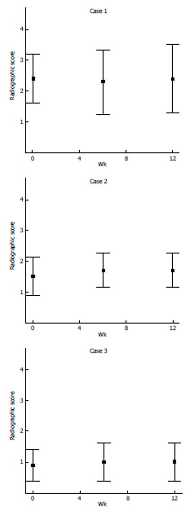
Figure 4 Radiographic evaluation.
X-ray images of the diseased stifle joint of each dog (cases 1 to 3) were evaluated by 5 investigators independently according to the scoring criteria listed in Table 3. Data presented are the average and range of these scores.
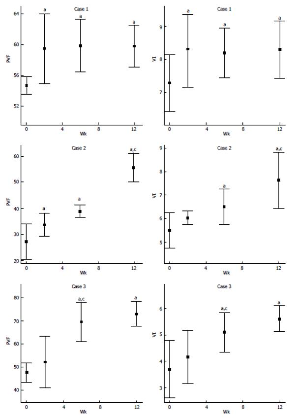
Figure 5 Force-plate evaluation.
Dogs (cases 1 to 3) were evaluated by force-plate gait analysis as shown in Figure 1. Values of PVF and VI were obtained from three walks with valid speed. Both values were normalized for body weight; VI was further normalized for time. Data presented are the average and range of values obtained from three walks with valid speed. Unit for PVF is N kg- in percentile (kg is dog’s body weight in kilogram). Unit for VI is N s kg- (s is time in second). aP < 0.05, the week after stem cells injection vs week 0 (pre-treatment); cP < 0.05, the week after stem cells injection vs the previous time point. Higher scores indicate better joint function. PVF: Peak vertical force; VI: Vertical impulse.
- Citation: Tsai SY, Huang YC, Chueh LL, Yeh LS, Lin CS. Intra-articular transplantation of porcine adipose-derived stem cells for the treatment of canine osteoarthritis: A pilot study. World J Transplant 2014; 4(3): 196-205
- URL: https://www.wjgnet.com/2220-3230/full/v4/i3/196.htm
- DOI: https://dx.doi.org/10.5500/wjt.v4.i3.196


