©The Author(s) 2020.
World J Transplant. Dec 28, 2020; 10(12): 381-391
Published online Dec 28, 2020. doi: 10.5500/wjt.v10.i12.381
Published online Dec 28, 2020. doi: 10.5500/wjt.v10.i12.381
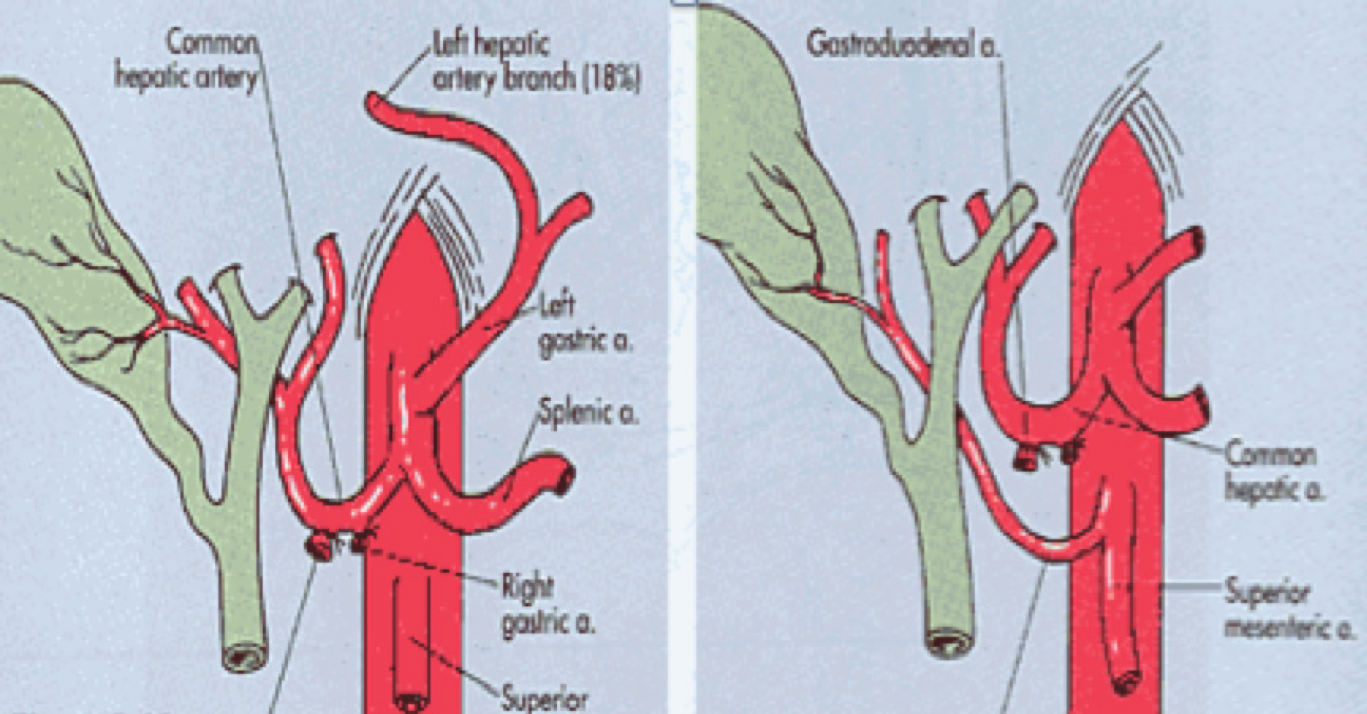
Figure 1 Right hepatic artery from mesenteric superior artery.
Figure 2 Right hepatic artery from mesenteric superior artery.
A: Intraoperative view; B: Overlayed with identifiers. SMA: Superior mesenteric artery; IPDA: Inferior pancreaticoduodenal artery; rRHA: Replaced right hepatic artery.
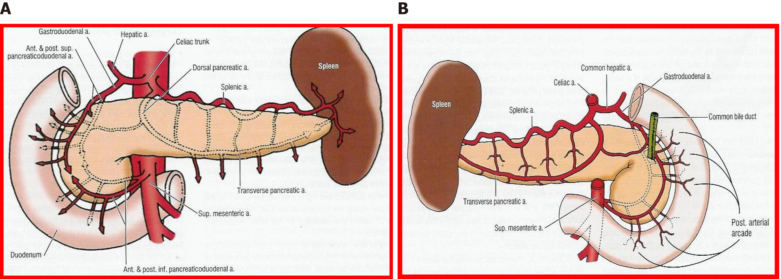
Figure 3 Arterial vasculature of the pancreas.
Illustrated from the view of contemporary surgery. A: Anterior; B: Posterior.
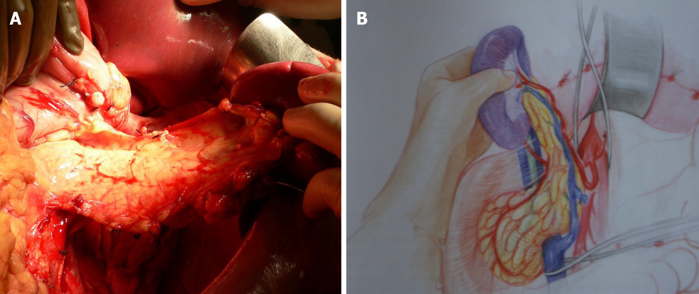
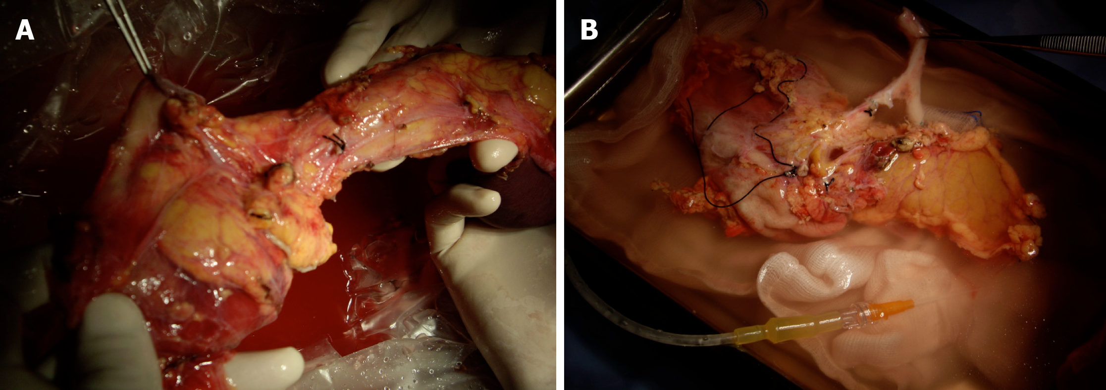
Figure 4 Pancreas graft mobilization.
A: Photo; B: Drawing from Martin Finch.
Figure 5 Retrieval of pancreas and liver.
A and B: Intraoperative views of the retrieval procedure, showing different aspects.
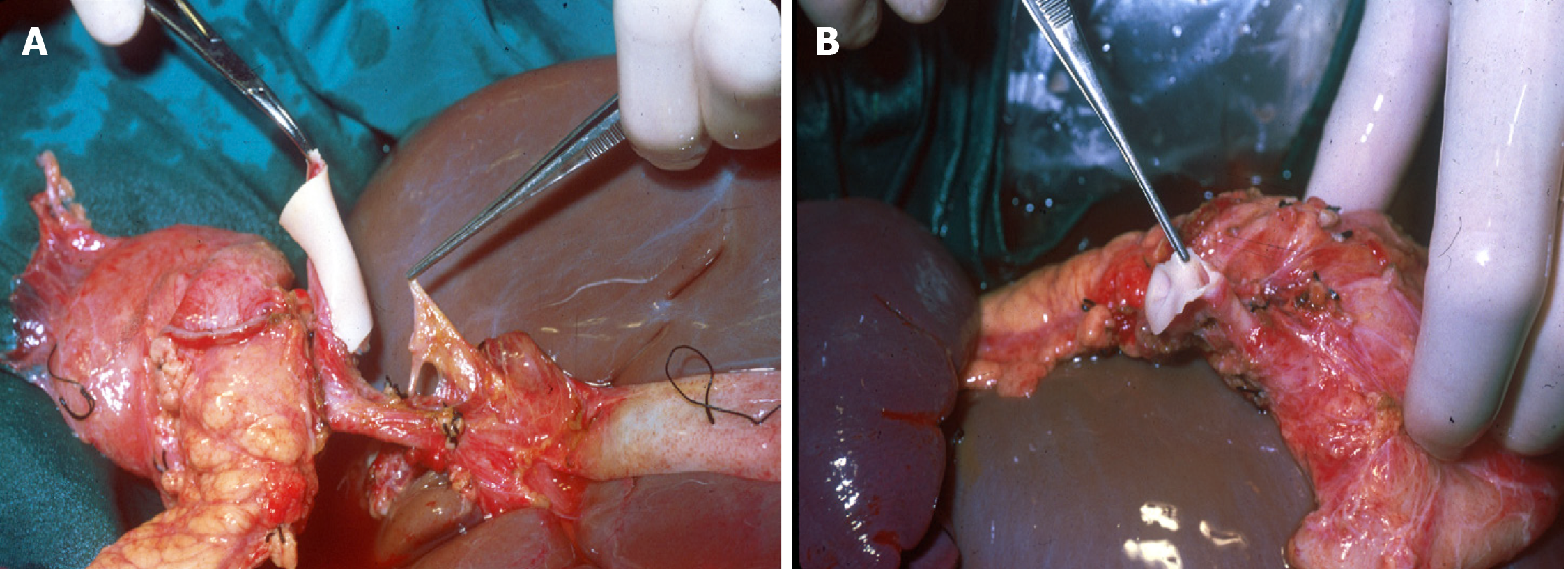
Figure 6 Pancreatico-duodenal graft.
A and B: Intraoperative views of the bench preparation procedure, showing different aspects.
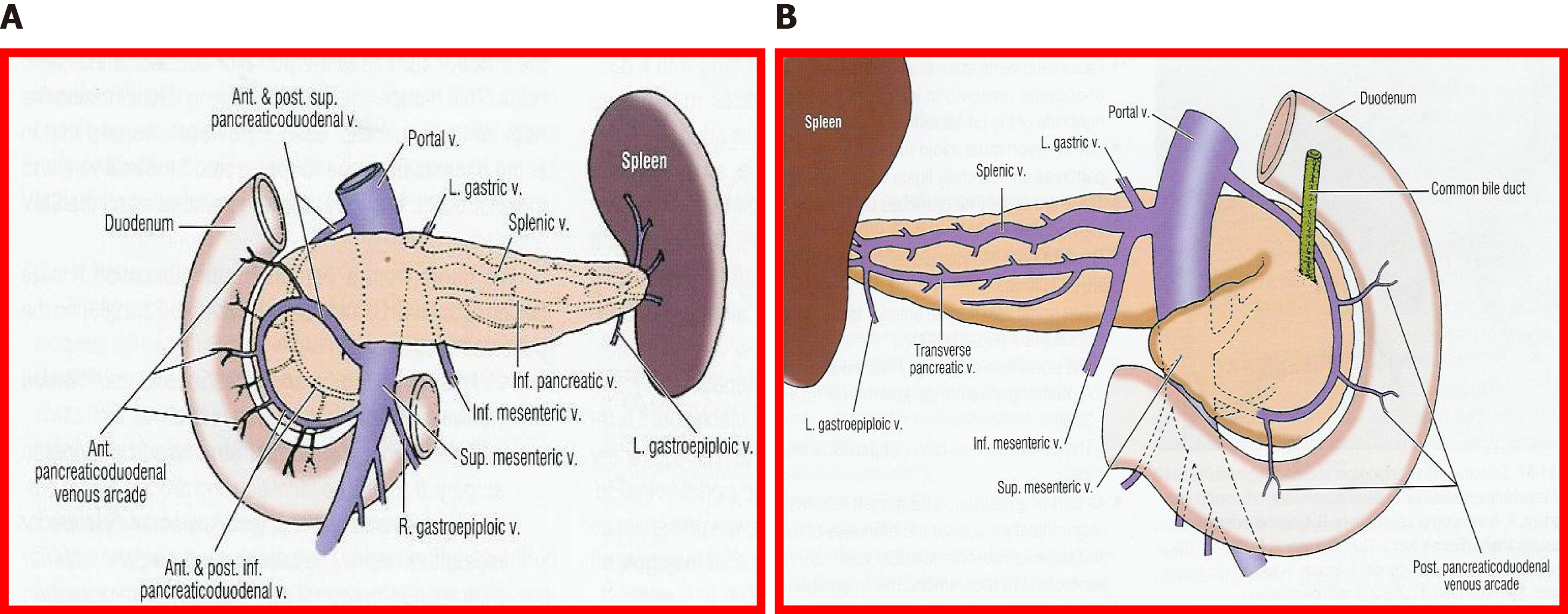
Figure 7 Venous vessels.
Illustrated from the view of contemporary surgery. A: Anterior view; B: Posterior view.
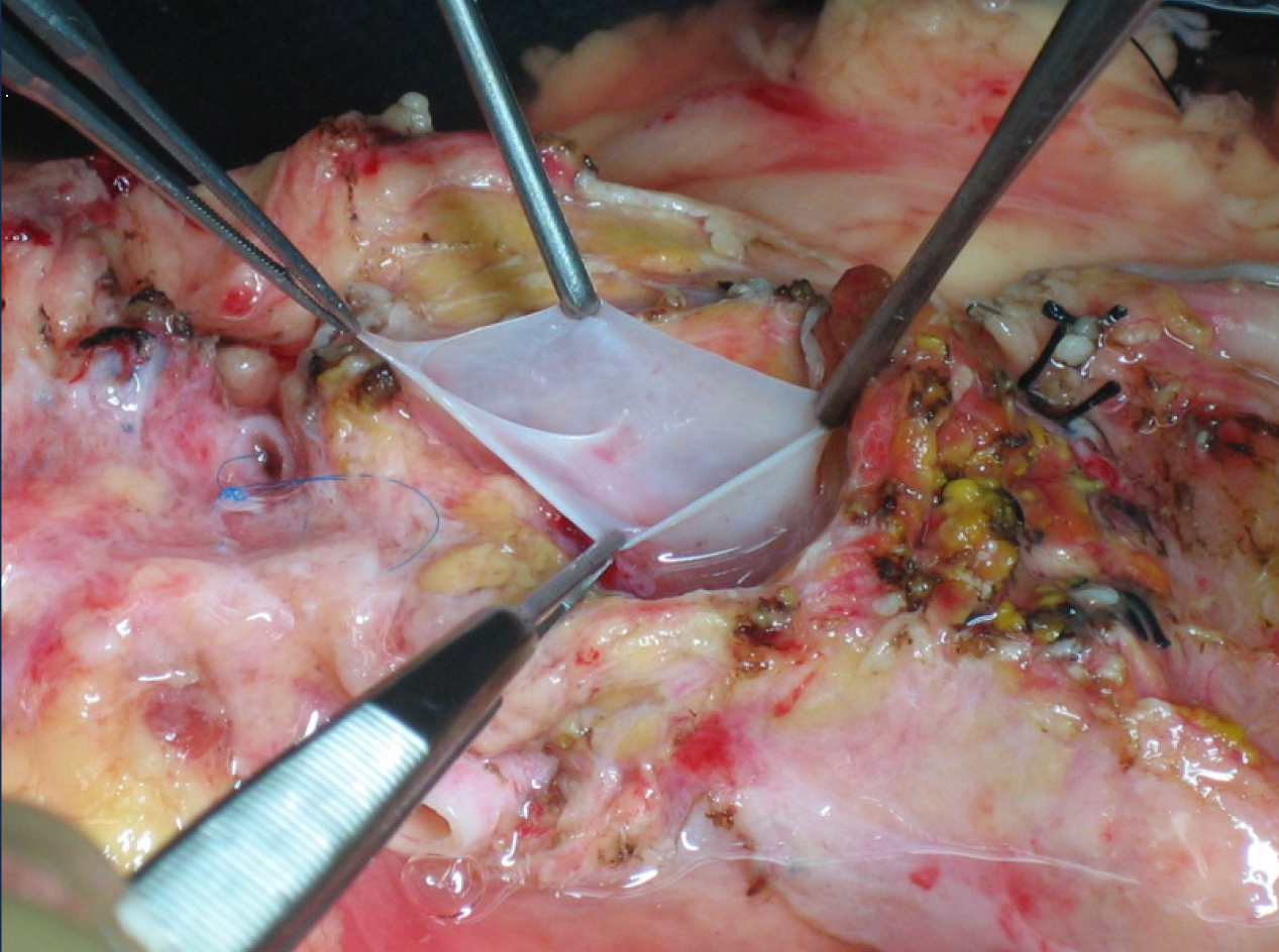
Figure 8 Portal vein.
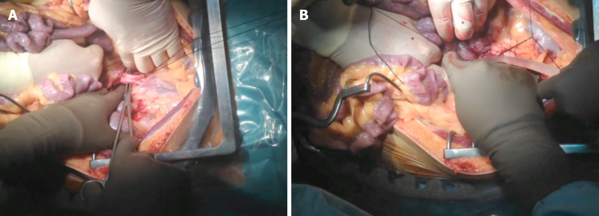
Figure 9 Aortic canula in super-fast retrieval.
A and B: Intraoperative views of the procedure, showing different aspects. Photos provided by Dr Perez Daga, pancreas transplant surgeon (Malaga, Spain).
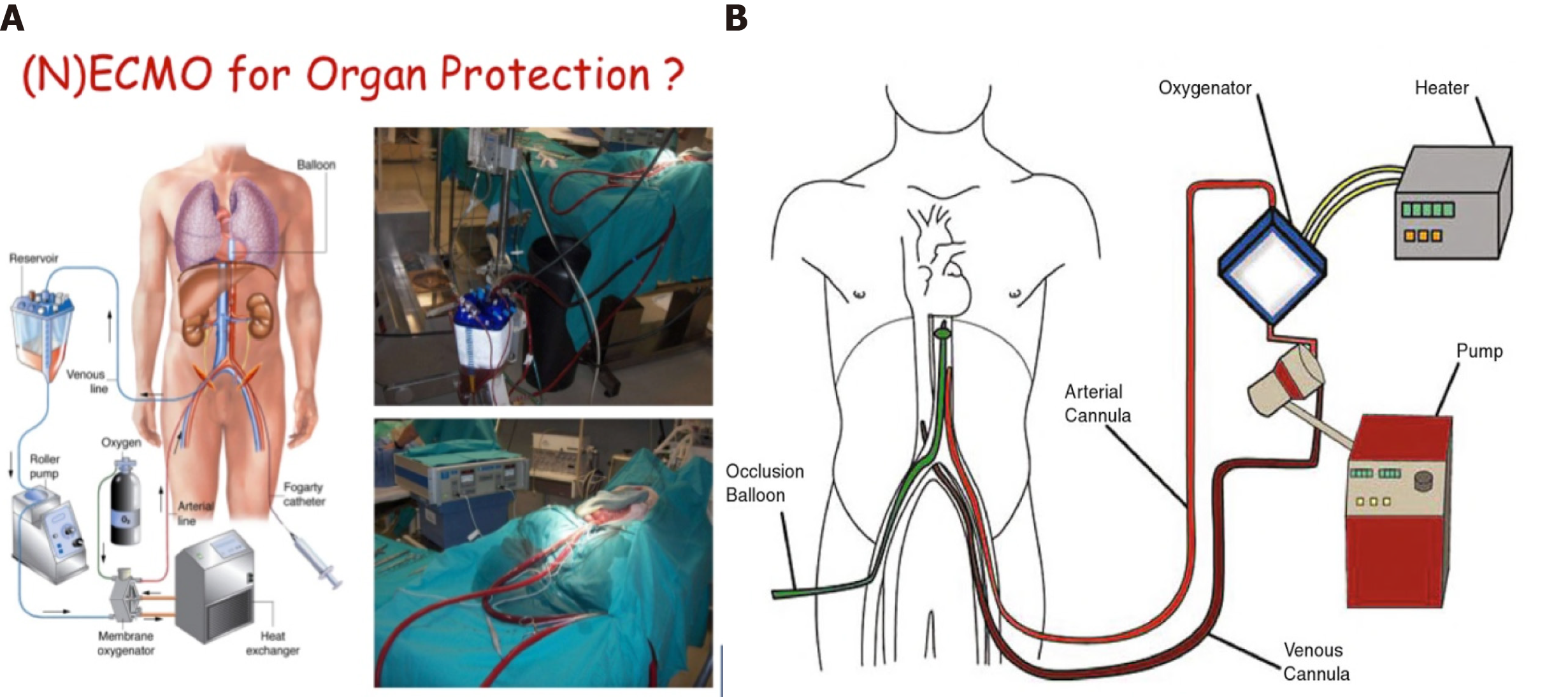
Figure 10 Normothermic circulation.
A: Picture of normothermic preservation machines (NECMO); B: Diagram of NECMO. NECMO: Normothermic preservation machines.
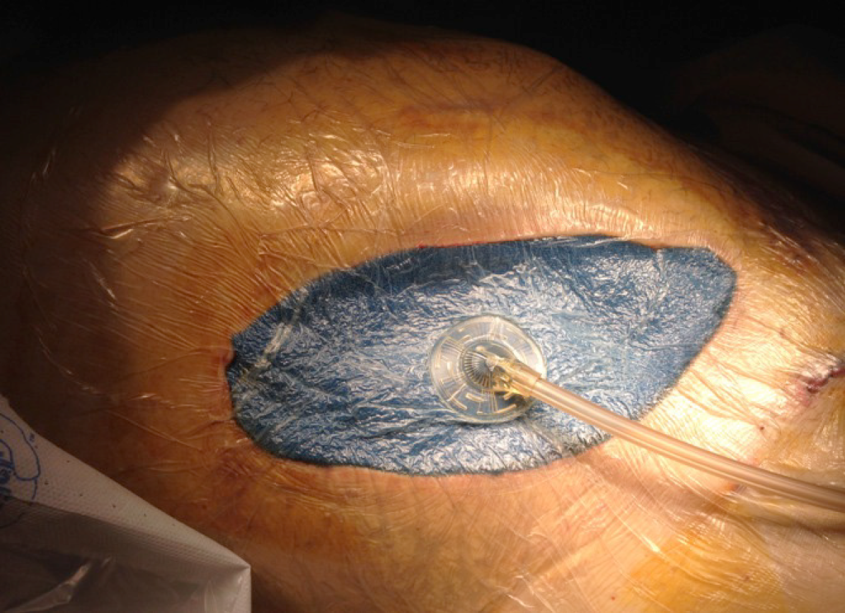
Figure 11 Negative-pressure wound therapy.
- Citation: Casanova D, Gutierrez G, Gonzalez Noriega M, Castillo F. Complications during multiorgan retrieval and pancreas preservation. World J Transplant 2020; 10(12): 381-391
- URL: https://www.wjgnet.com/2220-3230/full/v10/i12/381.htm
- DOI: https://dx.doi.org/10.5500/wjt.v10.i12.381


