Fan XW, Hou JZ, Wang YZ, Gao ML, Shen Y, Jian D, Zhou Q, Yang J, Zhao H, Zhang LJ. Divergent lymphocyte associations with depression severity and case status in first-episode, drug-naive adolescents: A multicenter study. World J Psychiatry 2026; 16(7): 119439 [DOI: 10.5498/wjp.119439]
Corresponding Author of This Article
Li-Juan Zhang, PhD, Health Management Center, Shanghai Yangzhi Rehabilitation Hospital (Shanghai Sunshine Rehabilitation Center), School of Medicine, Tongji University, No. 50 Chifeng Road, Yangpu District, Shanghai 201613, China. zhangxiaoyi@tongji.edu.cn
Research Domain of This Article
Psychiatry
Article-Type of This Article
research-article
Open-Access Policy of This Article
This article is an open-access article which was selected by an in-house editor and fully peer-reviewed by external reviewers. It is distributed in accordance with the Creative Commons Attribution Non Commercial (CC BY-NC 4.0) license, which permits others to distribute, remix, adapt, build upon this work non-commercially, and license their derivative works on different terms, provided the original work is properly cited and the use is non-commercial. See: http://creativecommons.org/licenses/by-nc/4.0/
Baishideng Publishing Group Inc, 7041 Koll Center Parkway, Suite 160, Pleasanton, CA 94566, USA
Share the Article
Fan XW, Hou JZ, Wang YZ, Gao ML, Shen Y, Jian D, Zhou Q, Yang J, Zhao H, Zhang LJ. Divergent lymphocyte associations with depression severity and case status in first-episode, drug-naive adolescents: A multicenter study. World J Psychiatry 2026; 16(7): 119439 [DOI: 10.5498/wjp.119439]
Xi-Wang Fan, Yi-Zhe Wang, Ming-Lin Gao, Hui Zhao, Clinical Research Center for Mental Disorders, Shanghai Pudong New Area Mental Health Center, School of Medicine, Tongji University, Shanghai 200124, China
Jia-Zhe Hou, Jing Yang, Li-Juan Zhang, Health Management Center, Shanghai Yangzhi Rehabilitation Hospital (Shanghai Sunshine Rehabilitation Center), School of Medicine, Tongji University, Shanghai 201613, China
Ying Shen, Psychosomatic Medicine, The Third People’s Hospital of Ganzhou, Ganzhou 341000, Jiangxi Province, China
Dai Jian, Department of Clinical Psychology, Jiangbin Hospital of Guangxi Zhuang Autonomous Region, Third People’s Hospital of Guangxi Zhuang Autonomous Region, Nanning 530021, Guangxi Zhuang Autonomous Region, China
Qin Zhou, The Affiliated Xuzhou Eastern Hospital of Xuzhou Medical University, Xuzhou Eastern People’s Hospital, Xuzhou 221004, Jiangsu Province, China
Author contributions: Fan XW contributed to methodology, formal analysis; Fan XW, Hou JZ, Wang YZ, Gao ML, and Yang J contributed to manuscript writing - review and editing; Fan XW, Hou JZ, Wang YZ, Yang J, Zhao H, and Zhang LJ participated in manuscript writing - original draft; Hou JZ was involved in data sorting, analysis, visualization; Wang YZ participated in validation; Shen Y contributed to investigation and resources; Shen Y, Jian D, and Zhou Q contributed to data curation; Jian D and Zhou Q were involved in investigation; Zhao H contributed to and supervised the study; Zhang LJ contributed to study conceptualization, supervision, project administration, and funding acquisition; Fan XW and Hou JZ contributed equally to this manuscript and are co-first authors.
AI contribution statement: During manuscript preparation, relevant language-editing tools were used solely for English-language polishing and grammatical refinement. The scientific content of the manuscript, including the study conception and design, data collection and analysis, interpretation of results, and formulation of conclusions, was completed independently by the authors and was not generated by artificial intelligence. These tools were used only to improve linguistic accuracy, clarity, and readability. They were not used for data processing, statistical analysis, result generation, or the development of academic content. Artificial intelligence tools had no role in the conception or design of the study, interpretation of findings, or any scholarly decision-making. No figures, images, or other visual materials in this manuscript were generated by artificial intelligence. We would like to emphasize that all scientific aspects of the study were conducted independently by the authors, and artificial intelligence tools were used strictly for language refinement purposes.
Supported by National Natural Science Foundation of China, No. 81872720; and the Shanghai Municipal Health Commission, No. 2023ZZ02027; and the National Clinical Key Specialty Construction Project of China, No. Z155080000004.
Institutional review board statement: The study protocol was reviewed and approved by the Ethics Committee of the Shanghai Pudong New Area Mental Health Center, Approval No.[2022] Research Review No. 011. All research activities were conducted in strict accordance with the principles of the Declaration of Helsinki and complied with relevant national and local regulatory standards to ensure the ethical conduct of research involving human participants.
Informed consent statement: Written informed consent was obtained from all participants aged 18 years or above. For participants younger than 18 years, written informed consent was obtained from a parent or legal guardian, and a written agreement for study participation was obtained from the adolescent when applicable.
Conflict-of-interest statement: All the authors report no relevant conflicts of interest for this article.
STROBE statement: The authors have read the STROBE Statement-checklist of items, and the manuscript was prepared and revised according to the STROBE Statement-checklist of items.
Data sharing statement: The datasets generated and/or analyzed during the current study are not publicly available but are available from the corresponding author upon reasonable request.
Corresponding author: Li-Juan Zhang, PhD, Health Management Center, Shanghai Yangzhi Rehabilitation Hospital (Shanghai Sunshine Rehabilitation Center), School of Medicine, Tongji University, No. 50 Chifeng Road, Yangpu District, Shanghai 201613, China. zhangxiaoyi@tongji.edu.cn
Received: January 29, 2026 Revised: February 22, 2026 Accepted: March 24, 2026 Published online: July 19, 2026 Processing time: 153 Days and 9.7 Hours
Abstract
BACKGROUND
Adolescent major depressive disorder (MDD) imposes an increasingly severe global health burden. Current diagnosis relies predominantly on subjective symptom reporting. Adolescents exhibit a unique immunobiological landscape, characterized by immune signatures that differ from those of adults. This feature may enable development of an immunopsychiatric framework for adolescent depression.
AIM
To investigate whether routinely measured peripheral immune parameters could both identify depression and reflect its severity in treatment-naive adolescents.
METHODS
We employed a two-stage, multicenter design. In stage I, a cross-sectional sample of 2115 first-episode, drug-naive adolescents with MDD was recruited from three regional centers in China. Associations between peripheral immune parameters and derived ratios with depression severity were assessed by the Self-Rating Depression Scale, with sex-stratified analyses. In stage II, a case-control study enrolled 41 adolescents with MDD and 41 controls. Multivariable logistic regression and receiver operating characteristic analyses were performed to evaluate the discriminatory capacity of candidate immune indicators.
RESULTS
Compared with healthy peers, adolescents with MDD had significantly lower lymphocyte counts [odds ratio (OR) = 0.27, 95% confidence interval (CI): 0.13-0.57] and lymphocyte-to-monocyte ratio (LMR; OR = 0.72, 95%CI: 0.56-0.92). Paradoxically, within the MDD group, higher lymphocyte count (OR = 1.10, 95%CI: 1.01-1.22) and LMR (OR = 1.06, 95%CI: 1.01-1.13) were independently associated with greater depression severity, an effect most accentuated in female participants (OR = 1.17, 95%CI: 1.01-1.35). receiver operating characteristic analyses confirmed moderate discriminative accuracy for lymphocyte count [area under the curve (AUC) = 0.76, 95%CI: 0.66-0.87] and LMR (AUC = 0.70, 95%CI: 0.59-0.81). A composite model integrating age, sex, and key immune parameters achieved good discrimination (bootstrap-corrected AUC = 0.79, 95%CI: 0.73-0.82).
CONCLUSION
Our findings reveal a bidirectional immunological pattern in adolescent MDD: Reduced lymphocyte count supports diagnostic identification, while elevated levels correlate with greater illness severity. These results support a stage-dependent immunopsychiatric model and lend support to precision mental health strategies in youth.
Core Tip: This two-stage multicenter study reveals a divergent immunopurified in first-episode, drug-naive adolescents with major depressive disorder. While patients exhibited significantly lower lymphocyte counts and lymphocyte-to-monocyte ratios than healthy controls, higher levels of these markers paradoxically correlated with greater disease severity, particularly in females. A composite model integrating these indices demonstrated robust diagnostic discrimination (area under the curve = 0.82). These findings support a stage-dependent immunopsychiatric paradigm, suggesting that routine peripheral immune metrics offer a practical, objective tool for identifying major depressive disorder and stratifying severity in youth.
Citation: Fan XW, Hou JZ, Wang YZ, Gao ML, Shen Y, Jian D, Zhou Q, Yang J, Zhao H, Zhang LJ. Divergent lymphocyte associations with depression severity and case status in first-episode, drug-naive adolescents: A multicenter study. World J Psychiatry 2026; 16(7): 119439
Adolescent major depressive disorder (MDD) is a leading cause of disability worldwide and an escalating public health concern in China, with profound impacts on social functioning, educational attainment, and suicide risk[1,2]. Existing diagnostic paradigms hinge on symptom-based criteria from Diagnostic and Statistical Manual of Mental Disorders, fifth edition or International Classification of Diseases, eleventh edition, alongside clinician rated scales. These approaches are susceptible to rater bias, limited by time constraints, and often insufficiently sensitive to developmental and psychosocial complexities unique to youth[3,4]. There is therefore an urgent need for objective, scalable biological indicators to augment conventional assessment and enable robust stratification of illness severity.
In adults, substantial evidence supports a role for immune dysregulation in the pathogenesis of depression, with heightened peripheral inflammatory activity linked to both symptomatic presentation and disease course[5-7]. However, translating of these insights to adolescents remains challenging. Studies in younger populations are limited in number, methodologically heterogeneous, and frequently underpowered, with inadequate attention to immunological variations related to age and sex[8,9]. Routine hematological indices derived from complete blood counts, including absolute lymphocyte, neutrophil, and monocyte counts and their ratios such as the lymphocyte-to-monocyte ratio (LMR), represent a feasible alternative[10]. These indices are economical, standardized, and widely available across clinical settings. Despite these advantages, their associations with clinically meaningful dimensions of adolescent depression, including both case ascertainment and severity profiling, remain insufficiently characterized[11,12].
A critical obstacle to extrapolating adult derived immunological models to adolescents lies in the distinct immunobiological characteristics of this developmental stage. Unlike the relatively stable immune parameters observed in adulthood, puberty is marked by dynamic neuroimmune remodeling driven by sharply rising sex hormone levels[13]. Physiological processes such as thymic involution and hormonally influenced lymphopoiesis contribute to substantial variability in lymphocyte counts and immune responsiveness[14,15]. These maturational changes suggest that adolescent depression may be accompanied by immune signatures distinct from those seen in adults, potentially characterized by temporal instability or stage-dependent directional shifts rather than the persistent low-grade inflammation typical of adult MDD.
To address these gaps, we employed a two-stage, multicenter framework integrating both discovery and validation phases. In stage I, we conducted a large multicenter cross-sectional study of treatment naive adolescents with first episode MDD across three regional centers in China to examine how routinely measured peripheral immune parameters relate to depression severity and whether these associations are modified by sex. In stage II, informed by severity-related immunological patterns identified in stage I, we performed a case-control analysis to evaluate the capacity of candidate immune markers to discriminate adolescents with MDD from healthy controls. By integrating severity-associated immune phenotypes with case status discrimination, this study aimed to identify biologically grounded and readily implementable adjuncts to symptom-based diagnosis and to advance an immunopsychiatric model to adolescent depression.
MATERIALS AND METHODS
Study design and participants
This study employed a two-stage design to evaluate the clinical utility of peripheral immune indicators in adolescent MDD. Stage I was a large multicenter cross-sectional study that recruited 2115 first-episode, drug-naive adolescents with MDD from three regional centers in China, namely Shanghai Pudong New Area Mental Health Center, the Third People’s Hospital of Ganzhou in Jiangxi Province, and the Third People’s Hospital of Guangxi Zhuang Autonomous Region. These regions correspond to eastern (Shanghai), central (Jiangxi Province), and western (Guangxi Zhuang Autonomous Region) China, with relatively higher, intermediate, and lower levels of economic development, respectively, thus enhancing geographic and socioeconomic diversity across mainland China. Recruitment took place between February 2023 and October 2024. The primary objectives of this stage were to determine whether peripheral immune indices varied across depression severity and to examine potential modifications by sex.
Building on severity related immunological findings from stage I, stage II comprised a case-control study conducted at Shanghai Pudong New Area Mental Health Center between December 2024 and July 2025. This stage enrolled 41 adolescents with MDD and 41 healthy controls. Controls were recruited from the same sampling geographic area during the same period and were free of current or lifetime psychiatric disorders, as assessed using the Mini International Neuropsychiatric Interview for Children and Adolescents; individuals who met diagnostic criteria for any psychiatric disorder were excluded[16]. The participant flowchart is presented in Figure 1.
Figure 1 Flowchart of the study design and subject screening procedure.
The study protocol was reviewed and approved by the Ethics Committee of the Shanghai Pudong New Area Mental Health Center, Approval No.[2022] Research Review No. 011. All procedures were conducted in accordance with the Declaration of Helsinki and relevant regulatory requirements. Written informed consent was obtained from all participants aged 18 years or above. For participants younger than 18 years, written informed consent was obtained from a parent or legal guardian, and assent was additionally obtained from the adolescent where appropriate.
Inclusion and exclusion criteria
Eligible participants met all of the following criteria: (1) Age 10 to 19 years; (2) Diagnosis of MDD according to Diagnostic and Statistical Manual of Mental Disorders, fifth edition criteria, confirmed by a board-certified psychiatrist; and (3) A first episode of depression and treatment naive to psychotropic medication.
Exclusion criteria were as follows: (1) History of immune dysfunction, endocrine disorders, or any significant systemic medical illness; (2) Presence of organic brain disease or other neurological conditions; (3) Psychiatric conditions judged to be secondary to severe somatic illness; (4) Substance use disorders; (5) Acute or chronic infectious disease, including the presence of active symptoms within the preceding 14 days; (6) Use of anti-inflammatory agents, immunomodulatory therapies, or hormonal treatments within the past month; and (7) History of psychotropic medication use, including antidepressants, antipsychotics, and mood stabilizers.
Laboratory testing and measures
Venous blood samples were collected into EDTA tubes at baseline and analyzed within two hours of collection. Complete blood counts, including absolute leukocyte, neutrophil, lymphocyte, and monocyte counts, were measured using automated hematology analyzers at each participating center in accordance with predefined institutional protocols. To minimize pre-analytic variability, blood draws were performed under standardized conditions during morning clinical hours between 7:00 and 10:00.
To enhance inter-site comparability across analyzer platforms, hematological parameters were harmonized using data from the Shanghai center as the reference. The parameters of linear transformation were calculated based on the original range and target range. The linear transformation formula is: . a = slope calculated from the original range and target (Shanghai) range; b = intercept calculated from the original range and target range; = calibrated value aligned to Shanghai reference; = raw measurement from each center.
Derived immune ratios were calculated from absolute counts, including lymphocyte-to-neutrophil ratio (LNR), neutrophil-to-monocyte ratio (NMR), and LMR. All assays were conducted in accredited laboratories affiliated with participating centers by using standardized operating procedures. Quality assurance measures were implemented to ensure analytical precision, with a coefficient of variation of less than 5%.
Depression severity was assessed using the Self-Rating Depression Scale (Cronbach’s α = 0.81). The Self-Rating Depression Scale includes 20 items scored on a 4-point Likert scale, with reverse scoring applied to negatively worded items. Raw scores, ranging from 20 to 80, were converted to standard scores by the formula standard score equals integer (raw score × 1.25)[17], resulting in an integer index based on the Chinese normative data[18]. Based on established cutoff values, severity was classified as mild (53-62), moderate (63-72), and severe (≥ 73). All assessments were administered by certified evaluators under the supervision of board-certified psychiatrists[19].
Statistical analysis
Sample size for stage II was estimated using G*Power 3.1, based on an effect size derived from prior meta-analytic evidence (Cohen’s d = 0.729)[20]. Assuming a two-sided α of 0.05 and 80% power for an independent samples t test, the minimum required sample size was 30 participants per group. To ensure > 90% power and accommodate potential attrition, 41 participants were enrolled in each group.
Distributional assumptions were evaluated using the Shapiro-Wilk test for normality, Levene’s test for homogeneity of variances between two groups and Bartlett’s test for comparisons involving more than two groups. Continuous variables are presented as mean ± SD if normally distributed or median (interquartile range) if non-parametric; categorical variables are n (%).
Stage I (severity analyses in the multicenter MDD cohort): Immune indices across depression severity strata (mild, moderate, and severe) were compared using the Kruskal-Wallis test, with Dunn’s post hoc tests and Bonferroni adjustment where appropriate. Ordinal logistic regression models, adjusted for age and sex, were used to evaluate monotonic associations between immune markers and depression severity. Sex-stratified analyses were performed to assess potential effect modification.
Stage II (case-control discrimination and risk association): Between-group comparisons (adolescents with MDD vs healthy controls) used independent samples t-tests for normally distributed variables, Mann-Whitney U tests for skewed variables, and χ2 tests with Yates’ continuity correction for categorical variables. Logistic regression models, adjusted for age and sex, were fitted to estimate associations between immune markers and depression status. Age-stratified analyses explored potential variation across developmental stages. Discriminative performance was evaluated by receiver operating characteristic (ROC) analysis. The area under the curve (AUC) value and 95% confidence intervals (CIs) were calculated using DeLong’s method[21].
All statistical analyses were performed using R software (version 4.4.1) with the MASS, lmtest and pROC packages. All tests were two-tailed, and P < 0.05 was considered statistically significant.
RESULTS
Immune indicators across depression severity in the multicenter study
A total of 2115 drug-naive adolescents with first-episode MDD were stratified according to depression severity into mild (n = 279, 13.19%), moderate (n = 588, 27.80%), and severe (n = 1248, 59.01%) groups (Table 1). Significant differences in immune parameters were observed across severity strata. Between-group comparisons showed statistically significant variation in white blood cell count (H = 9.31, P = 0.009), lymphocyte count (H = 8.85, P = 0.011), NMR (H = 5.99, P = 0.049), and LMR (H = 11.31, P = 0.003).
Table 1 Social demographic characteristics and immune markers across subgroups with different degrees of depression severity, n (%)/mean ± SD/median (interquartile rage).
Post hoc pairwise comparisons indicated that lymphocyte count and LMR were significantly higher in adolescents with severe compared to mild depression. Lymphocyte count was greater in the severe group than in the mild group (P = 0.0046), and LMR followed a similar pattern group (P = 0.0013; Figure 2). In ordinal logistic regression models adjusted for age and sex, higher lymphocyte count [odds ratio (OR) = 1.10, 95%CI: 1.01-1.22, P = 0.040] and LMR (OR = 1.06, 95%CI: 1.01-1.13, P = 0.033) were each independently associated with greater severity of depression (Figure 3A). Sex-stratified analyses showed that the association between lymphocyte count and depressive severity remained significant in female adolescents after adjustment for age (OR = 1.17, 95%CI: 1.01-1.35, P = 0.03; Figure 3B).
Figure 3 Ordered logistic regression analysis.
A: Ordered logistic regression analysis, adjusted for age and sex; B: Ordered logistic regression analysis between sexes. aP < 0.05. LNR: Lymphocyte-to-neutrophil ratio; NMR: Neutrophil-to-monocyte ratio; LMR: Lymphocyte-to-monocyte ratio; OR: Odds ratio; CI: Confidence interval.
Case-control comparison of sociodemographic and immune characteristics
In stage II, 41 adolescents with MDD and 41 healthy controls were included (Table 2). The two groups did not differ significantly in mean age (15.00 ± 2.06 years vs 15.29 ± 2.33 years, P = 0.619) or sex distribution (P = 0.165). Compared with healthy controls, adolescents with MDD had significantly lower white blood cell count (t = 2.73, P = 0.007), neutrophil count (Z = 7.79, P = 0.005), lymphocyte count (Z = 8.84, P = 0.002), NMR (Z = 6.25, P = 0.012), and LMR (Z = 4.03, P = 0.044). No significant differences were observed in monocyte count (P = 0.647) or LNR (P = 0.568).
Table 2 Social demographic and clinical immunological characteristics of each group, n (%)/mean ± SD/median (interquartile rage).
Associations between immune markers and MDD case status
Multivariable logistic regression models adjusted for age and sex demonstrated that several immune parameters were independently associated with MDD case status. Higher neutrophil count (OR = 0.62, 95%CI: 0.44-0.87, P = 0.006) and higher lymphocyte count (OR = 0.27, 95%CI: 0.13-0.57, P < 0.001) were both associated with reduced odds of MDD. Similarly, higher NMR (OR = 0.82, 95%CI: 0.70-0.96, P = 0.015) and higher LMR (OR = 0.72, 95%CI: 0.56-0.92, P = 0.009) were independently associated with lower odds of MDD. No significant associations were observed for monocyte count or LNR in either unadjusted or adjusted models (all P > 0.05; Table 3). To examine potential age-related differences, participants were stratified into early (10-13 years), middle (14-17 years), and late (18-19 years) adolescence. Among middle adolescents (n = 1492, 70.54%), higher lymphocytes (OR = 1.13, 95%CI: 1.01-1.27; P = 0.03) and LMR (OR = 1.09, 95%CI: 1.02-1.17; P = 0.01) were significantly associated with greater depression severity. No significant associations between immune markers and depression severity were observed in early or late adolescence (Table 4).
Table 3 Relationship between immunological markers and depression in adolescents with major depressive disorder.
Discriminative performance of immune indicators for adolescent MDD
ROC analyses adjusted for age and sex showed that lymphocyte count had the highest AUC values for discriminating adolescents with MDD from healthy controls (AUC = 0.76, 95%CI: 0.66-0.87), followed by neutrophil count (AUC = 0.71, 95%CI: 0.60-0.82). Both the NMR and LMR had similar, moderate discriminative ability (AUC = 0.70 for each; NMR: 95%CI: 0.59-0.81; LMR: 95%CI: 0.59-0.82). Monocyte count and LNR showed poorer discrimination, with an AUC value of 0.59 for both.
A composite model incorporating age, sex, neutrophil count, lymphocyte count, NMR, and LMR significantly improved discrimination (AUC = 0.82, 95%CI: 0.72-0.92, P = 0.014). After 1000 bootstrap resamples, the optimism-corrected AUC value was 0.79 (95%CI: 0.73-0.82; Figure 4).
Figure 4 Receiver operating characteristic curve analysis of inflammation indicators for predicting depression in adolescents.
ROC: Receiver operating characteristic; AUC: Area under the curve; CI: Confidence interval; NMR: Neutrophil-to-monocyte ratio; LMR: Lymphocyte-to-monocyte ratio.
DISCUSSION
This two-stage study provides convergent evidence that lymphocytic indices, particularly lymphocyte count and LMR, are associated with both depression severity and case status in treatment-naive adolescents with MDD. In a multicenter study, higher lymphocyte count and LMR correlated with greater symptom severity, an association that was more pronounced in female participants. Conversely, case-control analyses showed that adolescents with MDD had significantly lower lymphocyte count and LMR than healthy controls. Multivariable models confirmed that higher values of both indicators were independently associated with reduced odds of MDD, highlighting a bidirectional immunological pattern.
Individual immune markers yielded moderate discriminative performance, with AUC ranging from 0.70 to 0.76, whereas a composite model integrating immune indices achieved superior discrimination (AUC = 0.82). These findings underscore the value of integrating readily accessible immune indices rather than relying on single biomarkers. They align with emerging recognition that immune-related markers in adolescent depression are heterogeneous and should be interpreted as part of a broader biological profile rather than in isolation[22,23]. Consistent with recent evidence of immunometabolic heterogeneity in depression, our results further demonstrate that immune markers fluctuate with clinical severity and case status, highlighting the state-dependent nature of immune dysregulation in first-episode adolescent MDD[24].
Our observations extend current immunopsychiatric frameworks by revealing developmentally patterned variations in lymphocytic indices during adolescence. Puberty is characterized by concurrent neuroimmune and endocrine maturation, coinciding with heightened vulnerability to psychosocial stress[25]. According to stage-dependent models and recent developmental theories, inflammatory processes may disproportionately affect reward circuitry during this period, shaping immune signatures that co-vary with depressive phenotypes. Prior studies in adolescents have identified immune and inflammatory pathways implicated in depression risk or status, with evidence of sex-specific associations[26].
The stronger association between lymphocyte indices and depressive severity in females may reflect sex-specific immunomodulatory effects of gonadal steroids. Estradiol exerts proinflammatory influences and contributes to heightened basal inflammatory reactivity in females relative to males[27]. During puberty, increasing estradiol levels interact with stress-response systems to predict depressive symptoms, potentially through augmentation of lymphocytic stress signalling[28,29]. The estradiol surge characteristic of female adolescence may therefore amplify depression-related inflammatory activity, enhancing lymphocytic responses in severe cases to a greater extent than in males. Future studies should directly assess pubertal stage, menstrual cycle timing, or hormonal proxies to clarify the contribution of these variables to the observed sex differences[30].
A notable finding was the divergent directionality of immune alterations. Adolescents with MDD exhibited lower lymphocyte count and LMR than healthy controls, indicative of immune suppression. Yet within the MDD group, higher levels of these markers correlated with greater symptom severity. This dissociation reflects a stage-dependent evolution of neuroimmune regulation, possibly reflecting a transition from acute stress-induced immunosuppression to glucocorticoid resistance[31]. Early or acute phases of depression are often accompanied by hypothalamic pituitary adrenal axis activation, with resultant cortisol surges inducing lymphopenia through lymphocyte redistribution or apoptosis[32]. This may account for reduced immune indices in our first-episode, drug-naïve sample, who likely retained cortisol sensitivity. As illness severity increases, however, glucocorticoid resistance may emerge, blunting initial immunosuppressive effects and precipitating a rebound inflammatory response[32]. This model aligns with prior observations of elevated lymphocyte count only in recurrent, but not first-episode, depression[33], and supports the neuroprogression hypothesis of MDD, in which immune dysregulation evolves with disease stage[34]. Progression towards glucocorticoid insensitivity may compromise homeostatic immune control, triggering maladaptive peripheral immune activation[23].
Similar heterogeneity has been reported in recent depression studies, particularly in adolescents and early-stage illness. Emerging evidence indicates that inflammatory indices and immune cell count correlate with depressive symptom severity in adolescents, suggesting dynamic immune involvement in early-onset depression[35,36]. Mechanistic reviews have further demonstrated that glucocorticoid resistance may contribute to immune activation and increased lymphocyte-mediated inflammation as depression severity increases, supporting a stage-dependent model of immune dysregulation[37]. Immune-driven depression has been identified as a biologically distinct subtype characterized by systemic inflammation and heterogeneous immune profiles, indicating that immune alterations may evolve over time rather than remain static[38]. Given profound neuroimmune remodeling during adolescence, including hormonal fluctuations and immune maturation, adolescents may exhibit greater immune plasticity and stage-dependent immune responses than adults, potentially explaining the divergent associations observed in our study.
A post-hoc analysis of the Systemic Immune-Inflammation Index (SII) in a subset with available platelet data (n = 1299) revealed no significant association with depression severity. This divergence suggests that adolescent MDD may be driven predominantly by lymphocyte-mediated cellular immunity rather than the broad, systemic inflammation captured by the SII. This contrasts with adult populations, in whom elevated SII robustly correlates with depression severity[39]. This age-specific discrepancy may stem from several developmental and clinical factors: (1) Adolescent MDD might feature a more acute, lymphocyte-dominant dysregulation instead of the chronic low-grade inflammation typical of adults; (2) Pubertal hormones actively shape baseline hematopoiesis, potentially altering the predictive weight of platelets and neutrophils; and (3) Systemic inflammatory processes may only become prominent in later disease stages or with prolonged illness duration, unlike in our first-episode, drug-naive cohort[39].
The positive correlation between lymphocyte indices and depression severity in our multicenter study suggests that more severe cases may constitute a biologically distinct subgroup characterized by impaired immunoregulation. Although participants were treatment-naive and experienced a first episode, greater severity may act as a proxy for higher cumulative allostatic load or prolonged untreated illness duration. Such amplified stress exposure could accelerate the shift from hypothalamic pituitary adrenal-driven immunosuppression to glucocorticoid resistance, facilitating immune rebound in severe cases. However, given the cross-sectional design, we cannot definitively infer causality or staging effects. Longitudinal studies with serial immune profiling, detailed characterization of illness course, and harmonized laboratory protocols are needed to disentangle true biological staging from methodological or confounding influences[40].
From a clinical perspective, lymphocyte count and LMR are attractive markers owing to their low cost, widespread availability, and feasibility for standardization. Nonetheless, complete blood count-derived indices lack disease specificity and are susceptible to transient physiological variation. They should be therefore be considered adjunctive tools for risk stratification and severity profiling, particularly in research settings, rather than standalone diagnostic modalities. The improved performance of our composite model emphasizes that the modest effects of single markers may gain predictive utility when embedded within transparent multivariable frameworks. However, such models require rigorous internal validation, calibration, and external replication before clinical implementation. Reporting should follow contemporary guidelines for prediction modeling, with explicit evaluation of risk of bias and applicability[41,42]. Notably, the modest sample size in stage II limits the stability of observed AUC estimates, which may be overly optimistic and likely to overestimation in new samples. Prior studies of adolescent depression employing similar hematological indices have likewise highlighted their potential alongside limited specificity, reinforcing the need for cautious clinical interpretation[43].
Several limitations should be acknowledged. First, although we excluded individuals with systemic inflammatory conditions and recent immunomodulatory or psychotropic use, we did not adjust for potential confounders such as body mass index, smoking status, and physical activity, all of which may influence immune parameters[32,44,45]. Second, stage II involved a relatively small case-control sample, which we addressed through bootstrap resampling with 1000 iterations to enhance robustness of diagnostic models. We agree that external validation in an independent cohort is essential to confirm the generalizability of our findings[46,47]. Third, the cross-sectional design of the study precludes causal inference regarding the temporal relationship between immune markers and depression onset or progression. Prospective longitudinal studies are required to elucidate temporal dynamics of lymphocyte count and LMR across illness stages and in relation to treatment response[31].
CONCLUSION
This two-stage study identifies absolute lymphocyte count and LMR as readily accessible peripheral immune markers associated with both depression severity and MDD status in adolescents, with evidence of sex-specific effects. These findings support a developmentally informed, stage-dependent immunopsychiatric framework and demonstrate that routine hematological parameters can be pragmatically integrated into a multidimensional clinical assessment, particularly for severity stratification, case-control discrimination, and longitudinal monitoring of disease progression in research and clinical contexts. Prospective, multicenter validation and mechanistic investigations, including lymphocyte subset and cytokine profiling, are essential prerequisites for translating these markers into routine clinical decision-making tools.
ACKNOWLEDGEMENTS
The authors would like to express their gratitude to all participants and their families for their invaluable contributions to this study.
Mac Giollabhui N, Slaney C, Hemani G, Foley ÉM, van der Most PJ, Nolte IM, Snieder H, Davey Smith G, Khandaker GM, Hartman CA. Correction: Role of inflammation in depressive and anxiety disorders, affect, and cognition: genetic and non-genetic findings in the lifelines cohort study.Transl Psychiatry. 2025;15:437.
[RCA] [PubMed] [DOI] [Full Text] [Full Text (PDF)][Cited by in RCA: 1][Reference Citation Analysis (0)]
DeLong ER, DeLong DM, Clarke-Pearson DL. Comparing the areas under two or more correlated receiver operating characteristic curves: a nonparametric approach.Biometrics. 1988;44:837-845.
[PubMed] [DOI] [Full Text]
Furman D, Campisi J, Verdin E, Carrera-Bastos P, Targ S, Franceschi C, Ferrucci L, Gilroy DW, Fasano A, Miller GW, Miller AH, Mantovani A, Weyand CM, Barzilai N, Goronzy JJ, Rando TA, Effros RB, Lucia A, Kleinstreuer N, Slavich GM. Chronic inflammation in the etiology of disease across the life span.Nat Med. 2019;25:1822-1832.
[RCA] [PubMed] [DOI] [Full Text] [Full Text (PDF)][Cited by in Crossref: 4007][Cited by in RCA: 3542][Article Influence: 506.0][Reference Citation Analysis (5)]


















 . a = slope calculated from the original range and target (Shanghai) range; b = intercept calculated from the original range and target range;
 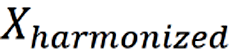 = calibrated value aligned to Shanghai reference;
 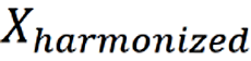 = raw measurement from each center.

