Copyright
©The Author(s) 2023.
World J Psychiatry. Nov 19, 2023; 13(11): 967-972
Published online Nov 19, 2023. doi: 10.5498/wjp.v13.i11.967
Published online Nov 19, 2023. doi: 10.5498/wjp.v13.i11.967
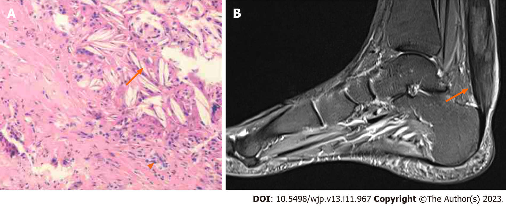
Figure 1 Imaging of left ankle joint and histopathological analysis of the resected specimen.
A: Histological findings of left Achilles tendon xanthoma shows foamy macrophages (arrowheads) and lipid crystal (arrows); B: Left ankle joint magnetic resonance imaging reveals a mild increase in signal intensity on the proton density-weighted image, accompanied by linear hypointensity within.
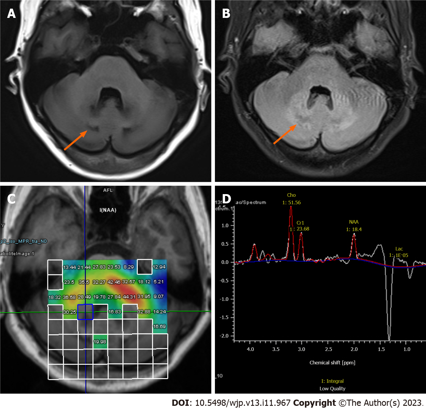
Figure 2 Brain imaging findings.
A: Brain magnetic resonance imaging reveals patchy symmetrical abnormal signals in the dentate nuclei and deep medulla of bilateral cerebellar hemispheres and a slight decrease in signal intensity on T1 weighted image; B: Slight increase in signal intensity on fluid attenuated inversion recovery; C: Magnetic resonance spectroscopy shows right cerebellar dentate nuclei; D: Decreases in N-acetylaspartate intensities and increases in lactate signals and lipid signals.
- Citation: Ling CX, Gao SZ, Li RD, Gao SQ, Zhou Y, Xu XJ. Cerebrotendinous xanthomatosis presenting with schizophrenia-like disorder: A case report. World J Psychiatry 2023; 13(11): 967-972
- URL: https://www.wjgnet.com/2220-3206/full/v13/i11/967.htm
- DOI: https://dx.doi.org/10.5498/wjp.v13.i11.967


