©The Author(s) 2021.
World J Psychiatr. Oct 19, 2021; 11(10): 681-695
Published online Oct 19, 2021. doi: 10.5498/wjp.v11.i10.681
Published online Oct 19, 2021. doi: 10.5498/wjp.v11.i10.681
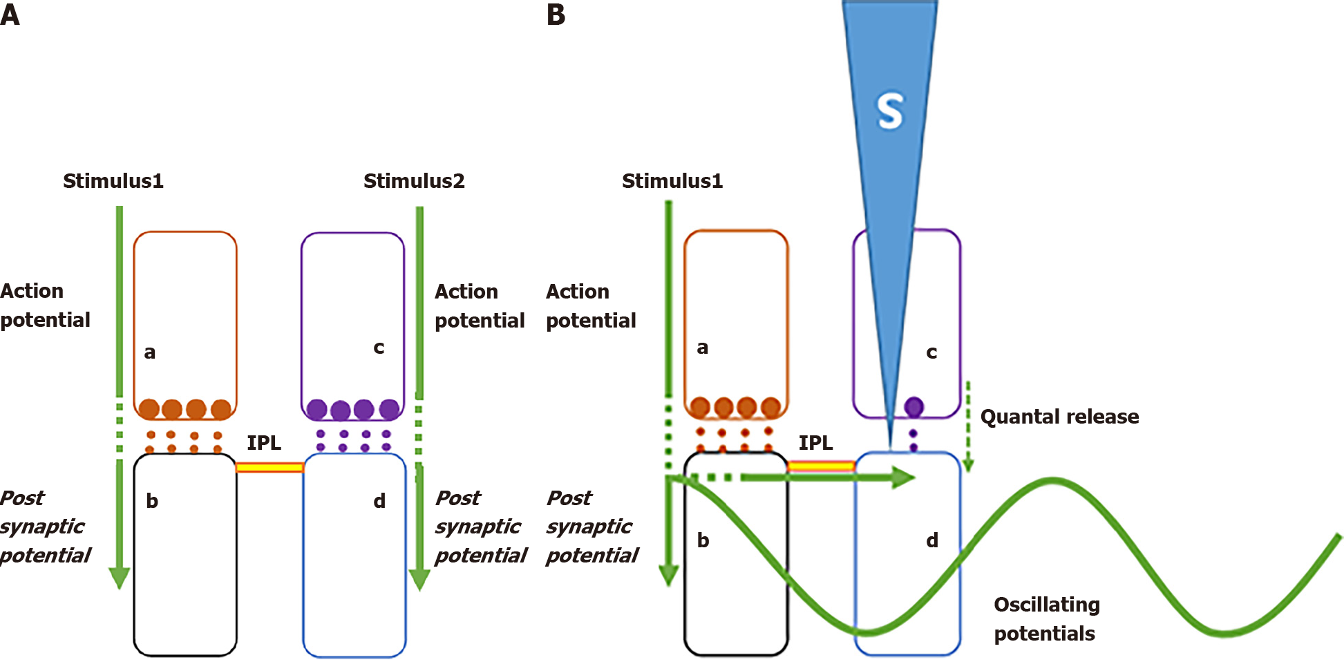
Figure 1 Generation of internal sensation of memory which is used as a reference mechanism to examine internal sensation of pleasure.
A: During associative learning between stimulus 1 and 2, signals from these stimuli propagate towards converging locations where inter-postsynaptic functional LINKs (IPLs) between postsynaptic terminals (spines) b and d occur. Spines b and d are depolarized intermittently when action potentials arrive at their presynaptic terminals a and c respectively, and the head regions of these spines are continuously being depolarized by quantally-released neurotransmitter molecules from their presynaptic terminals. These provide a dominant state that a spine is depolarized by its presynaptic terminal that in turn receive signals from stimuli from the environment and is a necessary background condition for generating internal sensation of memory; B: In the above mentioned background state, arrival of stimulus 1 reactivates IPL b-d to cause an incidental lateral activation of postsynaptic terminal d to spark a cellular hallucination (shown using a blue triangle marked S inside) of a sensory stimulus arriving from the environment through its presynaptic terminal c. Details of the method by which sensory qualia of semblions can be determined was described previously[3]. This matches with the expectation of a mechanism for memory[10]. Waveform: Synaptic transmission through synapse a and b and propagation of depolarization through IPL b-d contribute vector components of oscillating extracellular potentials whose frequency needs to be maintained in a narrow range for inducing internal sensation of memory. Specific electrophysiological findings in locations where sensory stimuli converge were found to correlate with behavioural motor actions indicative of specific brain functions. Long-term potentiation (LTP) that can be induced at locations where sensory stimuli converge is an example[87,88]. After application of a high-energy stimulus at a region rich in synapses and following a delay of at least 20 to 30 s[89,90], application of a regular stimulus at the same location generates a potentiated effect when recorded from the postsynaptic dendritic region or postsynaptic neuronal soma. Ability to induce LTP has shown several correlations with animals’ ability to learn. It was possible to explain how learning-induced formation of IPLs is artificially produced in a delayed scaled-up manner during experimental LTP induction[11]. By keeping correlation between the ability to generate internal sensation of memory that matches with sensory features of the item whose memory is retrieved and the ability to induce LTP at specific locations[11], specific electrophysiological changes that can be induced at these locations can be examined to arrive at a mechanistic explanation for internal sensation of pleasure. Since there are specific electrophysiological changes that can be induced at locations responsible for different brain functions, a comparative examination can be carried out to understand how different internal sensations are generated (modified from[3]).
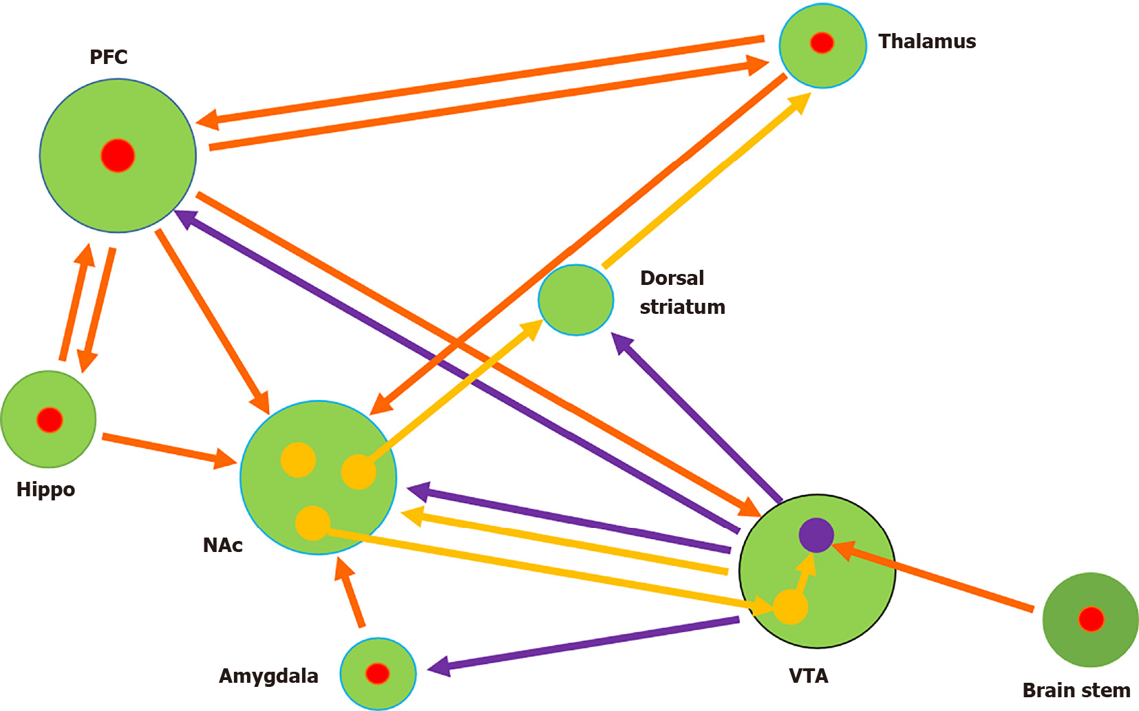
Figure 2 Input and output connections of nucleus accumbens.
One set of spines of medium spiny neurons (MSNs) in the nucleus accumbens receive excitatory inputs (red arrows) from several regions. The same spines of MSNs that receive excitatory inputs receive dopaminergic inputs (violet arrows) from ventral tegmental area (VTA). Another set of MSN spines receive inputs from inhibitory interneurons (orange arrow) in the VTA. Large green circles: Different brain regions. Small circles: Predominant cell types within brain regions (Red: Excitatory; Orange: Inhibitory; Violet: Dopaminergic). PFC: Prefrontal cortex; NAc: Nucleus accumbens; VTA: Ventral tegmental area.
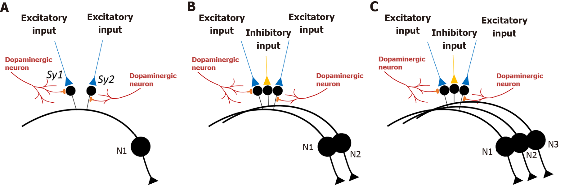
Figure 3 Interactions between spines of medium spiny neurons that synapse with excitatory inputs and spines of medium spiny neurons that synapse with inhibitory inputs.
A: Adjacent spines (small black circles) on the dendrite of a medium spiny neuron (MSN) (N1) (cell body is drawn in a large black circle) that synapse with two excitatory inputs (in blue) to form synapses Sy1 and Sy2). Golgi staining shows that spines are physically well separated from each other on the dendrites of MSNs[23,24] such that the inter-spine space is occupied by spines of other dendrites or processes of other neurons or glial cells. This increases the probability that the nearest spine to a spine on the dendrite of a MSN is most likely a spine that belongs to another neuron, or in rare cases belongs to another branch of the same neuron. Note that dopaminergic inputs synapse either onto the head or neck region of spines that synapse with excitatory inputs; B: In between two adjacent spines of MSN N1 shown in figure A, there is a spine that belongs to a second MSN (N2). This spine synapses with an inhibitory input (in orange). All the spines are electrically insulated from each other by fluid extracellular matrix. Natural stimulants or cocaine abuse causes release of dopamine that will cause enlargement of spines that synapse with excitatory inputs. Since the spine that synapses with the inhibitory input is spatially interposed between the expanding spines, inter-postsynaptic functional LINKs are formed between those three spines; C: Same configuration of two spines of MSNs that synapse with excitatory inputs and one middle spine synapsing with inhibitory input. Here, these spines belong to three different MSNs.
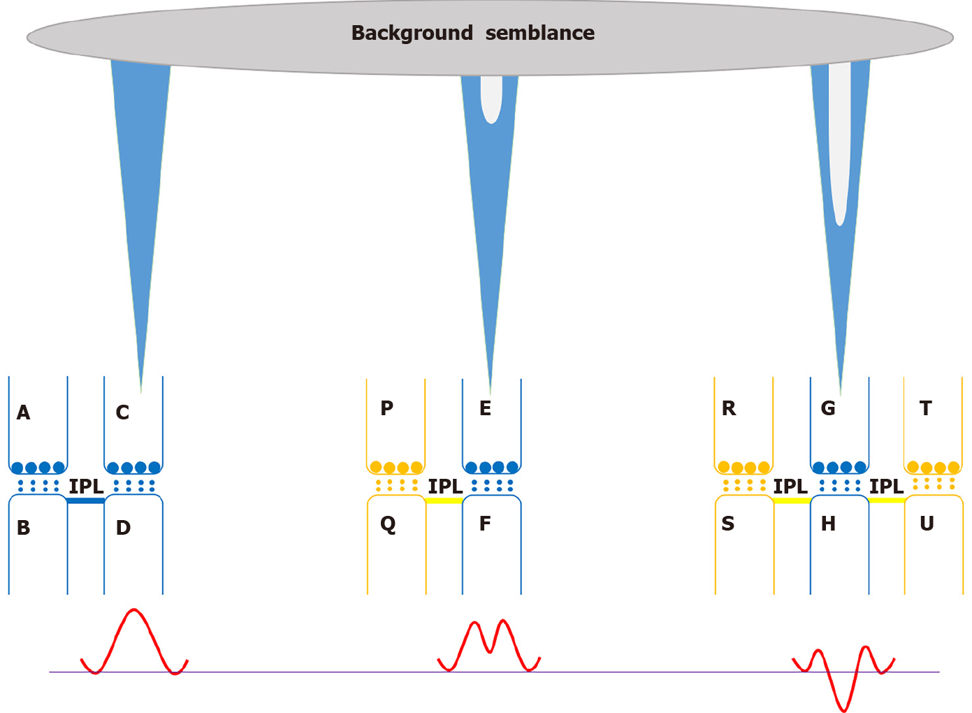
Figure 4 A schematic representation of units of internal sensations whose integral generates pleasure in a background net semblance of the system.
Left: Normal semblance as shown in Figure 1. Inter-postsynaptic functional LINK (IPL) between spines B and D that receive excitatory inputs (in blue). An action potential arriving at presynaptic terminal A from a stimulus depolarizes its postsynaptic terminal B, which in turn propagates through the IPL B-D and depolarizes (shown as a positive waveform) inter-LINKed spine D. This generates units of internal sensations (shown as a blue triangle projected upwards from presynaptic terminal C that denotes semblance). Middle: Reactivation of an IPL between spine Q of a medium spiny neuron (MSN) that synapse with an inhibitory input (in orange) and another spine F of a MSN that synapse with an excitatory input (in blue) results in spread of hyperpolarization from spine Q to spine F. This leads to changes in both the waveform of spine depolarization (red waveform) and conformation of semblance (shown as a dip in the blue triangle). Right: Here, spine H that synapses with an excitatory input (in blue) forms IPLs with spines S and U that synapse with inhibitory inputs (orange). Net effect of hyperpolarization results in profound changes in both waveform of spine depolarization (red waveform) and conformation of semblance (shown as a deep dip in the blue triangle). Net effect of changes in semblances from all the inter-LINKed spines of MSNs in nucleus accumbens (NAc) is expected to generate a special semblance of pleasure.
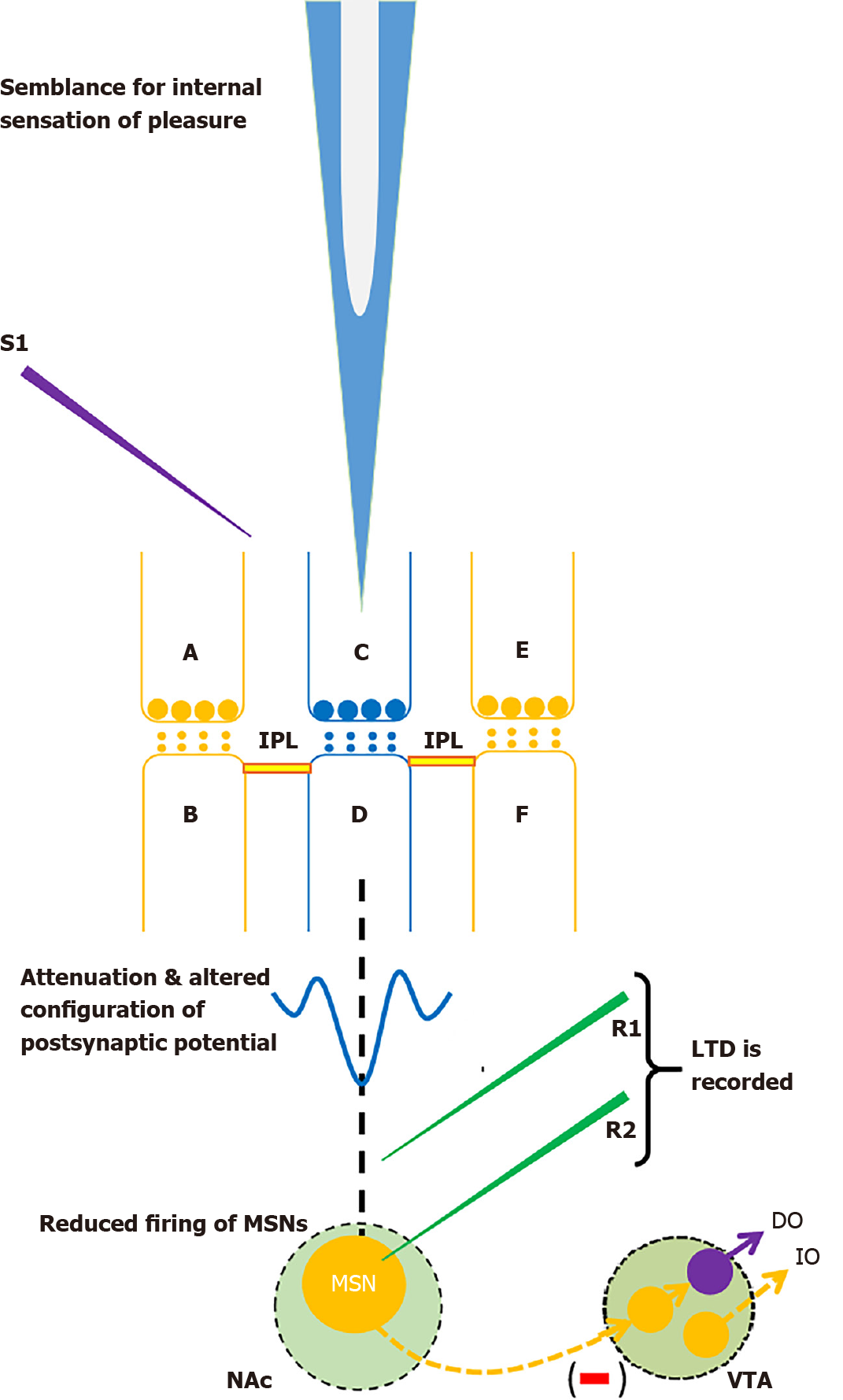
Figure 5 Nucleus accumbens circuitry that matches with constraints from several findings.
Spines B and F belonging to different medium spiny neurons (MSNs) synapse with inhibitory inputs arriving through presynaptic terminals A and E respectively. Spine D on a third MSN synapses with excitatory input arriving through presynaptic terminal C. Two inter-postsynaptic functional LINKs (IPLs) are formed between spines B, D and F. These IPLs between spines that synapse with excitatory and inhibitory inputs lead to mixing of depolarization on the spines that synapse with excitatory input and hyperpolarization on the spines that synapse with inhibitory inputs. This leads to alternation of configuration of the net postsynaptic potentials as shown in a trace. Net semblance from a large number of inter-LINKed spines is expected to generate a special semblance for internal sensation of pleasure. Due to propagation of hyperpolarization, sum of potentials reaching many MSNs may not cross the threshold for firing, which leads to reduced firing of MSNs. A specific stimulation pattern applied at the presynaptic region using stimulating electrode S1 results in the formation of a large number of the above-mentioned types of IPLs in a time-dependent manner (inferred from delay between stimulation and long-term depression (LTD) induction[17,18]) resulting in LTD recorded from either recording electrode R1 (extracellular field recording) or R2 (whole-cell recording). Two inhibitory inputs to MSN and one inhibitory output from MSN are shown in orange. Excitatory synapse is shown in blue. Dopaminergic neuron of ventral tegmental area is shown in violet. DO: Dopaminergic output; IO: Inhibitory output; VTA: Ventral tegmental area.
- Citation: Vadakkan KI. Framework for internal sensation of pleasure using constraints from disparate findings in nucleus accumbens. World J Psychiatr 2021; 11(10): 681-695
- URL: https://www.wjgnet.com/2220-3206/full/v11/i10/681.htm
- DOI: https://dx.doi.org/10.5498/wjp.v11.i10.681


