Copyright
©The Author(s) 2015.
World J Exp Med. Aug 20, 2015; 5(3): 160-163
Published online Aug 20, 2015. doi: 10.5493/wjem.v5.i3.160
Published online Aug 20, 2015. doi: 10.5493/wjem.v5.i3.160
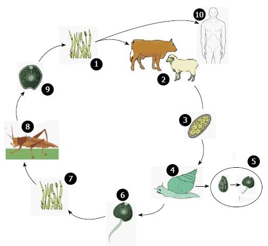
Figure 1 Eurytrema spp.
life cycle. 1: Infection by grazing on infected grass; 2: Vertebrate host (cow or sheep); 3: Unembryonated eggs released in feces; 4: Ingestion of eggs by Bradybaena sp.; 5: Sporocyst mother in sporocyst son in the digestive tube between 90 and 350 d, according to envionmental conditions. Sporocyst sons containing cercarea are released in the environment few hours before dawn; 6: Cercarea released in the environment; 7: Ingestion of cercarea by Conocephalus sp.; 8: Development of infective metacercarea inside grasshopper; 9: Infective metacercarea released in the environment; 10: Possible human infection by consumption of infected salad. One egg of Eurytrema spp. produces 100 sporocyst sons which turn into 200 cercareas and latter 20000 infective metacercarea. Adapted from Tessele, CDC and Bennett[6,15,16]. CDC: Centers for Disease Control and Prevention.
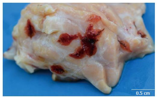
Figure 2 Eurytrema sp.
in bovine pancreas. The fluke is red, oval shaped and measuring between 8 to 13 mm in length by 6 to 7 mm width. The pancreas of infected animals has a harder consistency and hyperplasic ducts partially filled with adult parasites are observed.
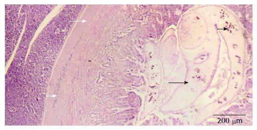
Figure 3 Histopathology of pancreas with Eurytrematosis.
Moderate proliferation of connective tissue (white large arrow), severe hyperplasic ducts containing adult parasites (black large arrow), parasite eggs (black short arrow) and mild inflammatory infiltrates of lymphocytes, eosinophils, plasma cells and macrophages (white short arrow). The islets of Langerhans are usually not affected but in most severe cases, they are replaced by connective tissue.
- Citation: Schwertz CI, Lucca NJ, Silva ASD, Baska P, Bonetto G, Gabriel ME, Centofanti F, Mendes RE. Eurytrematosis: An emerging and neglected disease in South Brazil. World J Exp Med 2015; 5(3): 160-163
- URL: https://www.wjgnet.com/2220-315X/full/v5/i3/160.htm
- DOI: https://dx.doi.org/10.5493/wjem.v5.i3.160


