Published online May 9, 2022. doi: 10.5492/wjccm.v11.i3.192
Peer-review started: October 24, 2021
First decision: December 2, 2021
Revised: December 8, 2021
Accepted: March 15, 2022
Article in press: March 15, 2022
Published online: May 9, 2022
Processing time: 195 Days and 4 Hours
Renal cell carcinoma (RCC) is an aggressive tumor, with an incidental discovery in most patients. Classic presentation is rare, and it has a high frequency of local and distant metastasis at the time of detection.
We present a rare case of a 58-year-old man with a ball-shaped thrombus in the right atrium at the time of first incidental identification of RCC in the emergency department. Cardiac metastasis, especially thrombus in the right atrium, is rare. It could either be a bland thrombus or a tumor thrombus, and physicians should consider this potentially fatal complication of RCC early at the time of initial presentation.
Ball-shaped lesions in the right atrium are rare, and bland thrombus should be differentiated from tumor thrombus secondary to intracardiac metastasis.
Core Tip: The classic presentation of renal cell carcinoma is rare, and patients can present with atypical symptoms and local or distant metastasis at the time of initial detection. Cardiac metastasis, especially thrombus in the right atrium, is rare and emergency physicians should consider it early at the time of presentation. Detection of a ball-shaped lesion in the right atrium is rare, and the patient should undergo appropriate evaluation with the aim to differentiate bland thrombus from a tumor thrombus secondary to intracardiac metastasis, as it aids in therapeutic management and prognosis.
- Citation: Pothiawala S, deSilva S, Norbu K. Ball-shaped right atrial mass in renal cell carcinoma: A case report. World J Crit Care Med 2022; 11(3): 192-197
- URL: https://www.wjgnet.com/2220-3141/full/v11/i3/192.htm
- DOI: https://dx.doi.org/10.5492/wjccm.v11.i3.192
Renal cell carcinoma (RCC) is an aggressive tumor constituting about 3% of all adult malignancies, with a peak incidence in the sixth and seventh decades of life. The classic triad of flank pain, abdominal mass and hematuria is seen in 10% of the cases. Most cases have an incidental discovery[1,2] with local or distant metastases in 25% of the cases at the time of detection. About 10% of these patients have tumor extension into the renal vein and inferior vena cava[3], while only 1% of the total cases have the tumor extending into the right atrium[4]. We present a rare case of a 58-year-old man with a right atrial ball thrombus secondary to metastasis at the time of first incidental identification of RCC in the emergency department (ED).
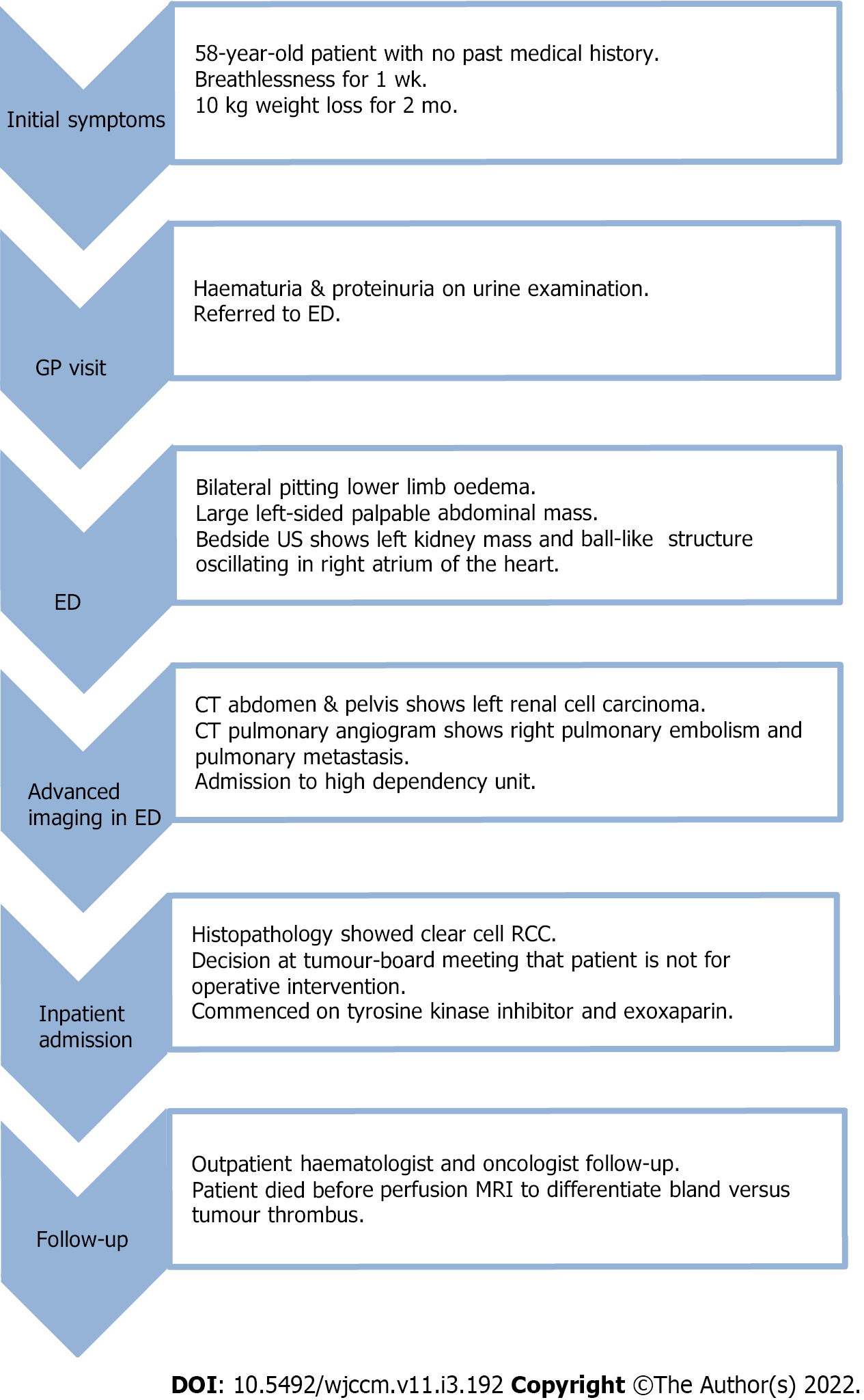
A 58-year-old man presented to the ED with complaints of breathlessness and reduced effort tolerance for 1 wk.
A 58-year-old man presented to the ED with complaints of breathlessness and reduced effort tolerance for 1 wk. He denied chest pain, orthopnea or paroxysmal nocturnal dyspnea. He also noted a 10 kg weight loss over the last 2 mo. He went to a family physician where he was found to have hematuria and proteinuria on urine examination, and hence referred to the ED. He denied hematuria, lower urinary tract symptoms or fever.
He had no past medical history and was not on any long-term medication.
On presentation, his vital signs were stable but he appeared pale and had bilateral pitting lower limb edema up to the knees. Abdominal examination revealed a left sided large, palpable abdominal mass, but there was no rectal bleeding or melena. Examination of respiratory, cardiac and neurological systems was normal.
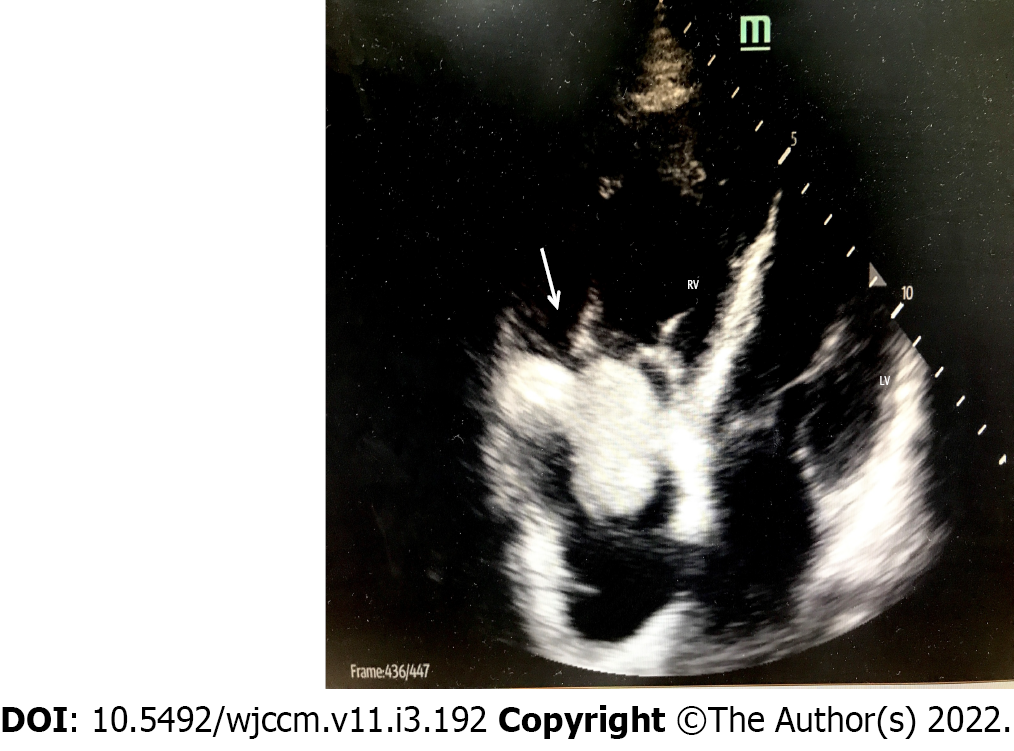
His electrocardiogram showed normal sinus rhythm with T-wave inversions and ST-segment flattening in all leads, along with deep T inversions in leads V2-V4. Bedside ultrasound showed a large heterogeneous mass arising from the left kidney suspicious of RCC. Bedside echocardiogram showed a ball-like structure in the right atrium (Figure 1), oscillating between the right atrium and right ventricle intermittently during cardiac cycles, suspicious for a tumor thrombus. There was no pericardial effusion but the right ventricle appeared larger than the left ventricle, suggestive of signs of right heart strain. Blood investigations showed hemoglobin 7.3 g/dL, elevated serum creatinine 155 mol/L (1.75 mg/dL) and N-terminal pro-B type natriuretic peptide 7325 pg/mL. The remaining blood tests, including liver panel, troponin-T, prothrombin time/activated partial thromboplastin time and severe acute respiratory syndrome coronavirus 2 polymerase chain reaction, were normal.
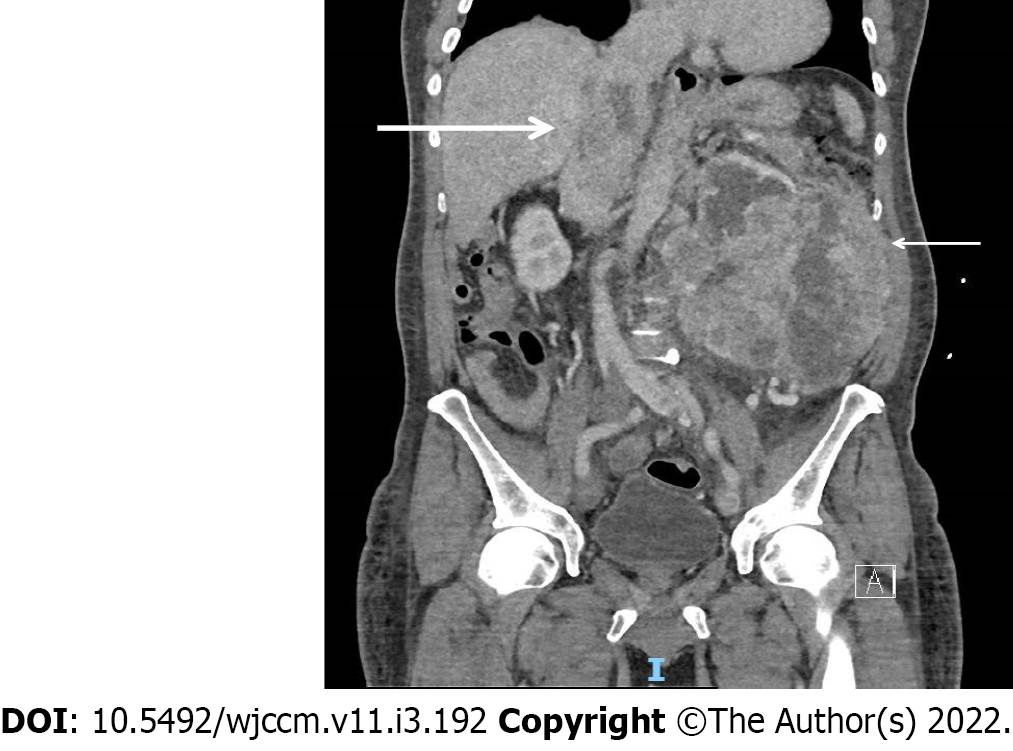
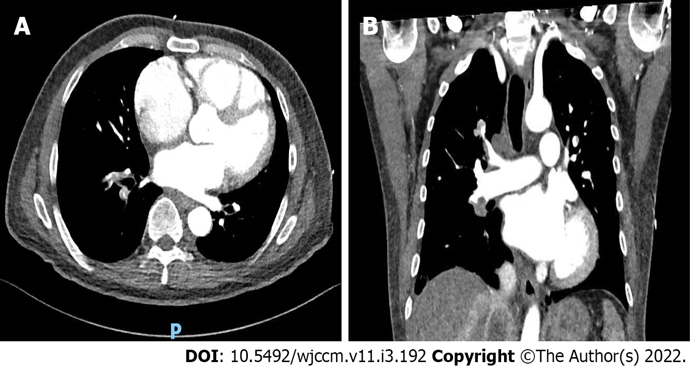
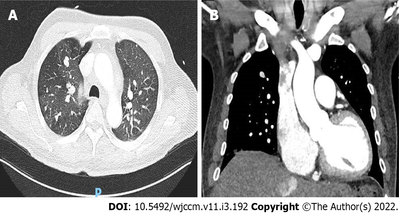
Chest X-ray revealed mild pulmonary venous congestion. Computed tomography (CT) of the abdomen and pelvis (Figure 2) revealed a 13.9 cm × 15.8 cm × 16 cm irregular, heterogeneous left renal mass, suspicious of RCC, with extension of tumor into left renal vein and inferior vena cava (IVC) and metastasis to the liver. CT pulmonary-angiogram showed extensive right pulmonary embolism (Figure 3) with evidence of right heart strain and pulmonary arterial hypertension, as well as pulmonary metastasis (Figure 4). A thrombus was noted in the enlarged right ventricle and right atrial appendage.
The patient was commenced on subcutaneous enoxaparin 80 mg and admitted to a high dependency unit. Histopathology after imaging-guided biopsy of the left renal tumor revealed clear cell RCC. After discussion at the multidisciplinary tumor board meeting, he was not scheduled for cytoreductive nephrectomy and thrombectomy in view of metastatic burden.
He was diagnosed to have left RCC with ball-shaped thrombus in the right atrium, with associated right pulmonary embolism as well as pulmonary metastasis.
He was treated with enoxaparin 80 mg twice daily and tyrosine kinase inhibitor (TKI) pazopanib 800 mg once daily.
He was discharged and scheduled for outpatient follow-up with a hematologist and oncologist (Figure 5).
RCC can present as a solitary metastatic lesion or as a widespread systemic disease, but cardiac metastasis from RCC is extremely rare. The incidence of a thrombus in the right atrium is 0.75%, which is significantly lower than that that of a thrombus in the left atrium[5]. Thrombus in the right atrium is usually located at the atrial appendage or atrial wall. A ball thrombus in the right atrium is even rarer[5].
The ball-shaped lesion in our patient’s right atrium could either have been a bland thrombus or a tumor thrombus that spread along the IVC. In patients with malignancy, bland thrombus is usually a venous thrombus. Pathogenesis of ball thrombus is still unclear and it can be difficult to make a diagnosis. The challenge is to correctly differentiate bland thrombus from a tumor thrombus secondary to intracardiac metastasis, as it aids in appropriate therapeutic management as well as prognosis. On perfusion magnetic resonance imaging (MRI), heterogeneous enhancement of this ball-shaped lesion and presence of blood products within it suggests a tumor thrombus secondary to RCC metastases. On the contrary, a bland thrombus shows unrestricted diffusion and complete nulling of the mass on MR perfusion imaging[6]. Bland thrombus may resolve after thrombolytic and anticoagulant therapy, unlike tumor thrombus. Our patient unfortunately died before further evaluation could be conducted.
Combining cytoreductive nephrectomy and/or metastasectomy with chemotherapy helps in palliation. The possible surgical option for metastatic RCC extending into the right atrium and causing pulmonary embolism, in this patient, was cardiopulmonary bypass with deep hypothermic circulatory arrest, which is a safe and efficient method for removal of the tumor and thrombus[7]. Manual repositioning of the tumor thrombus out of the right atrium into the IVC on the beating heart is also a safe approach with low risk of tumor thrombembolization[8]. In recent times, the progression free survival has improved due to advances in chemotherapy, immunotherapy and TKI[9]. The overall long-term prognosis of patients with metastatic RCC is poor, with a median survival of 6-12 mo.
The classic presentation of RCC is rare, and patients can present with atypical symptoms and local or distant metastasis at the time of initial detection. Cardiac metastasis, especially thrombus in the right atrium, is rare and emergency physicians should consider it early at the time of presentation. Detection of a ball-shaped lesion in the right atrium is rare, and the patient should undergo appropriate evaluation with an aim to differentiate bland thrombus from a tumor thrombus secondary to intracardiac metastasis, as it aids in therapeutic management and prognosis.
| 1. | Sountoulides P, Metaxa L, Cindolo L. Atypical presentations and rare metastatic sites of renal cell carcinoma: a review of case reports. J Med Case Rep. 2011;5:429. [RCA] [PubMed] [DOI] [Full Text] [Full Text (PDF)] [Cited by in Crossref: 103] [Cited by in RCA: 137] [Article Influence: 9.1] [Reference Citation Analysis (0)] |
| 2. | Doshi D, Saab M, Singh N. Atypical presentation of renal cell carcinoma: a case report. J Med Case Rep. 2007;1:26. [RCA] [PubMed] [DOI] [Full Text] [Full Text (PDF)] [Cited by in Crossref: 7] [Cited by in RCA: 9] [Article Influence: 0.5] [Reference Citation Analysis (0)] |
| 3. | Hunsaker RP, Stone JR. Images in clinical medicine. Renal-cell carcinoma extending into the vena cava and right side of the heart. N Engl J Med. 2001;345:1676. [RCA] [PubMed] [DOI] [Full Text] [Cited by in Crossref: 8] [Cited by in RCA: 9] [Article Influence: 0.4] [Reference Citation Analysis (0)] |
| 4. | Lu HT, Chong JL, Othman N, Vendargon S, Omar S. An uncommon and insidious presentation of renal cell carcinoma with tumor extending into the inferior vena cava and right atrium: a case report. J Med Case Rep. 2016;10:109. [RCA] [PubMed] [DOI] [Full Text] [Full Text (PDF)] [Cited by in Crossref: 4] [Cited by in RCA: 5] [Article Influence: 0.5] [Reference Citation Analysis (0)] |
| 5. | Zhang B, Tian ZW, Zhang J, Yuan KM, Yu H. Calcified ball thrombus in the right atrium: A case report. Iran J Radiol. 2020;17:e95169. [RCA] [DOI] [Full Text] [Cited by in Crossref: 1] [Cited by in RCA: 1] [Article Influence: 0.1] [Reference Citation Analysis (0)] |
| 6. | Elgendy A. Intracardiac metastasis from known renal cell cancer. Available from: https://radiopaedia.org/cases/intracardiac-metastasis-from-known-renal-cell-cancer. |
| 7. | Posacioglu H, Ayik MF, Zeytunlu M, Amanvermez D, Engin C, Apaydin AZ. Management of renal cell carcinoma with intracardiac extension. J Card Surg. 2008;23:754-758. [RCA] [PubMed] [DOI] [Full Text] [Cited by in Crossref: 10] [Cited by in RCA: 10] [Article Influence: 0.6] [Reference Citation Analysis (0)] |
| 8. | Schneider M, Hadaschik B, Hallscheidt P, Jakobi H, Fritz M, Motsch J, Pahernik S, Hohenfellner M. Manual repositioning of intra-atrial kidney cancer tumor thrombus: a technique reducing the need for cardiopulmonary bypass. Urology. 2013;81:909-914. [RCA] [PubMed] [DOI] [Full Text] [Cited by in Crossref: 13] [Cited by in RCA: 11] [Article Influence: 0.8] [Reference Citation Analysis (0)] |
| 9. | Barata PC, Rini BI. Treatment of renal cell carcinoma: Current status and future directions. CA Cancer J Clin. 2017;67:507-524. [RCA] [PubMed] [DOI] [Full Text] [Cited by in Crossref: 391] [Cited by in RCA: 625] [Article Influence: 69.4] [Reference Citation Analysis (0)] |
Open-Access: This article is an open-access article that was selected by an in-house editor and fully peer-reviewed by external reviewers. It is distributed in accordance with the Creative Commons Attribution NonCommercial (CC BY-NC 4.0) license, which permits others to distribute, remix, adapt, build upon this work non-commercially, and license their derivative works on different terms, provided the original work is properly cited and the use is non-commercial. See: https://creativecommons.org/Licenses/by-nc/4.0/
Provenance and peer review: Invited article; Externally peer reviewed.
Peer-review model: Single blind
Specialty type: Critical care medicine
Country/Territory of origin: Singapore
Peer-review report’s scientific quality classification
Grade A (Excellent): 0
Grade B (Very good): B
Grade C (Good): C
Grade D (Fair): D
Grade E (Poor): 0
P-Reviewer: Benekli M, Turkey; Yang RM, China S-Editor: Guo XR L-Editor: Kerr C P-Editor: Guo XR


