Copyright
©The Author(s) 2024.
World J Crit Care Med. Mar 9, 2024; 13(1): 86866
Published online Mar 9, 2024. doi: 10.5492/wjccm.v13.i1.86866
Published online Mar 9, 2024. doi: 10.5492/wjccm.v13.i1.86866
Figure 1 The leg.
A: Right thigh raw areas with necrosis of surrounding soft tissues and slough; B: Raw areas over left thigh and leg.
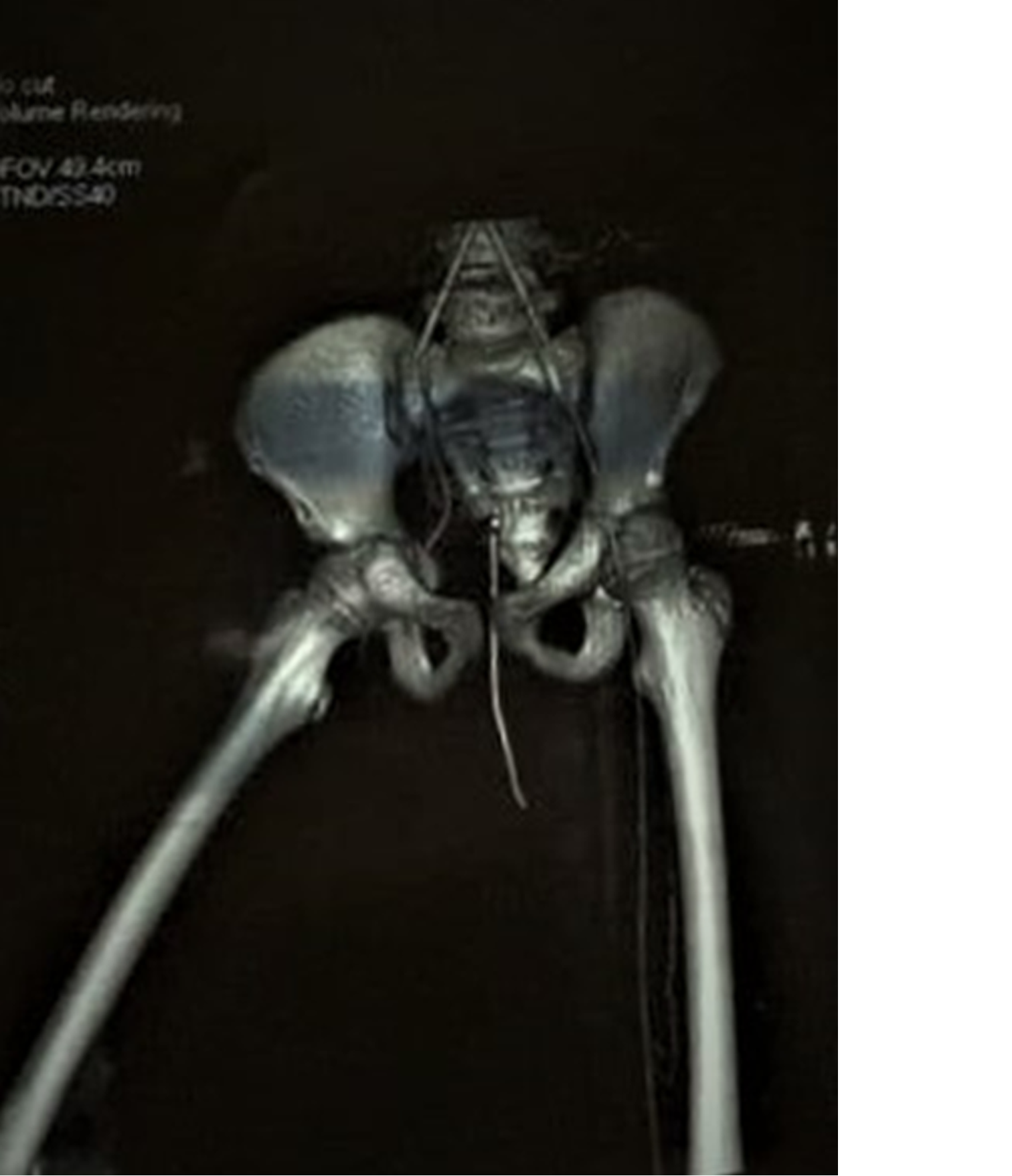
Figure 2 Computed tomography angiography showing non-opacification of the right external iliac artery.
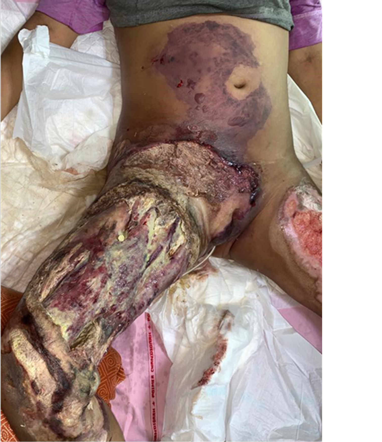
Figure 3 Rapidly progressive necrosis of the anterior abdominal wall.
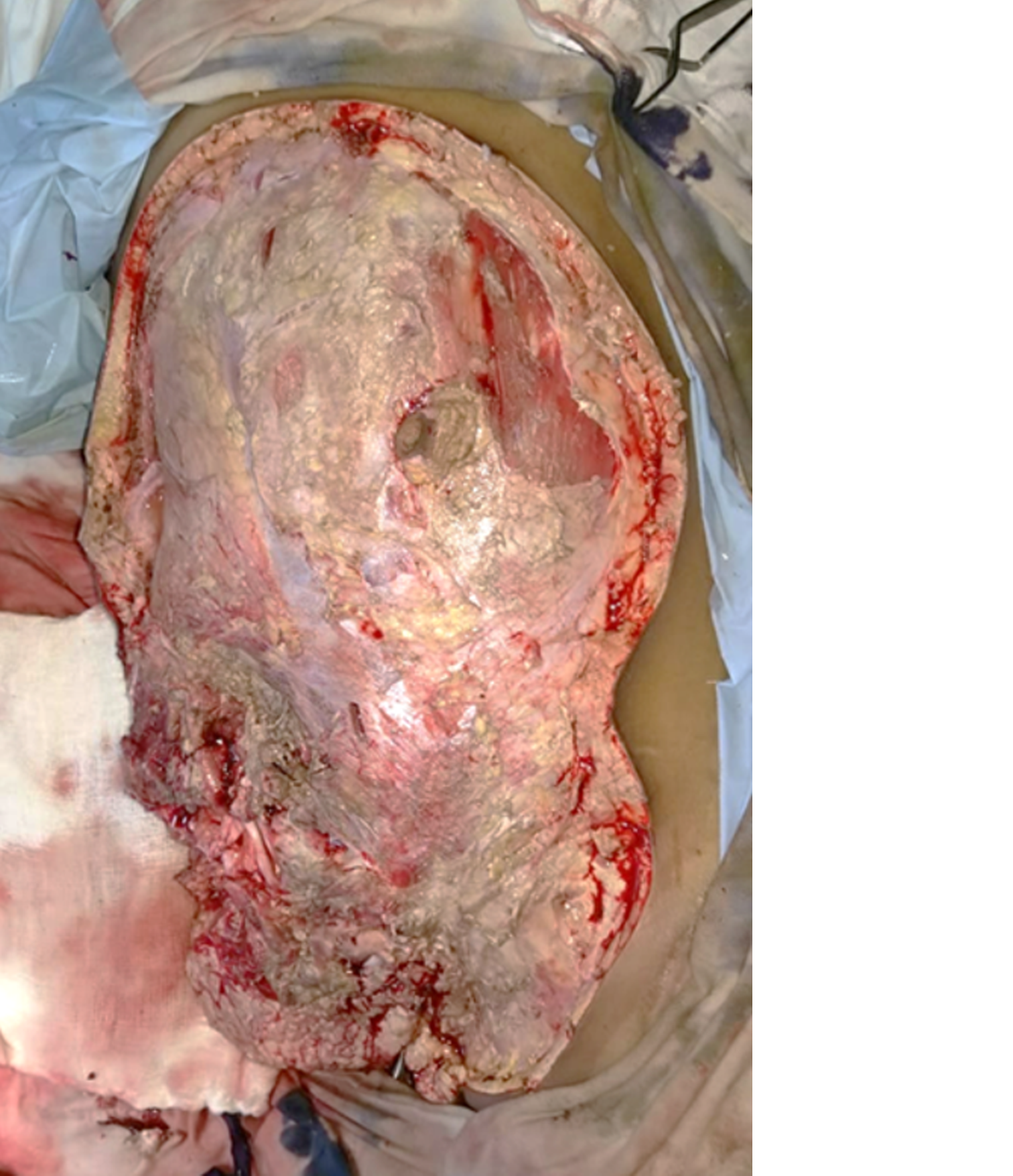
Figure 4 Postoperative wound status after right hip disarticulation and debridement of the anterior abdominal wall and perineum.
- Citation: Parashar A, Singh C. Angioinvasive mucormycosis in burn intensive care units: A case report and review of literature. World J Crit Care Med 2024; 13(1): 86866
- URL: https://www.wjgnet.com/2220-3141/full/v13/i1/86866.htm
- DOI: https://dx.doi.org/10.5492/wjccm.v13.i1.86866


