©The Author(s) 2022.
World J Crit Care Med. Dec 9, 2023; 12(5): 248-253
Published online Dec 9, 2023. doi: 10.5492/wjccm.v12.i5.248
Published online Dec 9, 2023. doi: 10.5492/wjccm.v12.i5.248
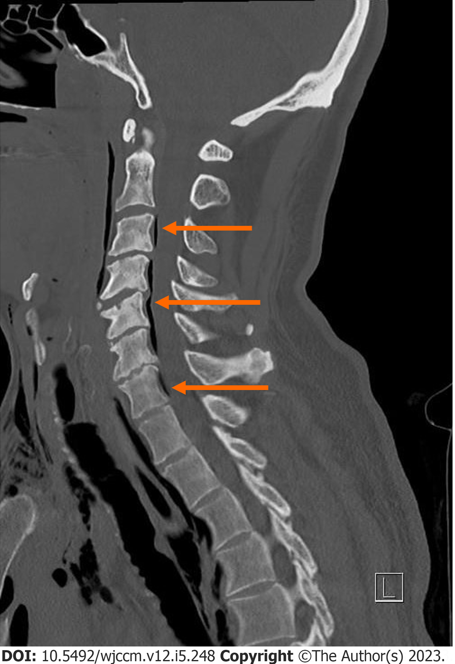
Figure 1
Sagittal computed tomography scan demonstrating peripheral pneumorrhachis in the spinal canal from the level of C3 to C7 (marked by arrows), suggestive of epidural pneumorrhachis.
Figure 2 Axial computed tomography scan demonstrating epidural pneumorrhachis (marked by arrows) in the anterior spinal canal, left lateral spinal canal and both anterior and lateral part of spinal canal.
A: Anterior spinal canal; B: Left lateral spinal canal; C: Both anterior and lateral part of spinal canal.
- Citation: Pothiawala S, Civil I. Narrative review of traumatic pneumorrhachis. World J Crit Care Med 2023; 12(5): 248-253
- URL: https://www.wjgnet.com/2220-3141/full/v12/i5/248.htm
- DOI: https://dx.doi.org/10.5492/wjccm.v12.i5.248


