©The Author(s) 2022.
World J Crit Care Med. May 9, 2022; 11(3): 192-197
Published online May 9, 2022. doi: 10.5492/wjccm.v11.i3.192
Published online May 9, 2022. doi: 10.5492/wjccm.v11.i3.192
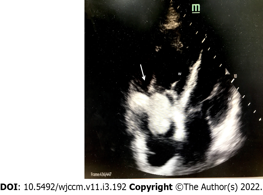
Figure 1 Bedside ultrasound showing ball-shaped thrombus in the right atrium (arrow).
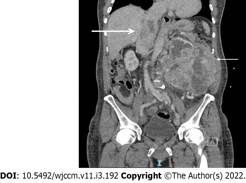
Figure 2 Computerized tomography scan of abdomen and pelvis showing left renal cell carcinoma (thin arrow) invading in to the hepatic portion of inferior vena cava (thick arrow).
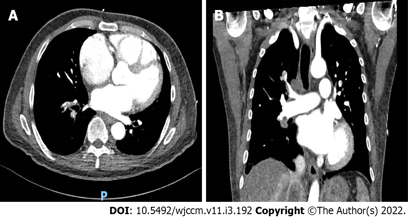
Figure 3 Large filling-defects in the right segmental and subsegmental branches and right lobar and interlobar arteries suggestive of right-sided pulmonary embolism.
A: Right segmental and subsegmental branches; B: Right lobar and interlobar arteries suggestive of right-sided pulmonary embolism.
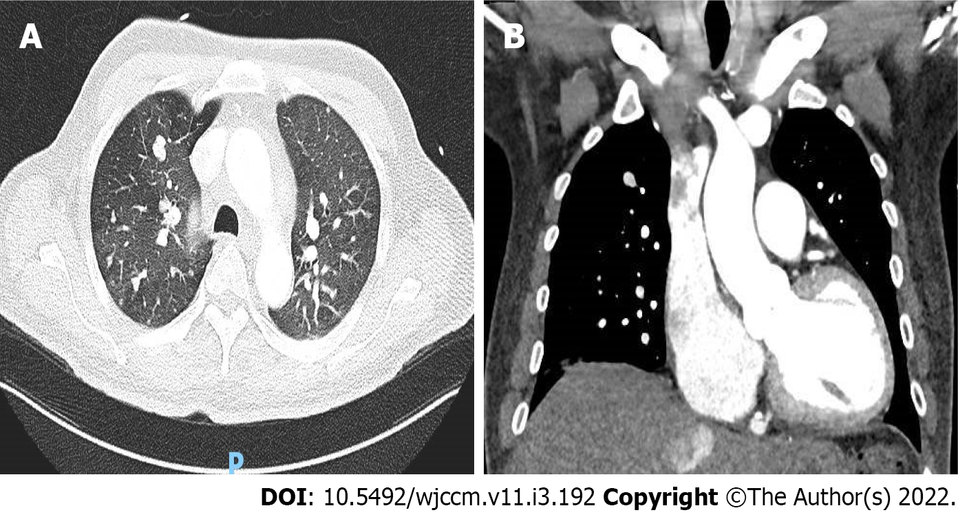
Figure 4 Multiple pulmonary nodules of varying sizes in the lungs suggestive of pulmonary metastasis.
A: Multiple bilateral pulmonary nodules; B: Prominent right sided pulmonary metastasis.
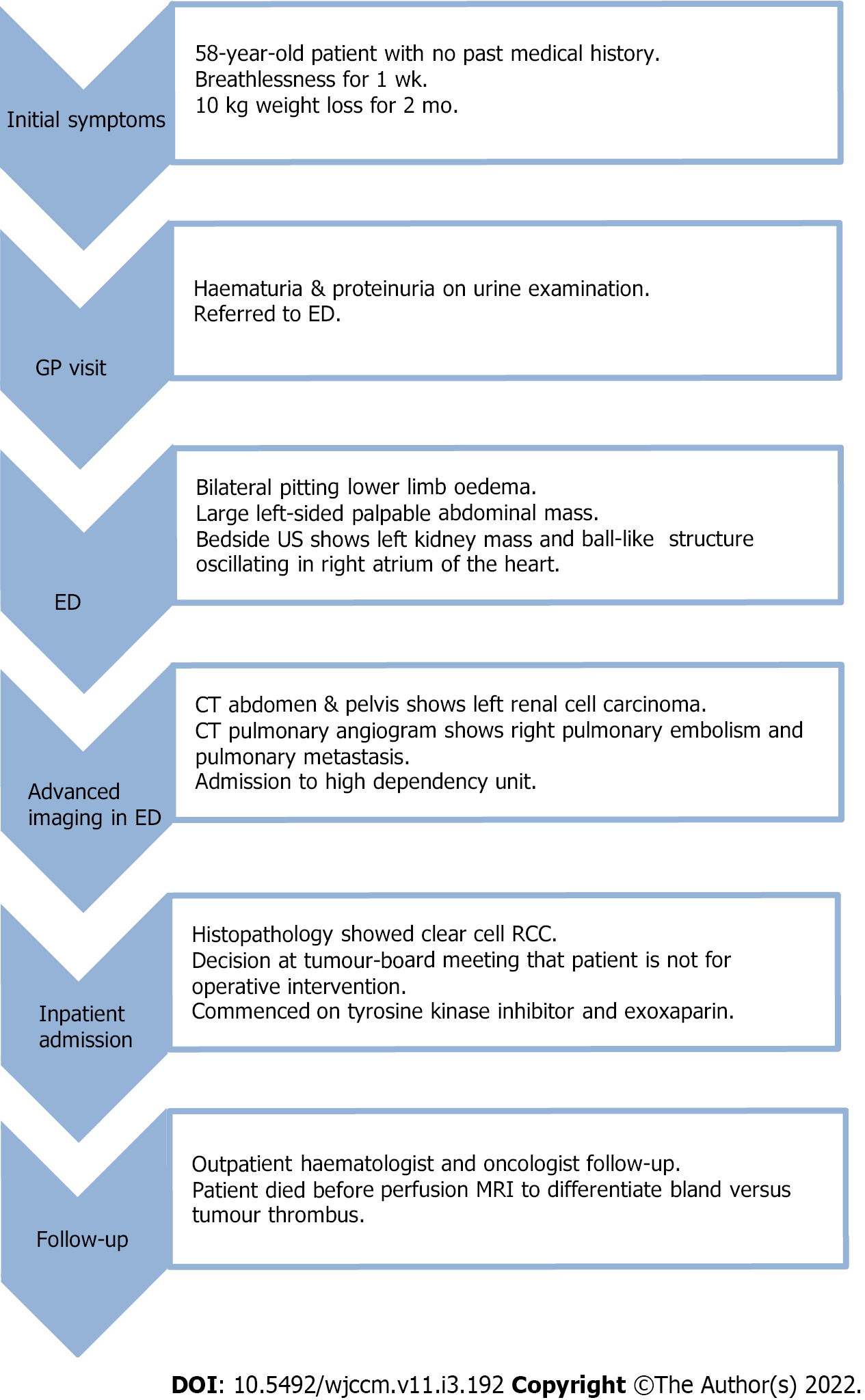
Figure 5 Timeline following case report guidelines.
ED: Emergency department; CT: Computerized tomography; RCC: Renal cell carcinoma; MRI: Magnetic resonance imaging.
- Citation: Pothiawala S, deSilva S, Norbu K. Ball-shaped right atrial mass in renal cell carcinoma: A case report. World J Crit Care Med 2022; 11(3): 192-197
- URL: https://www.wjgnet.com/2220-3141/full/v11/i3/192.htm
- DOI: https://dx.doi.org/10.5492/wjccm.v11.i3.192


