©2012 Baishideng.
World J Crit Care Med. Apr 4, 2012; 1(2): 50-60
Published online Apr 4, 2012. doi: 10.5492/wjccm.v1.i2.50
Published online Apr 4, 2012. doi: 10.5492/wjccm.v1.i2.50
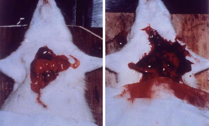
Figure 1 The gross inspection of the lung following cerebral compression in anesthetized rats.
The normal lung in a rat without cerebral compression appears pink color and small size (left); Severe pulmonary edema and hemorrhage occur after cerebral compression. The lung is swollen and dark red in color (right).
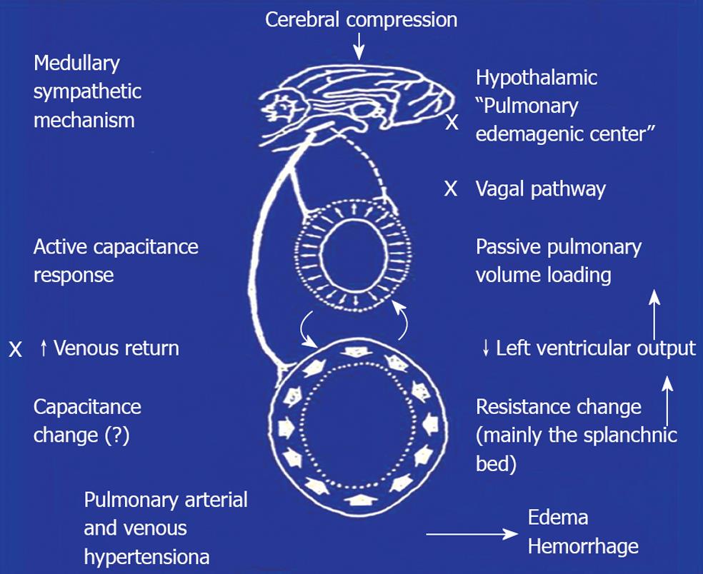
Figure 2 Schematical representation of the neural and hemodynamic mechanisms involved in the pulmonary edema and hemorrhage following cerebral compression.
The hypothalamic “pulmonary edemagenic center” and vagal pathway are not important. Activation of the medullary sympathetic mechanism causes vasoconstriction of the systemic resistance and capacitance vessels, resulting in blood shift from the systemic to pulmonary circulation. A dramatic decrease in the left ventricular output produces pulmonary volume loading. Subsequently, pulmonary arterial and venous hypertension ensue, and finally, severe lung edema and hemorrhage.
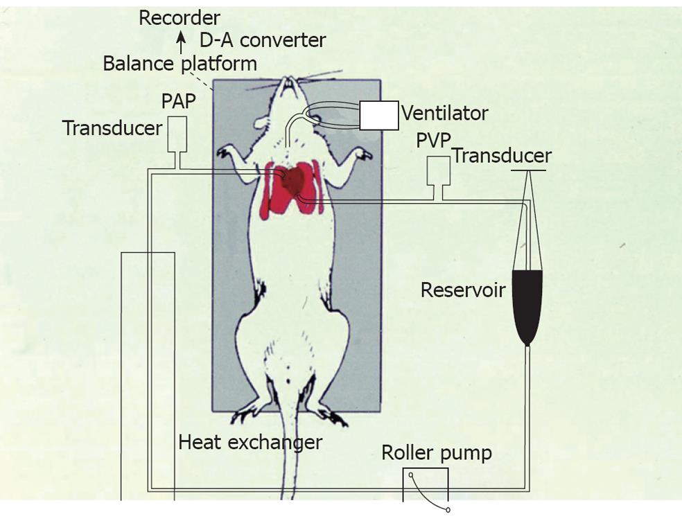
Figure 3 The isolated perfused lung in situ preparation.
A roller pump provides constant flow. The pulmonary arterial pressure (PAP) and venous pressure (PVP) are monitored by pressure transducers. The change in body weight is determined by a balance platform. The increase in body weight reflects the lung weight gain.
- Citation: Su CF, Kao SJ, Chen HI. Acute respiratory distress syndrome and lung injury: Pathogenetic mechanism and therapeutic implication. World J Crit Care Med 2012; 1(2): 50-60
- URL: https://www.wjgnet.com/2220-3141/full/v1/i2/50.htm
- DOI: https://dx.doi.org/10.5492/wjccm.v1.i2.50


