©2014 Baishideng Publishing Group Inc.
World J Surg Proced. Jul 28, 2014; 4(2): 23-32
Published online Jul 28, 2014. doi: 10.5412/wjsp.v4.i2.23
Published online Jul 28, 2014. doi: 10.5412/wjsp.v4.i2.23
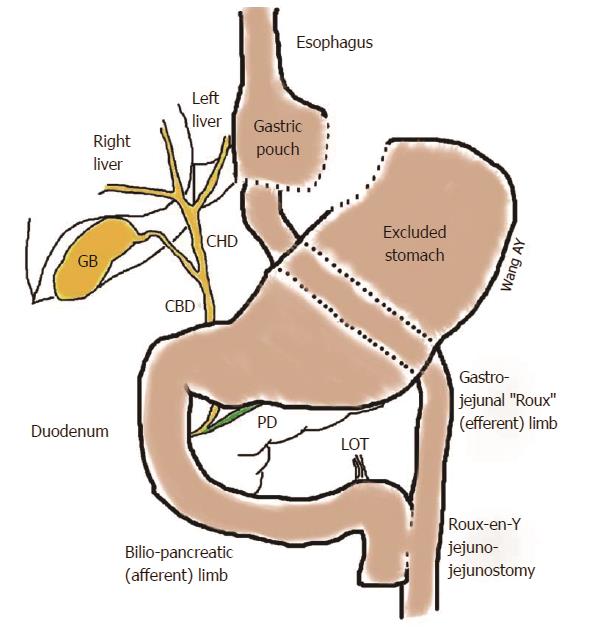
Figure 1 Surgically altered gastroduodenal anatomy found after Roux-en-Y gastric bypass.
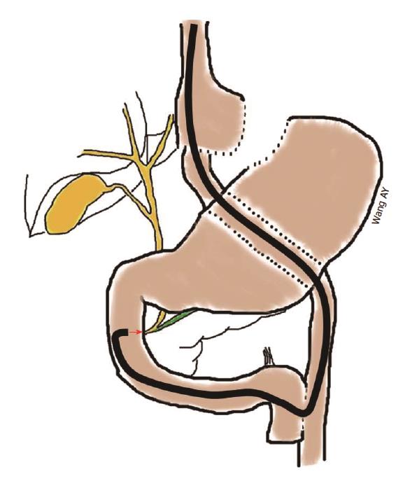
Figure 2 Using a long forward-viewing endoscope, such as an enteroscope with or without a spiral- or balloon-overtube or a pediatric colonoscope, endoscopic retrograde cholangiopancreatography can be performed in patients with Roux-en-Y gastric bypass anatomy or other surgically altered gastroduodenal anatomies.
However, this technique is challenging due to forward-viewing optics, lack of an elevator, a smaller accessory channel, and need for specialized long catheters and guidewires in order to accomplish endoscopic retrograde cholangiopancreatography.
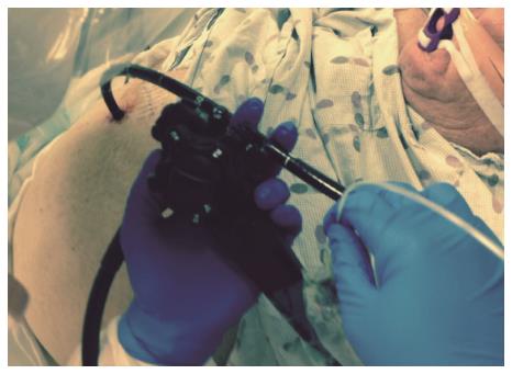
Figure 3 Low-profile, soft, distal attachment cap (D-201-10704, Olympus America, Center Valley, PA) is shown affixed to an enteroscope (SIF-Q180, Olympus America) for use in single-balloon-enteroscopy-assisted endoscopic retrograde cholangiopancreatography.
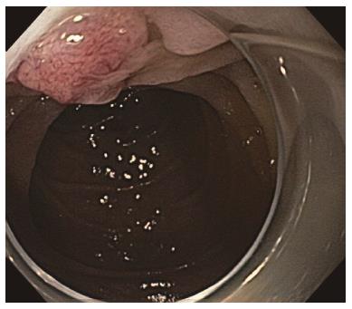
Figure 4 Low-profile, distal attachment cap was used in this case to push back duodenal folds to enable better visualization of the Ampulla of Vater during single-balloon-enteroscopy-assisted endoscopic retrograde cholangiopancreatography.
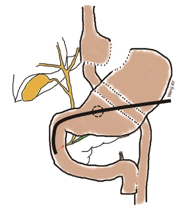
Figure 5 Endoscopic retrograde cholangiopancreatography using a duodenoscope can be performed in patients with Roux-en-Y gastric bypass anatomy by using a gastrostomy, which can be created surgically or endoscopically, to access the remnant stomach.
Depending on the manner in which the gastrostomy is created, immediate or delayed endoscopic retrograde cholangiopancreatography can be performed.
Figure 6 Example of transgastric endoscopic retrograde cholangiopancreatography in a patient with Roux-en-Y gastric bypass anatomy who underwent laparoscopic cholecystectomy and had an intraoperative cholangiogram that was suspicious for small, non-obstructing, bile duct stones.
A 36-Fr Malecot tube had been left across a surgical Stamm gastrostomy. Endoscopic retrograde cholangiopancreatography in the supine position (under general anesthesia) was performed two weeks after surgical gastrostomy using a therapeutic duodenoscope. Despite an awkward scope position requiring the stabilization of the duodenoscope shaft using the left hand (as might be seen during complex colonoscopic polypectomy), biliary sphincterotomy and stone removal were successful.
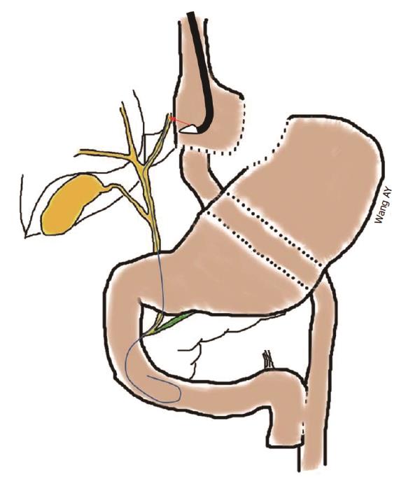
Figure 7 Using a therapeutic linear-array echoendoscope, a 19 G fine-needle-aspiration needle can be directed into dilated intrahepatic bile ducts in the left lobe of the liver.
Once biliary access is established, up to an 0.035” guidewire can be passed antegrade across the extrahepatic bile duct and into the duodenum so as to facilitate rendezvous endoscopic retrograde cholangiopancreatography or antegrade bile duct therapy, such as large papillary balloon dilation to create sufficient space to push stones out of the bile duct and into the duodenum.
- Citation: Cosgrove ND, Wang AY. Endoscopic approaches to biliary intervention in patients with surgically altered gastroduodenal anatomy. World J Surg Proced 2014; 4(2): 23-32
- URL: https://www.wjgnet.com/2219-2832/full/v4/i2/23.htm
- DOI: https://dx.doi.org/10.5412/wjsp.v4.i2.23


