©The Author(s) 2015.
World J Clin Pediatr. Nov 8, 2015; 4(4): 160-166
Published online Nov 8, 2015. doi: 10.5409/wjcp.v4.i4.160
Published online Nov 8, 2015. doi: 10.5409/wjcp.v4.i4.160
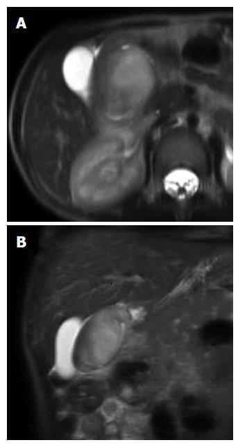
Figure 1 T2- weighted magnetic resonance image in axial (A) and coronal (B) scans, showing the solid mass arising from the duodenum.
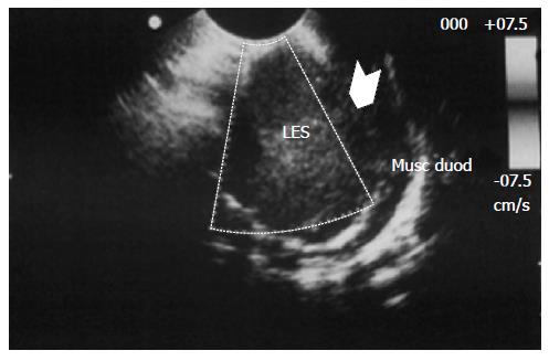
Figure 2 Endoscopic ultrasound showed an homogeneous, hypoechoic mass of 36 mm located into the echo-layer 3 (submucosa), but not completely separated from the muscularis propria (echo-layer 4); in some endoscopic ultrasound scans, the lesion showed features suggesting origin from the muscular layer, thus mimicking gastrointestinal stromal tumors features.
The wall of the lesion was homogeneous and the external part showed hyperechoic spots, suggestive of microcalcifications.
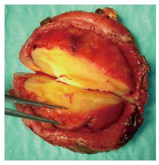
Figure 3 Gross examination: Firm round yellowish mass without necrosis and with an overlying area of mucosal ulceration.
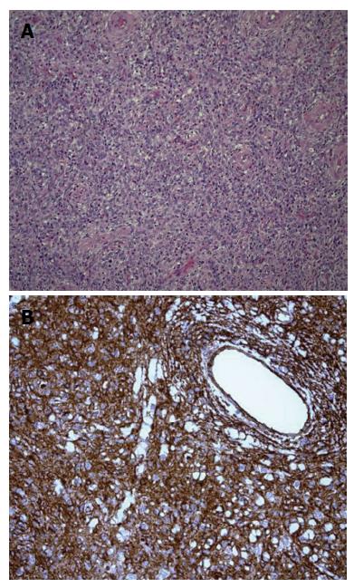
Figure 4 Stromal looking cells has no presence of mitosis or necrotic areas and the lesion was immunoistochemically reactive to vimentin.
A: Stromal looking cells intermixed with marked inflammatory infiltrates rich in eosinophils granulocytes (40 ×); B: Marked immunohistochemical positivity to vimentin.
- Citation: Righetti L, Parolini F, Cengia P, Boroni G, Cheli M, Sonzogni A, Alberti D. Inflammatory fibroid polyps in children: A new case report and a systematic review of the pediatric literature. World J Clin Pediatr 2015; 4(4): 160-166
- URL: https://www.wjgnet.com/2219-2808/full/v4/i4/160.htm
- DOI: https://dx.doi.org/10.5409/wjcp.v4.i4.160


