
















Copyright: ©Author(s) 2026.
World J Clin Pediatr. Sep 9, 2026; 15(3): 118237
Published online Sep 9, 2026. doi: 10.5409/wjcp.118237
Published online Sep 9, 2026. doi: 10.5409/wjcp.118237
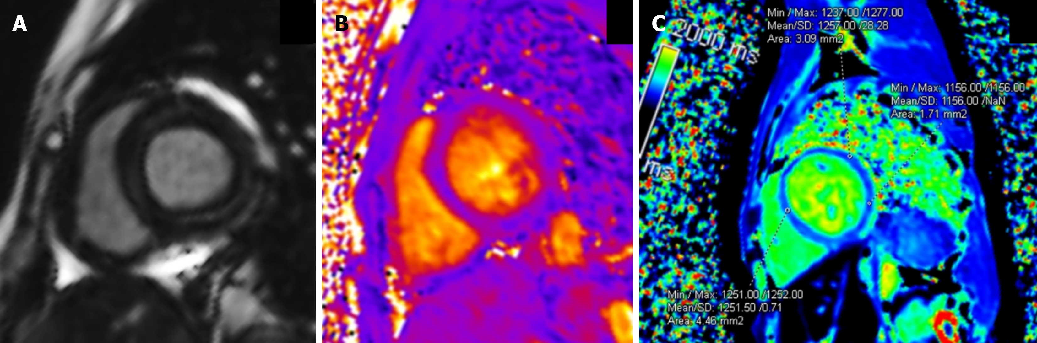
Figure 1 Cardiac magnetic resonance imaging short-axis views at mid-ventricular level in a representative patient at follow-up.
A: Late gadolinium enhancement image showing no myocardial enhancement; B: T2 parametric map demonstrating normal myocardial T2 values, indicating the absence of active myocardial edema; C: Native T1 parametric map demonstrating elevated T1 values, suggesting diffuse myocardial changes. These findings illustrate subclinical myocardial abnormalities detected on cardiac magnetic resonance imaging despite normal echocardiographic findings.
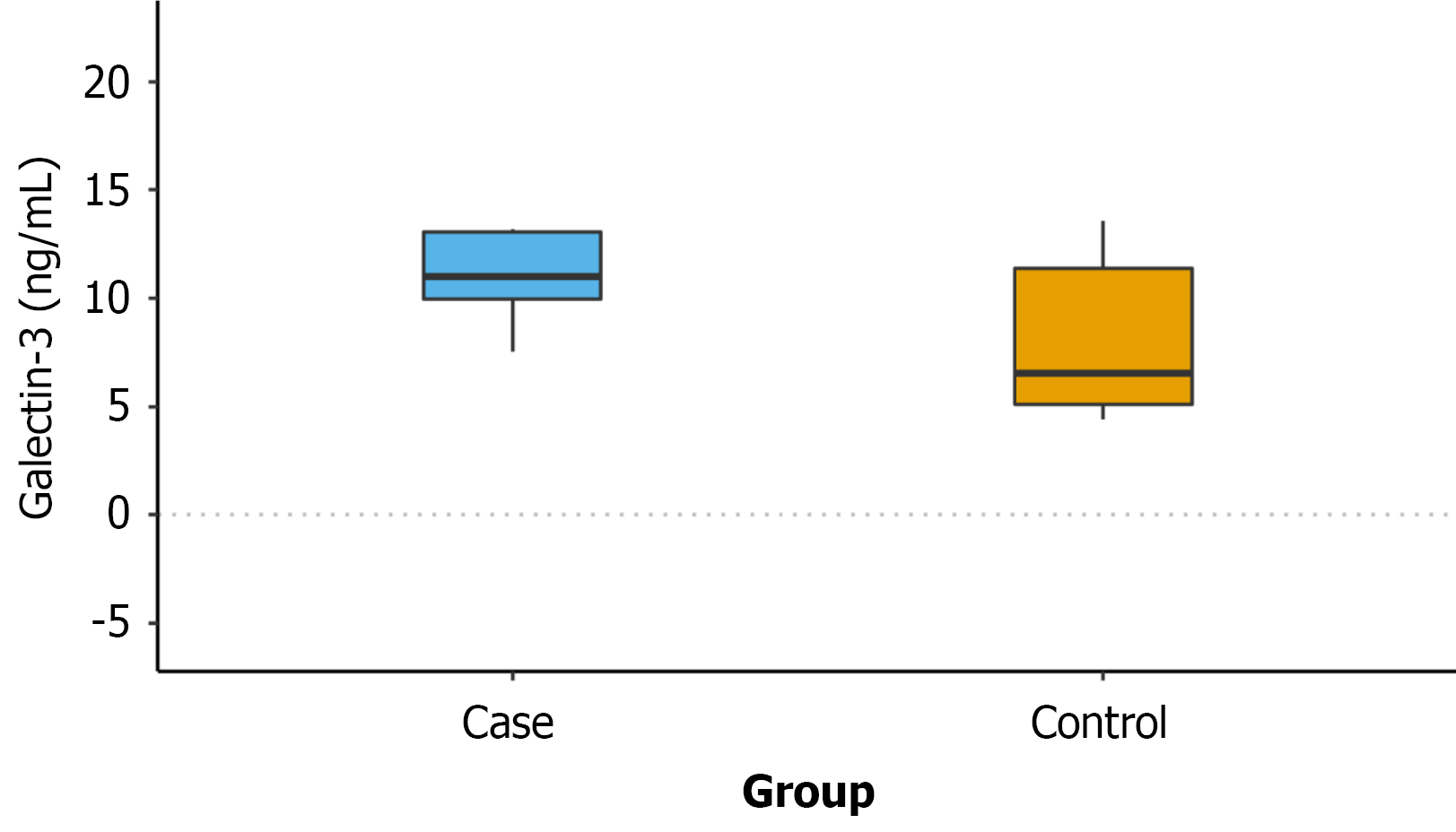
Figure 2 Comparison of serum galectin-3 levels (ng/mL) in patients with prior multisystem inflammatory syndrome in children at a mean follow-up of 23.
58 months and in healthy controls. Although galectin-3 levels were higher in the multisystem inflammatory syndrome in children group, the difference did not reach statistical significance (P = 0.063).
- Citation: OV SK, Pilania RK, Semwal R, Sri G, Kaur T, Subramanian P, Jindal A, Naganur S, Singh S, Singhal M. Persistent long-term subclinical myocardial changes in multisystem inflammatory syndrome in children revealed by cardiac magnetic resonance and galectin-3. World J Clin Pediatr 2026; 15(3): 118237
- URL: https://www.wjgnet.com/2219-2808/full/v15/i3/118237.htm
- DOI: https://dx.doi.org/10.5409/wjcp.118237