©The Author(s) 2025.
World J Clin Pediatr. Dec 9, 2025; 14(4): 107075
Published online Dec 9, 2025. doi: 10.5409/wjcp.v14.i4.107075
Published online Dec 9, 2025. doi: 10.5409/wjcp.v14.i4.107075
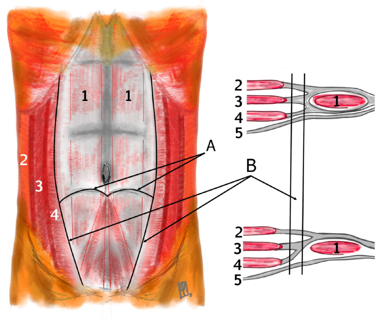
Figure 1 Anatomy of the anterior abdominal wall.
1: m. rectus abdominis; 2: m. obliquus externus; 3: m. obliquus internus; 4: m. transversus abdominis; 5: f. transversalis; A: Semilunar line; B: Spigelian line.
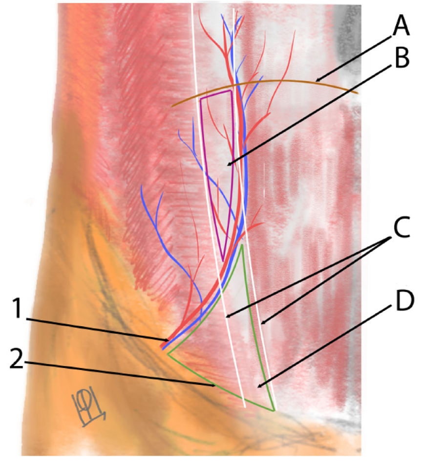
Figure 2 Anatomy of the Spigelian line.
1: Hypogastric vessels; 2: Hesselbach's triangle; A: Semilunar line; B: Upper Spigelian hernia; C: Spigelian line; D: Lower Spigelian hernia.
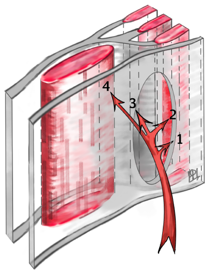
Figure 3 Variants of hernia protrusion location.
1: Subfascial; 2: Within the thickness of the transverse muscle; 3: Beneath the aponeurosis of the external oblique muscle; 4: Subcutaneous.
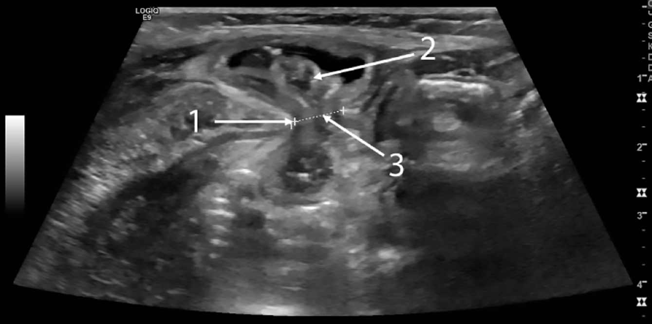
Figure 4 Ultrasound image of hernia protrusion at the time of strangulation.
1: Muscle edge; 2: Loop of intestine; 3: Hernia gates.
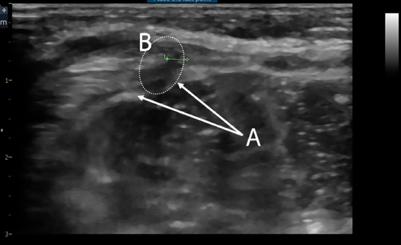
Figure 5 Ultrasound image of the area of interest in the absence of hernia protrusion during examination.
A: Edges of the transverse fascia defect; B: Zone of muscle layer hypoechogenicity.
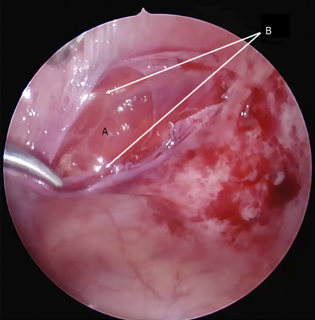
Figure 6 Laparoscopic view of Spigelian hernia.
A: Internal oblique muscle; B: Edges of the transverse fascia defect.
- Citation: Shchapov NF, Kulikov DV, Viborniy MI, Bullikh PV, Keshishian ES, Degtyarev AS. Spigelian hernia in children: A systematic review. World J Clin Pediatr 2025; 14(4): 107075
- URL: https://www.wjgnet.com/2219-2808/full/v14/i4/107075.htm
- DOI: https://dx.doi.org/10.5409/wjcp.v14.i4.107075


