Copyright
©The Author(s) 2015.
World J Obstet Gynecol. Feb 10, 2015; 4(1): 16-23
Published online Feb 10, 2015. doi: 10.5317/wjog.v4.i1.16
Published online Feb 10, 2015. doi: 10.5317/wjog.v4.i1.16
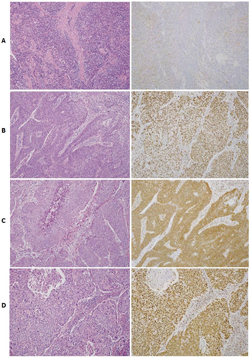
Figure 1 Hematoxylin and eosin staining (left lane) and hepatoma-derived growth factor immunohistochemistry (right lane).
A: CC with HDGF index level 0. The majority of tumor cells were negatively stained for HDGF; B: CC with HDGF index level 1. More than 80% of tumor cells were positively stained in the nucleus, but negatively stained in the cytoplasm; C: CC with HDGF index level 1. More than 80% of tumor cells were positively stained in the cytoplasm, but negatively stained in the nucleus; D: CC with HDGF index level 2. More than 80% of tumor cells were positively stained in both the nucleus and cytoplasm (magnification × 50). CC: Cervical cancer of the uterus; HDGF: Hepatoma-derived growth factor.
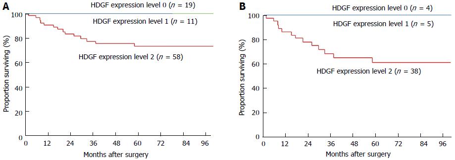
Figure 2 Overall survival curve of patients.
A: Overall survival curve of patients with CC showing HDGF index levels 0, 1 and 2. Significant difference was observed between patients with HDGF index level 2 and other expression levels (P < 0.0001); B: Overall survival curve of patients with pT2-4 CC showing HDGF index levels 0, 1 and 2. Significant difference was observed between patients with HDGF index level 2 and other expression levels (P = 0.0463). CC: Cervical cancer of the uterus; HDGF: Hepatoma-derived growth factor.
- Citation: Song M, Tomoeda M, Jin YF, Kubo C, Yoshizawa H, Kitamura M, Nagata S, Ohta Y, Kamiura S, Nakamura H, Tomita Y. Hepatoma-derived growth factor expression as a prognostic marker in cervical cancer. World J Obstet Gynecol 2015; 4(1): 16-23
- URL: https://www.wjgnet.com/2218-6220/full/v4/i1/16.htm
- DOI: https://dx.doi.org/10.5317/wjog.v4.i1.16


