
















Revised: July 5, 2026
Accepted: July 17, 2026
Published online: August 18, 2026
Processing time: 87 Days and 16.6 Hours
In recent years, many developed countries have implemented prevention pro
To identify clinically significant dysplastic signs of predisposition to bone injuries and to assess their diagnostic value in verifying individuals prone to bone damage.
This cross-sectional study was performed at Sechenov University in accordance with STROBE guidelines. A total of 538 young adults (18-35 years) involved in sports were examined. Participants were divided into a main group with repeated bone injuries and a control group without such history. Screening for more than 80 dysplastic signs was performed through questionnaires, diagnostic tests, anthropometric and phenotypic assessments, and instrumental studies (esophagogastroduodenoscopy, ultrasound, X-ray examination/computed tomography). Statistical analyses were conducted using StatTech v. 4.8.11 software. Significance was set at P < 0.05.
The results indicated that the dysplastic phenotype in young athletes is characterized primarily by external dysplastic stigmata, with osteoarticular manifestations being the most prevalent. Repeated sports-related bone injuries were observed in 6.7% of young adults involved in mass sports (95% confidence interval [CI]: 4.59%-8.81%). The bone fractures/cracks were more frequently associated with sports involving high mechanical load on bones (wrestling, weightlifting). CTD syndrome plays a significant role in bone re-injury genesis. The dysplastic severity (total CTD score) determines the bone re-injury risk (area under the curve = 0.759; 95%CI: 0.665-0.852, P < 0.001). The level of CTD severity, which indicates a predisposition to bone injuries, was defined as the dysplastic re-injury threshold (DRIT) for bone re-injury incidence. The value of this indicator was 44.2 CTD scores. An approach for identifying predisposition to bone injury based on the total CTD score was established. The dysplastic phenotype of individuals predisposed to recurrent bone injury is characterized predominantly by an asthenic body type, dolichostenomelia, arachnodactyly, chest deformities (pectus carinatum or pectus excavatum), a narrow facial skeleton, and a high-arched (gothic) palate. The most common complaints in this group were paresthesia and muscle spasms/cramps. It was demonstrated that none of the dysplastic features associated with recurrent bone injury is sufficient on its own to identify individuals predisposed to bone injury. However, the presence of each of these characteristic dysplastic features is clinically significant and is associated with a substantially increased risk of bone injury.
The high incidence of recurrent bone injuries among young adults participating in recreational sports underscores the importance of its preventive strategies. Screening for connective tissue abnormalities during the initial medical evaluation, with consideration of the characteristic dysplastic features identified in this study, may help identify athletes at increased risk who could benefit from closer monitoring and individualized injury prevention strategies. Determining the DRIT for recurrent bone injury enables early risk stratification and the implementation of targeted preventive measures, supporting personalized strategies to preserve musculoskeletal health in young athletes. The dysplastic phenotype associated with susceptibility to bone injury is characterized by a distinct constellation of dysplastic features that, when considered collectively, may provide the basis for the development of a rapid screening method to identify individuals at increased risk of bone injury.
Core Tip: Recurrent bone injuries in young athletes may reflect underlying connective tissue dysplasia (CTD) rather than isolated traumatic events. Although individual dysplastic features [e.g., arachnodactyly, chest deformities, and a high-arched (gothic) palate] lack sufficient diagnostic accuracy, their cumulative severity defines a DRIT threshold of 44.2 points on the CTD scale, representing a novel indicator of susceptibility to recurrent bone injury. This threshold enables rapid risk stratification without the need for molecular genetic testing, supporting individualized sports participation and targeted injury prevention strategies. A composite phenotype comprising ten readily identifiable external dysplastic features provides a practical screening tool for sports physicians to identify at-risk athletes and help preserve musculoskeletal health.
- Citation: Sankova MV, Achkasov EE, Nikolenko VN, Oganesyan MV, Rizaeva NA, Sankov AV, Pires LAS, Zharikova TS, Zharikov YO. New paradigms for identifying predisposition to bone injuries in young athletes: Significant keys for rapid diagnostics. World J Orthop 2026; 17(8): 123460
- URL: https://www.wjgnet.com/2218-5836/full/v17/i8/123460.htm
- DOI: https://dx.doi.org/10.5312/wjo.123460
In recent years, many developed countries have implemented preventive programs to promote regular physical activity among young people and foster a health-promoting sports environment[1-3]. In this context, a primary objective for sports physicians is the prevention of sports-related musculoskeletal injuries, which are the leading cause of primary health care visits among athletes[4,5]. The prevention of bone injuries is of particular importance, as these injuries are a leading cause of early disability and reduced work capacity among young people[5-7]. Early identification of an un
This cross-sectional study was conducted at Sechenov University in accordance with the STROBE guidelines to ensure methodological transparency and reproducibility. A total of 538 young adults (18-35 years of age) participating in recreational sports were enrolled between December 2024 and August 2025. Participants were excluded if they had differentiated CTD, including osteogenesis imperfecta, Marfan syndrome, Ehlers-Danlos syndrome, Shprintzen-Goldberg syndrome, or Stickler syndrome; cardiovascular diseases (ischemic heart disease, arterial hypertension, myocarditis, primary or secondary cardiomyopathies, congenital or acquired heart disease, or chest trauma); central nervous system disorders (stroke, traumatic brain injury, tumors, or infections); endocrine disorders (diabetes mellitus, hypothyroidism, pheochromocytoma, or primary aldosteronium [Conn syndrome]); or were pregnant or in the postpartum period. These exclusion criteria were applied to ensure a homogenous study population and to minimize potential confounding when evaluating the association between CTD severity and the risk of recurrent bone injury.
All participants training at World Class fitness clubs 2-3 times per week for 2-3 hours per session, following standardized World Class Training System programs designed to improve muscular strength and endurance, coordi
A sports history was obtained to identify the participants’ primary sport and training frequency, as these factors may influence the incidence of sport-related bone injuries. Most participants engaged in endurance-based (cyclic) sports, including running, swimming, race walking, skiing, and cycling. The second and third most common categories were complex coordination sports (e.g., acrobatics, gymnastics, and dance) and speed-strength sports (e.g., track and field events and weightlifting), respectively. Team sports (e.g., football, volleyball, basketball, and hockey) ranked fourth in popularity. The fewest participants were involved in combat sports, including boxing and various forms of wrestling (Figure 1).

Participants were divided into a main group comprising individuals with a history of recurrent bone injuries and a control group comprising those without such a history. The groups were matched for age and sex (Table 1) to minimize potential confounding. Recurrent bone injury was defined as a documented history of two or more sports-related bone injuries (fractures or stress fractures) occurring at the same anatomical site within the musculoskeletal system[19]. Bone injuries were confirmed using medical records from sports physician or orthopedic trauma specialists and radiographic findings, including X-ray and computed tomography (CT) imaging. All recorded injuries were non-contact in nature, occurring during activities such as twisting or turning in the absence of external contact, or resulting from altered biomechanics, abnormal movement patterns, inappropriate training loads, or improper movement technique[20,21]. Injuries involved the bones of the upper or lower extremities and consisted of either stress fractures or fractures classified as type A or type B according to the Arbeitsgemeinschaft für Osteosynthesefragen/Association for the Study of Internal Fixation (AO/OTA) fracture classification system[22].
| Variables | Persons with repeated bone injuries (main group) | Persons without repeated bone injuries (control group) | P value |
| Total number | 36 | 502 | |
| Female number | 26 (72.2) | 362 (72.1) | Р = 0.946 |
| Male number | 10 (27.8) | 140 (27.9) | |
| Age | 24.50 (19.00-27.25) | 23.50 (19.25-27.00) | Р = 0.523 |
The study sample size was calculated using G*Power software, version 3.1.9.4 (Christian-Albrechts-Universität, Kiel, Germany)[23], assuming a moderate effect size (f = 0.3), a statistical power of 85%, and a two-tailed significance level of α ≤ 0.05. The minimum required sample size for each group was determined using the following formula:

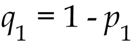
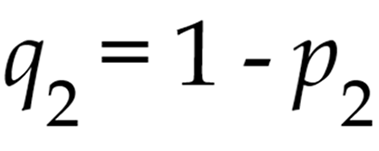
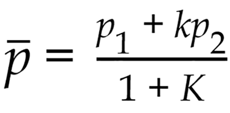


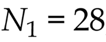

where p1 and p2 represent the proportions in groups N1 and N2, respectively; Δ = |p2 - p1| is the absolute difference between the two proportions; N1 is the sample size of the control group; N2 is the sample size of the main group; α (type I error probability) = 0.05; and β (type II error probability) = 0.2. Thus, a minimum of 28 participants was required for each group. With one control group and one main group, the minimum total sample size required for the study was 56 participants.
This study was conducted in accordance with the clinical guidelines issued by the Russian Scientific Medical Society of Therapists[9]. A comprehensive clinical evaluation was performed using questionnaires, diagnostic tests, detailed anthropometric measurements, calculation of diagnostic ratios and indices, instrumental examinations (esophagogastroduodenoscopy, ultrasound, X-ray examination/computed tomography) and specialist consultations in orthopedics, dentistry, otolaryngology, and ophthalmology. During the structured interview, information was collected on participants’ primary sports activity, training regimen, and history of recurrent sports-related bone injuries.
Anthropometric assessment included measurements of body weight, height, arm span, chest circumference, facial height, bizygomatic width, wrist circumference, hand length, middle finger length, lower body segment length, and foot length. Somatotype was classified using the Verveck index [height (cm) divided by the sum of twice the body weight (kg) and chest circumference (cm)] and the Pignet index [height (cm) minus the sum of body weight (kg) and chest circumference (cm)]. Morphological indicators of dolichostenomelia were defined as an arm length-to-height ratio > 11%, a foot length-to-height ratio > 15%, an arm span-to-height ratio > 1.05, and an upper-to-lower body segment ratio < 0.85. Arachnodactyly was diagnosed when the middle finger length exceeded 10 cm. Joint hypermobility was assessed using the Beighton score. Facial skeletal morphology was evaluated by calculating the facial index as the ratio of morphological facial height to bizygomatic width multiplied by 100[9].
Finally, a standardized phenotypic examination was performed to identify the maximum number of CTD manifestations. According to clinical guidelines, more than 80 characteristic morphometric dysplastic features have been described and are classified into two groups: External dysplastic features, including osteoarticular, cutaneous, muscular, and ophthalmologic manifestations; and visceral dysplastic features, which reflect abnormalities of the internal organs. The diagnostic significance of each external and visceral dysplastic feature is quantified using a standardized diagnostic scoring system[9].
All identified dysplastic features were systematically recorded using a customized data collection form developed in Google Forms (Alphabet Inc., Mountain View, CA, United States). Dysplastic severity was quantified using the total СTD score, calculated as the sum of the diagnostic scores assigned to all dysplastic features identified in each participant. A total CTD score ≥ 17 points was considered diagnostic of CTD syndrome[9].
The study protocol was reviewed and approved by the Local Ethics Committee of I.M. Sechenov First Moscow State Medical University (Sechenov University) (Approval No. 29-24, December 5, 2024) and was conducted in accordance with the principles of the Declaration of Helsinki. All participants were enrolled voluntarily and provided written informed before participation. No financial or material incentives were provided.
Statistical analyses were performed using StatTech version 4.8.11 (StatTech LLC, Russia). Data distribution was assessed for normality using the Kolmogorov-Smirnov test. Continuous variables are presented as the median and interquartile range (IQR). Between-group differences in continuous variables were evaluated using the non-parametric Mann-Whitney U test, whereas categorical variables were compared using Pearson’s χ2 test or Fisher’s exact test, as appropriate. Comparisons among multiple groups were performed using the Kruskal-Wallis test for nonparametric data and one-way analysis of variance (ANOVA) for normally distributed data.
Correlations between variables were assessed using Spearman’s rank correlation coefficient. The strength of associations was estimated using odds ratios (ORs) with 95% confidence intervals (Cis). A prognostic model describing the relationship between a quantitative outcome and predictive factors was developed using linear regression analysis. The discriminative performance of quantitative variables for identifying specific outcomes was evaluated using receiver operating characteristic (ROC) curve analysis, and the optimal cut-off values were determined by maximizing the Youden index. The performance of the CTD score model was further evaluated by assessing calibration using the Hosmer-Lemeshow goodness-of-fit test and a calibration plot derived from a logistic regression model relating recurrent bone injury to the total CTD score. Internal validation was performed using bootstrap resampling (3000 replicates) with Efron-Gong optimism correction. Non-parametric bootstrap resampling (3000 replicates) was also used to estimate the 95%CI for the threshold value and its corresponding diagnostic indicator. In addition, the diagnostic performance of the characteristic dysplastic features was evaluated by calculating sensitivity, specificity, positive and negative predictive values, positive and negative likelihood ratios, and overall diagnostic accuracy. Statistical significance was defined as P < 0.05. All statistical analyses were independently verified by two researchers.
The results showed that the dysplastic phenotype in young athletes was characterized predominantly by external dysplastic feature (Figure 2), with osteoarticular manifestations being significantly more prevalent than other categories of dysplastic features (Table 2).

Recurrent sports-related bone injuries were observed in 6.7% of participants (95%CI: 4.59%-8.81%) with a markedly higher prevalence among individuals participating in high bone-loading sports (e.g., strength and combat sports) (Figure 3). In more than half of these participants (55.6%), recurrent bone injuries were accompanied by recurrent ligament injuries, recurrent joint dislocations or subluxations, or both (Figure 4).


CTD syndrome was found to play a significant role in the development of recurrent bone injuries in young adults. The incidence of recurrent sports-related bone injuries (fractures and stress fractures) was significantly higher among participants with CTD syndrome than among those without CTD [9.0% (95%CI: 6.4%-12.3%) and 0.0% (95%CI: 0.0%-0.0%), respectively; P < 0.001]. The presence of underlying CTD was associated with an approximately 19-fold increase in the risk of recurrent bone injury (OR: 18.68; 95%CI: 11.45-24.56).
The incidence of recurrent bone injury was associated with a greater accumulation of dysplastic features and increased CTD severity. Compared with the control group, which had no history of recurrent musculoskeletal injury, young athletes in the main group demonstrated a significantly higher number of dysplastic features and higher total CTD scores (Table 3). Furthermore, all participants (100%) in the main group met the diagnostic criteria for CTD syndrome.
| Variables | Control group (n = 502) | Main group (n = 36) |
| Prevalence of CTD syndrome (%) | 66.2 (61.4-70.9) | 100.0 (90.3-100.0)a |
| Number of dysplastic signs (%) | 7.00 (5.00-9.00) | 10.00 (8.00-12.00) |
| Dysplastic severity, CTD scores | 22.90 (14.46-32.73) | 44.77 (30.25-53.40) |
For the first time, ROC curve analysis (Table 4) demonstrated that dysplastic severity quantified by the total CTD score, predicts the risk of recurrent bone injury [area under the curve (AUC) = 0.759; 95%CI: 0.665-0.852, P < 0.001] and can be used to identify individuals predisposed to bone injury (Figure 5). An AUC between 0.7 to 0.8 indicates moderate discriminative ability. The model showed a statistically significant overall fit (P < 0.001). Internal validation using non-parametric bootstrap resampling (3000 replicates) with Efron–Gong optimism correction demonstrated negligible optimism in model discrimination (ΔAUC = 0.001; optimism-corrected AUC 0.758 vs apparent AUC, 0.759), indicating minimal overfitting. Model calibration as further evaluated using the Hosmer-Lemeshow goodness-of-fit test and a calibration plot. The Hosmer-Lemeshow test demonstrated good agreement between predicted and observed risk (χ2 = 3.42, df = 3, P = 0.331), and the calibration plot closely followed the line of identity across the range of predicted probabilities (Figure 6).


| Threshold | Sensitivity (%) | Specificity (%) | Positive predictive value | Negative predictive value |
| 44.62 | 88.2 | 52.8 | 96.3 | 24.4 |
| 44.43 | 87.3 | 52.8 | 96.3 | 22.9 |
| 44.20 | 87.3 | 55.6 | 96.5 | 23.8 |
| 44.10 | 87.1 | 55.6 | 96.5 | 23.5 |
| 43.75 | 86.7 | 55.6 | 96.5 | 23.0 |
| 37.53 | 76.3 | 55.6 | 96.0 | 14.4 |
| 37.50 | 76.3 | 58.3 | 96.2 | 15.0 |
| 36.42 | 74.5 | 58.3 | 96.1 | 14.1 |
| 36.00 | 74.1 | 58.3 | 96.1 | 13.9 |
| 35.68 | 73.3 | 58.3 | 96.1 | 13.5 |
| 35.66 | 72.9 | 58.3 | 96.1 | 13.4 |
| 35.46 | 72.5 | 58.3 | 96.0 | 13.2 |
| 35.40 | 72.5 | 61.1 | 96.3 | 13.8 |
| 34.38 | 70.1 | 61.1 | 96.2 | 12.8 |
| 34.37 | 70.1 | 63.9 | 96.4 | 13.3 |
| 34.16 | 69.9 | 63.9 | 96.4 | 13.2 |
| 33.91 | 69.9 | 66.7 | 96.7 | 13.7 |
| 33.64 | 69.5 | 66.7 | 96.7 | 13.6 |
| 32.73 | 66.7 | 66.7 | 96.5 | 12.6 |
| 32.63 | 66.3 | 66.7 | 96.5 | 12.4 |
| 31.42 | 63.3 | 66.7 | 96.4 | 11.5 |
| 31.41 | 63.3 | 69.4 | 96.7 | 12.0 |
| 31.34 | 63.1 | 69.4 | 96.6 | 11.9 |
| 31.25 | 63.1 | 72.2 | 96.9 | 12.3 |
| 31.20 | 62.9 | 72.2 | 96.9 | 12.3 |
| 30.96 | 62.9 | 75.0 | 97.2 | 12.7 |
| 28.14 | 57.0 | 75.0 | 96.9 | 11.1 |
| 28.11 | 56.6 | 77.8 | 97.3 | 11.4 |
| 27.91 | 56.0 | 77.8 | 97.2 | 11.2 |
| 27.87 | 56.0 | 80.6 | 97.6 | 11.6 |
| 26.15 | 50.8 | 80.6 | 97.3 | 10.5 |
| 26.10 | 50.4 | 80.6 | 97.3 | 10.4 |
The threshold level of dysplastic severity associated with susceptibility to bone fractures was defined as the dysplastic re-injury threshold (DRIT) for recurrent bone injury. This threshold corresponds to the total CTD score at the cut-off point that maximizes the Youden index. The DRIT for recurrent bone injury was calculated to be 44.20 CTD points, with a sensitivity of 87.3% and specificity of 55.6% (Figure 7). The 95%CI for the DRIT was 40.5-46.2 CTD points, indicating that the threshold estimate was stable. A DRIT of ≥ 44.2 CTD points is appropriate for confirming an increased risk of recurrent bone injury (rule-in). For screening purposes, where the priority is to identify athletes at low risk of recurrent bone injury (rule-out), a lower cut-off value of 27.87 CTD points may be used. This threshold corresponds to a specificity of 80.6% and a sensitivity of 56.0% (Table 4). The 95%CI for this cut-off was 24.5-31.3 CTD points, indicating that this estimate was also stable.

Thus, an approach for identifying predisposition to bone injury was established based on assessment of the total CTD score, which incorporates all dysplastic morphometric features identified in an individual. A total CTD score of ≥ 44.20 points indicates a predisposition to bone injury. Based on the established DRIT for recurrent bone injury, 15.6% of participants (95%CI: 12.53%-18.67%), approximately one in six individuals, were classified as being predisposed to bone fractures.
For the rapid identification of individuals predisposed to bone injury, external dysplastic morphometric features that are readily identifiable during routine medical examination are of particular importance. In the present cohort of young athletes, individuals predisposed to bone fractures were predominantly characterized by an asthenic body type and increased longitudinal body dimensions (Table 5). Their dysplastic phenotype was distinguished primarily by osteoarticular manifestations. Specifically, a high-arched (gothic) palate, a narrow facial skeleton, dolichostenomelia, and arachnodactyly were observed significantly more frequently in this group. In addition, chest deformities (pectus ex

| Somatometric and osteoarticular dysplastic signs | Persons with repeated bone injuries (main group, n = 36) (%) | Persons without repeated bone injuries (control group, n = 502) (%) |
| Asthenic body type | 52.8 (35.5-69.6)a | 26.4 (22.2-31.1) |
| Underweight | 5.6 (0.7-18.7) | 14.1 (10.8-17.9) |
| Severe underweight | 5.6 (0.7-18.7) | 8.3 (5.8-11.5) |
| Dolichostenomelia | 47.2 (30.4-64.5)b | 16.4 (12.9-20.4) |
| Arachnodactyly | 66.7 (49.0-81.4)b | 30.2 (25.7-35.0) |
| Shoulder blade asymmetry | 30.6 (16.3-48.1) | 35.8 (31.0-40.7) |
| Pelvic bone asymmetry | 19.4 (8.2-36.0) | 9.6 (6.9-12.9) |
| Pterygoid shoulder blades | 19.4 (8.2-36.0) | 14.1 (10.8-17.9) |
| Scoliosis | 36.1 (20.8-53.8) | 27.2 (22.9-31.9) |
| Kyphotic spine deformity | 8.3 (1.8-22.5) | 6.3 (4.1-9.2) |
| Kyphoscoliosis | 8.3 (1.8-22.5) | 1.3 (0.4-2.9) |
| Flat spine | 0.0 (0.0) | 0.3 (0.0-1.4) |
| Hyperlordosis | 2.8 (0.1-14.5) | 2.3 (1.0-4.3) |
| Infundibular chest | 25.0 (12.1-42.2)b | 2.5 (1.2-4.6) |
| Keel-shaped chest | 16.7 (6.4-32.8)b | 2.0 (0.9-3.9) |
| Acetabular protrusion | 2.8 (0.1-14.5) | 1.3 (0.4-2.9) |
| O-shaped legs | 16.7 (6.4-32.8) | 11.8 (8.8-15.4) |
| X-shaped legs | 11.1 (3.1-26.1) | 8.6 (6.0-11.8) |
| Valgus foot position | 16.7 (6.4-32.8) | 6.3 (4.1-9.2) |
| Flatfoot | 50.0 (32.9-67.1) | 47.6 (42.6-52.6) |
| First toe macrodactyly | 2.8 (0.1-14.5) | 0.0 (0.0) |
| Narrow facial skeleton | 86.1 (70.5-95.3)a | 57.7 (52.7-62.6) |
| Gothic palate | 41.7 (25.5-59.2)b | 8.6 (6.0-11.8) |
| Malocclusion | 41.7 (25.5-59.2) | 46.6 (41.6-51.6) |
| Deviated nasal septum | 22.2 (10.1-39.2) | 28.0 (23.6-32.7) |
| Joint hypermobility | 47.2 (30.4-64.5) | 56.9 (51.9-61.9) |
| Joint hypermobility degree (score) | 3.00 (2.00-4.25)a | 4.00 (2.00-6.00) |
| Joint crunching | 63.9 (46.2-79.2) | 68.0 (63.2-72.6) |
| Temporomandibular joint crunching | 33.3 (18.6-51.0) | 35.5 (30.8-40.4) |
| Skin-muscle and ophthalmological dysplastic signs | Persons with repeated bone injuries (main group, n = 36) (%) | Persons without repeated bone injuries (control group, n = 502) (%) |
| Thin skin | 25.0 (12.1-42.2) | 33.5 (28.9-38.4) |
| Skin hyperextensibility | 11.1 (3.1-26.1) | 3.3 (1.8-5.5) |
| Hyperelastic auricles | 16.7 (6.4-32.8) | 10.6 (7.7-14.0) |
| Atrophic striae | 36.1 (20.8-53.8) | 30.7 (26.2-35.5) |
| Keloid scars | 47.2 (30.4-64.5)a | 25.7 (21.5-30.3) |
| Recurrent hernia | 5.6 (0.7-18.7) | 2.5 (1.2-4.6) |
| Abdominal muscle diastasis | 2.8 (0.1-14.5) | 2.3 (1.0-4.3) |
| Nails (exfoliated/fragile) | 41.7 (25.5-59.2) | 32.0 (27.4-36.8) |
| Hair (brittle/excessive hair loss) | 19.4 (8.2-36.0) | 24.9 (20.8-29.5) |
| Lower extremity varicose veins | 5.6 (0.7-18.7) | 4.8 (2.9-7.4) |
| Petechia | 25.0 (12.1-42.2) | 29.2 (24.8-34.0) |
| Myopia | 30.6 (16.3-48.1) | 25.4 (21.2-30.0) |
| Myopia degree, diopters | -3.25 (-2.88 to -6.88) | -4.00 (-2.00 to -6.00) |
| Retinal detachment in anamnesis | 5.6 (0.7-18.7) | 2.5 (1.2-4.6) |
| Blue sclera | 19.4 (8.2-36.0) | 10.8 (8.0-14.3) |
| Muscle pain | 69.4 (51.9-83.7) | 64.2 (59.3-69.0) |
| Нand and foot coldness | 61.1 (43.5-76.9) | 55.9 (50.9-60.9) |
| Paresthesia | 72.2 (54.8-85.8)a | 43.6 (38.6-48.6) |
| Muscle spasms/сramps | 88.9 (73.9-96.9)a | 70.0 (65.3-74.5) |
| Feet pastosity | 27.8 (14.2-45.2) | 13.1 (9.9-16.8) |
The diagnostic performance of each characteristic dysplastic feature for identifying predisposition to bone injury is presented in Tables 7 and 8.
| Characteristic dysplastic signs | Sensitivity (%) | Specificity (%) | Diagnostic accuracy (%) |
| Asthenic body type | 52.8 | 74.9 | 73.4 |
| Dolichostenomelia | 47.2 | 82.3 | 79.9 |
| Arachnodactyly | 66.7 | 68.5 | 68.4 |
| Narrow facial skeleton | 86.1 | 42.2 | 45.2 |
| Infundibular chest | 25.0 | 97.8 | 92.9 |
| Keel-shaped chest | 16.7 | 97.6 | 92.2 |
| Gothic palate | 41.7 | 91.4 | 88.1 |
| Keloid scars | 47.2 | 69.9 | 68.4 |
| Paresthesia | 72.2 | 54.6 | 55.8 |
| Muscle spasms/сramps | 88.9 | 27.5 | 31.6 |
| Characteristic dysplastic signs | Positive predictive value | Negative predictive value | Positive likelihood ratio (%) | Negative likelihood ratio (%) |
| Asthenic body type | 2.1 | 0.6 | 43.8 | 81.1 |
| Dolichostenomelia | 2.7 | 0.6 | 39.5 | 86.4 |
| Arachnodactyly | 2.1 | 0.5 | 52.0 | 80.1 |
| Narrow facial skeleton | 1.5 | 0.3 | 68.8 | 67.2 |
| Infundibular chest | 11.4 | 0.8 | 30.5 | 97.1 |
| Keel-shaped chest | 7.0 | 0.9 | 19.2 | 97.2 |
| Gothic palate | 4.9 | 0.6 | 37.1 | 92.8 |
| Keloid scars | 1.6 | 0.8 | 41.6 | 74.5 |
| Paresthesia | 1.6 | 0.5 | 58.7 | 68.7 |
| Muscle spasms/cramps | 1.2 | 0.4 | 77.4 | 47.0 |
As shown in Table 7, most characteristic dysplastic features demonstrate relatively high diagnostic accuracy for identifying predisposition to bone injury. However, none of these features alone is suitable as an independent diagnostic marker, as each exhibited either low sensitivity or low specificity. This finding is further supported by the positive and negative predictive values presented in Table 8.
At the same time, the positive and negative likelihood ratios indicate that the presence of each of these characteristic dysplastic features is clinically meaningful and is associated with an increased risk of predisposition to bone injury. The strongest associations were observed for pectus excavatum, pectus carinatum, a high-arched (gothic) palate, and dolichostenomelia (Figure 9).

Thus, the dysplastic phenotype associated with a predisposition to bone injury is characterized by a distinct set of dysplastic features that, when considered collectively, may provide the basis for the development of a rapid screening method to identify individuals at increased risk of bone injury.
In recent years, CTD syndrome has become the focus of extensive clinical research across a wide range of medical specialties. The growing scientific and clinical interest in this condition among sports physicians in driven by its adverse effects on physiological adaptation and tolerance to physical exertion in young individuals[8,9,11]. The increasing prevalence of CTD syndrome among young people, together with its substantial impact on musculoskeletal structure and function, underscores the need to investigate the prevalence and clinical significance of dysplastic features in young athletes. The present findings demonstrate that the dysplastic phenotype of young athletes is characterized predominantly by external dysplastic features, with osteoarticular manifestations being significantly more prevalent than other categories of dysplastic features. Alterations in the bones and joints have the greatest impact on musculoskeletal resilience to physical loading and contribute substantially to the development of a predisposition to bone injury[8,11].
Consistent with these findings, 6.7% of young athletes participating in recreational sports (95%CI: 4.59%-8.81%) reported a history of recurrent sports-related bone injuries (fractures and stress fractures). The high prevalence of recurrent sports-related bone injuries in this population underscores the importance of effective prevention strategies. We found that the type of sports activity influenced injury patterns, with bone fractures and stress fractures occurring more frequently in high bone-loading sports, such as strength sports (e.g., weightlifting), and combat sports (e.g., boxing and various forms of wrestling). These findings are consistent with those reported in previous studies[9]. Our results also highlight the importance of a personalized approach to sports selection based on an individual’s predisposition to injury. In contrast, complex coordination sports (e.g., acrobatics, gymnastics, and dance) and endurance-based (cyclic) sports (e.g., general physical training, swimming, race walking, skiing, table tennis, badminton, and cycling) were associated with a lower of bone injury and may therefore be preferable for individuals predisposed to recurrent bone injury.
A history of recurrent fractures or stress fractures suggests an underlying impairment in bone formation[5,8,9,11]. Bone strength depends on adequate mineralization, which is mediated by several non-collagenous macromolecules within the bone matrix. In particular, phosphoproteins promote calcium deposition, alkaline phosphatase facilitates mineralization, proteoglycans mediate the binding of mineral components to collagen, and osteonectin supports the growth of mineral crystals and their aggregation. Defects in the synthesis or remodeling of these structural proteins and proteoglycan complexes are associated with progressive bone loss and deterioration of bone microarchitecture, resulting in increased bone fragility and a higher risk of bone injury during sports participation[24-27]. The coexistence of recurrent bone ligament injuries, recurrent joint dislocations or subluxations, or both in more than half of the participants further supports the polygenic nature of connective tissue dysplasia and suggests concurrent abnormalities affecting multiple connective tissue types[8,9,11].
The present study demonstrated a significant association between CTD syndrome and the incidence of recurrent bone injury incidence in young adults. CTD is a constitutional condition with an effect that is largely independent of external confounding factors[8,9-11]. Notably, CTD was strongly associated with recurrent bone injury (OR: 18.68; 95%CI: 11.45-24.56). Furthermore, recurrent bone injury was associated with a greater accumulation of dysplastic features and increased dysplastic severity.
In this study, dysplastic severity, quantified by the total CTD score, represents a multidimensional, integrative measure. It is calculated as a weighted sum of more than 80 phenotypic dysplastic features spanning multiple connective tissue domains, including somatometric indicators (reflecting body proportions and weight-to-height ratio), osteoarticular features (reflecting musculoskeletal integrity, biomechanics, and resistance to physical loading), cutaneous and muscular features (reflecting connective tissue structure and musculoskeletal biomechanics), ophthalmological features (reflecting connective tissue involvement affecting biomechanics), and visceral manifestations (reflecting internal organ involvement and physiological adaptation to physical activity). This, the total CTD score integrates not only the cumulative effects of polygenic variation but also multifactorial influences, including nutritional status and vitamin and micronutrient levels[8,9-11].
For the first time, the total CTD score, a measure of dysplastic severity, was identified as a predictor of recurrent bone injury (AUC = 0.759; 95%CI: 0.665-0.852, P < 0.001). An AUC of 0.759 falls within the range generally considered to represent acceptable discrimination, supporting its use as a first-line screening and risk-stratification tool. A total CTD score of ≥ 44.20 points, defined as the dysplastic re-injury threshold (DRIT), indicates an increased risk of recurrent bone injury ( rule-in threshold), whereas a total CTD score of ≤ 27.87 points identifies athletes at low risk of recurrent bone injury (rule-out threshold). This two-threshold approach (screening rule-out and confirmatory rule-in) is illustrated in the clinical algorithm shown in Figure 10. Thus, calculation of the total CTD score provides a practical tool for sports medicine practitioners to identify individuals at increased risk of bone injury, exclude those at low risk, and implement targeted preventive strategies. Despite its high diagnostic performance, the routine use of this method during a standard medical examination is challenging because assessment of more than 80 dysplastic morphometric features is time-consuming and requires comprehensive clinical evaluation[9].

External clinical features that can be readily identified during routine medical examination are particularly valuable for rapid screening[10,28-31]. Accordingly, most contemporary studies aimed at developing rapid methods for identifying predisposition to bone injury have focused on readily identifiable external dysplastic features. Several studies have demonstrated that the presence of six or more skeletal dysplastic features in young individuals is associated with reduced bone mineral density and, consequently, an increased risk of osteoporosis and fractures[32]. Similarly, other investigators have reported arachnodactyly, dolichostenomelia, and a high-arched (gothic) palate are highly specific skeletal features associated with an increased risk of osteopenia in young adults[33]. Previous studies have reported a higher incidence of musculoskeletal injuries among athletes with pronounced lower limb asymmetry[34]. In addition, a low body mass index has been identified as a risk factor for musculoskeletal injuries in athletes[5,35]. However, to date, a comprehensive evaluation of all external dysplastic features described in the clinical guidelines[9] for assessing the risk of recurrent bone injury has not been performed.
For the first time, a large clinical cohort was used to comprehensively evaluate the prevalence of all external dysplastic featured described in the clinical guidelines in young athletes with recurrent sports-related bone injuries (main group) compared with a control group. This analysis identified a distinct pattern of dysplastic features, characteristic of the main group. Specifically, the dysplastic phenotype associated with predisposition to bone injury was characterized by the predominance of 10 morphometric dysplastic features. These features primarily reflect abnormalities in the synthesis and/or remodeling of cartilage and bone matrix components. In particular, dolichostenomelia and arachnodactyly are skeletal abnormalities resulting from disproportionate longitudinal growth of the long bones, accompanied by alterations in bone microarchitecture and mineral density[8,9,11,36]. This disproportionate longitudinal growth also contributes to the higher prevalence of an asthenic body type observed in this group. Of particular diagnostic importance for iden
Our findings demonstrate that none of the dysplastic features associated with recurrent bone injury is sufficient on its own to serve as a diagnostic marker for identifying predisposition to bone injury. Although many of the features exhibited relatively high diagnostic performance, each was limited by either low sensitivity or low specificity. Ne
In clinical practice, this screening approach can be incorporated into the standard pre-participation or initial medical examination. The sports physician performs a focused assessment of the ten external dysplastic features that constitute the dysplastic features that constitute the bone injury predisposition phenotype - asthenic body type, dolichostenomelia, arachnodactyly, narrow facial skeleton, pectus excavatum or pectus carinatum, high-arched (gothic) palate, keloid scars, exercise-induced limb paresthesia, and post-exertional muscle cramps (Figure 8). These findings are recorded on a standardized one-page scoring sheet, and corresponding diagnostic scores to summed to calculate the total CTD score. The assessment requires approximately 5-10 minutes, does not require laboratory or molecular testing, and only necessitates brief standardized training in the recognition of dysplastic features. Athletes are then stratified using two thresholds: A lower screening cut-off (27.87 CTD points; higher sensitivity, intended to rule out increased risk) and the DRIT cutoff (≥ 44.2 CTD points; higher specificity, intended to rule in increased risk). Individuals identified as being at increased risk can then be referred for individualized preventive strategies and personalized recommendations regarding sports participation and training programs This workflow is summarized in the clinical algorithm (Figure 10).
This study has several limitations. First, no correction for multiple comparisons was applied. However, the primary conclusions are based on the total CTD score and the combined dysplastic phenotype rather than on any individual feature, as the cumulative dysplastic burden appears to have greater diagnostic relevance for identifying individuals at risk of recurrent bone injury. Second, multivariable regression analysis incorporating potential confounding factors for bone injury was not performed. Quantifying the independent contribution of these factors within an adequately powered multivariable framework is a key objective of planned multicenter studies. Another limitations of this study is the absence of adolescent participants. Their inclusion is particularly important because adolescence is the period during which most athletes begin systematic, increasingly intensive training while simultaneously undergoing peak bone accrual and remodeling. Consequently, dysplastic predisposition to bone injury is likely to manifest earliest and have its greatest clinical impact during this stage, making adolescents the highest-priority population for future screening and preventive interventions. The absence of adolescent participants therefore limits the generalizability of our findings to this clinically important population. Accordingly, adolescents will be a primary focus of the planned multicenter studies. Another limitation of this study is the absence of a cohort of professional athletes. According to the literature, professional athletes exhibit a higher prevalence of certain dysplastic features relevant to specific sports and are exposed to high-intensity, high-volume training loads that may unmask an underlying predisposition to bone injury. Future studies will include independent, multicenter cohorts to validate and extend these findings. External validation in such cohorts represents the primary direction of our future research. Consequently, the present study represents an initial step toward the deve
The high incidence of recurrent bone injuries among young adults participating in in recreational sports underscores the importance of effective preventive strategies. Screening for connective tissue dysplasia during the initial medical examination, based on characteristic external dysplastic features, may help identify athletes at increased risk who could benefit from closer monitoring and individualized preventive interventions. The proposed DRIT for recurrent bone injury provides a practical approach for early risk stratification and the implementation of targeted preventive measures, supporting personalized strategies to preserve musculoskeletal health in young athletes. Furthermore, the characteristic combination of dysplastic features identified in this study may provide the foundation for a rapid screening method to identify individuals at increased risk of recurrent bone injury.
| 1. | Oldridge-Turner K, Kokkorou M, Sing F, Klepp KI, Rutter H, Helleve A, Sinclair B, Meincke L, Mitrou G, Wiseman M, Allen K. Promoting Physical Activity Policy: The Development of the MOVING Framework. J Phys Act Health. 2022;19:292-315. [RCA] [PubMed] [DOI] [Full Text] [Cited by in RCA: 12] [Reference Citation Analysis (0)] |
| 2. | Oldridge-Turner K, Kokkorou M, Vlad I, Klepp KI, Rutter H, Helleve A, Fanian D, Sing F, O’Mara J, Mitrou G, Wiseman M, Allen K. Scan of physical activity policy actions in Europe: Lessons learned from populating the MOVING database. Obes Rev. 2023;24 Suppl 1:e13523. [RCA] [PubMed] [DOI] [Full Text] [Cited by in RCA: 4] [Reference Citation Analysis (0)] |
| 3. | Whiting S, Abu-Omar K, Gelius P, Firmino-Machado J, Rakovac I, Mendes R. Physical activity policy implementation and physical activity levels in the European Union: Are we on track to close the gap between policy and practice? Health Policy. 2025;151:105200. [RCA] [PubMed] [DOI] [Full Text] [Cited by in Crossref: 4] [Cited by in RCA: 2] [Article Influence: 2.0] [Reference Citation Analysis (0)] |
| 4. | Gimigliano F, Resmini G, Moretti A, Aulicino M, Gargiulo F, Gimigliano A, Liguori S, Paoletta M, Iolascon G. Epidemiology of Musculoskeletal Injuries in Adult Athletes: A Scoping Review. Medicina (Kaunas). 2021;57:1118. [RCA] [PubMed] [DOI] [Full Text] [Full Text (PDF)] [Cited by in Crossref: 35] [Cited by in RCA: 52] [Article Influence: 10.4] [Reference Citation Analysis (0)] |
| 5. | Hoenig T, Hollander K, Popp KL, Fredericson M, Kraus EA, Warden SJ, Tenforde AS; Bone Stress Injury Authorship Group. International Delphi consensus on bone stress injuries in athletes. Br J Sports Med. 2025;59:78-90. [RCA] [PubMed] [DOI] [Full Text] [Cited by in RCA: 22] [Reference Citation Analysis (0)] |
| 6. | Gray C, Pizzari T, Murphy MC, Schache AG, Breidahl WH, Klinken S, Bell T, Green B. Gastrocnemius Muscle Strain Injury Characteristics in Elite Male Australian Football Players: A 10-Year Longitudinal Cohort Study. J Orthop Sports Phys Ther. 2025;55:681-688. [RCA] [PubMed] [DOI] [Full Text] [Cited by in Crossref: 1] [Cited by in RCA: 2] [Article Influence: 2.0] [Reference Citation Analysis (1)] |
| 7. | Orchard JW, Meeuwisse W, Derman W, Hägglund M, Soligard T, Schwellnus M, Bahr R. Sport Medicine Diagnostic Coding System (SMDCS) and the Orchard Sports Injury and Illness Classification System (OSIICS): revised 2020 consensus versions. Br J Sports Med. 2020;54:397-401. [RCA] [PubMed] [DOI] [Full Text] [Cited by in Crossref: 36] [Cited by in RCA: 98] [Article Influence: 16.3] [Reference Citation Analysis (0)] |
| 8. | Sankova M, Nikolenko V, Achkasov E, Schamas-esposel M, Sankov A, Orliuk M, Redina S, Oganesyan M, Zharikova T, Pontes-silva A, Zharikov Y. Connective tissue dysplasia and pathogenetic mechanisms as a factor in impaired musculoskeletal adaptation in youth: a sports medicine review. Sport Sci Health. 2025;21:2391-2406. [RCA] [DOI] [Full Text] [Cited by in RCA: 1] [Reference Citation Analysis (0)] |
| 9. | Akatova AE, Arutyunov AG, Baranov BA, Butolin BE, Vereshchagina VG, Vershinina VM, Viktorova VI, Voevoda VI, Garganeeva GN, Gladkikh GN, Gnusaev GS, Gorbunova GM, Grinberg GL, Gromova GO, Domnitskaya DT, Drokina DO, Druk DI, Dubiley DG, Izmozherova IN, Evsevieva EM, Ivanova ID, Kadurina KT, Kanunnikova KO, Karpov KS, Kildiyarova KR, Konev KV, Konenkov KV, Kononov KA, Kononova KI, Korennova KO, Kudinova KE, Loginova LE, Lyalukova LE, Maksimov MV, Martynov M, Miller MO, Momot MA, Nadya N, Nesterenko NV, Nechaeva NG, Nikulina N, Pervichko PE, Plotnikova P, Popov PV, Potapov PV, Rozhkova RM, Rudoy RA, Savelyeva SI, Semenkin SA, Semenova SE, Semyachkina SA, Smetanin SM, Smolnova ST, Torshin TI, Trisvetova TE, Tyurin TA, Usacheva UV, Khusainova KR, Chernova CA, Chernyavsky CA, Chernyshova CT, Shambatov SM, Shupina SM, Yagoda YA. Clinical guidelines. Undifferentiated connective tissue disease. Therapy. 2024;5 suppl:1-43. [DOI] [Full Text] |
| 10. | Nikolenko VN, Oganesyan MV, Vovkogon AD, Cao Y, Churganova AA, Zolotareva MA, Achkasov EE, Sankova MV, Rizaeva NA, Sinelnikov MY. Morphological signs of connective tissue dysplasia as predictors of frequent post-exercise musculoskeletal disorders. BMC Musculoskelet Disord. 2020;21:660. [RCA] [PubMed] [DOI] [Full Text] [Full Text (PDF)] [Cited by in Crossref: 3] [Cited by in RCA: 9] [Article Influence: 1.5] [Reference Citation Analysis (0)] |
| 11. | Sankova MV, Nikolenko VN, Gridin LA. Changes in musculoskeletal adaptation to physical activity in connective tissue dysplasia: analyzing some triggering factors. Sports Med: Res Pract. 2025;14:55-64. [DOI] [Full Text] |
| 12. | Ivyansky SA, Balykova LA, Shchekina NV, Aritkina AA, Varlashina KA, Shirokova AA, Gromova EV. Connective Tissue Disorders in Children and Adolescents Engaged in Sports. Pediatr Cons Med. 2016;4:94-101. |
| 13. | Akhmetzhanova D, Akhmetkaliyeva S, Turakhanova B, Kazangapova A, Imangazinova S, Kazangapov R, Omarov N, Masalova Z. The relationship between connective tissue dysplasia and childhood osteoporosis. Georgian Med News. 2025;68-74. [PubMed] |
| 14. | Borzemska B, Cięszczyk P, Żekanowski C. The Genetic Basis of Non-Contact Soft Tissue Injuries-Are There Practical Applications of Genetic Knowledge? Cells. 2024;13:1828. [RCA] [PubMed] [DOI] [Full Text] [Full Text (PDF)] [Cited by in RCA: 8] [Reference Citation Analysis (0)] |
| 15. | Pfab F, Sieland J, Haser C, Banzer W, Kocher T. [Genetics in sports-muscle injuries]. Orthopadie (Heidelb). 2023;52:889-896. [RCA] [PubMed] [DOI] [Full Text] [Cited by in RCA: 2] [Reference Citation Analysis (0)] |
| 16. | Kumagai H, Miller B, Kim SJ, Leelaprachakul N, Kikuchi N, Yen K, Cohen P. Novel Insights into Mitochondrial DNA: Mitochondrial Microproteins and mtDNA Variants Modulate Athletic Performance and Age-Related Diseases. Genes (Basel). 2023;14:286. [RCA] [PubMed] [DOI] [Full Text] [Cited by in Crossref: 12] [Cited by in RCA: 26] [Article Influence: 8.7] [Reference Citation Analysis (0)] |
| 17. | Leońska-Duniec A. Genetic Susceptibility to Sport-Related Muscle Injuries: Insights from the Literature and Novel Gene Candidates. Int J Mol Sci. 2025;26:11175. [RCA] [PubMed] [DOI] [Full Text] [Full Text (PDF)] [Cited by in RCA: 7] [Reference Citation Analysis (0)] |
| 18. | Anastasiou K, Morris M, Akam L, Mastana S. The Genetic Profile of Combat Sport Athletes: A Systematic Review of Physiological, Psychological and Injury Risk Determinants. Int J Environ Res Public Health. 2024;21:1019. [RCA] [PubMed] [DOI] [Full Text] [Full Text (PDF)] [Cited by in Crossref: 21] [Cited by in RCA: 15] [Article Influence: 7.5] [Reference Citation Analysis (0)] |
| 19. | Hamilton GM, Meeuwisse WH, Emery CA, Shrier I. Subsequent injury definition, classification, and consequence. Clin J Sport Med. 2011;21:508-514. [RCA] [PubMed] [DOI] [Full Text] [Cited by in Crossref: 41] [Cited by in RCA: 53] [Article Influence: 3.5] [Reference Citation Analysis (0)] |
| 20. | Bahr R, Clarsen B, Derman W, Dvorak J, Emery CA, Finch CF, Hägglund M, Junge A, Kemp S, Khan KM, Marshall SW, Meeuwisse W, Mountjoy M, Orchard JW, Pluim B, Quarrie KL, Reider B, Schwellnus M, Soligard T, Stokes KA, Timpka T, Verhagen E, Bindra A, Budgett R, Engebretsen L, Erdener U, Chamari K. International Olympic Committee consensus statement: methods for recording and reporting of epidemiological data on injury and illness in sport 2020 (including STROBE Extension for Sport Injury and Illness Surveillance (STROBE-SIIS)). Br J Sports Med. 2020;54:372-389. [RCA] [PubMed] [DOI] [Full Text] [Full Text (PDF)] [Cited by in Crossref: 759] [Cited by in RCA: 655] [Article Influence: 109.2] [Reference Citation Analysis (0)] |
| 21. | Boden BP, Sheehan FT. Mechanism of non-contact ACL injury: OREF Clinical Research Award 2021. J Orthop Res. 2022;40:531-540. [RCA] [PubMed] [DOI] [Full Text] [Cited by in Crossref: 5] [Cited by in RCA: 89] [Article Influence: 22.3] [Reference Citation Analysis (0)] |
| 22. | Ba C, Ni XH, Zhu GP, Yu JL, Cheng JJ, Wu JS, Xu RS, Guo T, Li YK, Zhao QM. Clinical effect of open reduction and internal fixation with a steel plate through the triceps approach in the treatment of fractures of the middle and lower 1/3 of the humerus. Eur Rev Med Pharmacol Sci. 2022;26:7413-7419. [RCA] [PubMed] [DOI] [Full Text] [Cited by in RCA: 1] [Reference Citation Analysis (0)] |
| 23. | Faul F, Erdfelder E, Lang AG, Buchner A. G*Power 3: a flexible statistical power analysis program for the social, behavioral, and biomedical sciences. Behav Res Methods. 2007;39:175-191. [RCA] [PubMed] [DOI] [Full Text] [Cited by in Crossref: 51559] [Cited by in RCA: 39171] [Article Influence: 2061.6] [Reference Citation Analysis (1)] |
| 24. | Hart NH, Newton RU, Tan J, Rantalainen T, Chivers P, Siafarikas A, Nimphius S. Biological basis of bone strength: anatomy, physiology and measurement. J Musculoskelet Neuronal Interact. 2020;20:347-371. [PubMed] |
| 25. | Iaquinta MR, Montesi M, Mazzoni E. Advances in Bone Biology. Int J Mol Sci. 2024;25:6892. [RCA] [PubMed] [DOI] [Full Text] [Cited by in RCA: 5] [Reference Citation Analysis (0)] |
| 26. | Sillence DO. A Dyadic Nosology for Osteogenesis Imperfecta and Bone Fragility Syndromes 2024. Calcif Tissue Int. 2024;115:873-890. [RCA] [PubMed] [DOI] [Full Text] [Full Text (PDF)] [Cited by in Crossref: 28] [Cited by in RCA: 23] [Article Influence: 11.5] [Reference Citation Analysis (0)] |
| 27. | Huybrechts Y, Mortier G, Boudin E, Van Hul W. WNT Signaling and Bone: Lessons From Skeletal Dysplasias and Disorders. Front Endocrinol (Lausanne). 2020;11:165. [RCA] [PubMed] [DOI] [Full Text] [Full Text (PDF)] [Cited by in Crossref: 38] [Cited by in RCA: 90] [Article Influence: 15.0] [Reference Citation Analysis (0)] |
| 28. | Sankova MV, Nikolenko VN. Diagnostically significant dysplastic feature identification as a tool for managing connective tissue risks in sports medicine. Sports Med: Res Prac. 2023;13:78-87. [DOI] [Full Text] |
| 29. | Sankova M, Nikolenko V. Identifying connective tissue dysplasia signs as one of the factors in the prevention of chronic pain syndrome formation in the post-traumatic period. Rus J Pain. 2022;20:27. [DOI] [Full Text] |
| 30. | Sankova MV, Nikolenko VN, Oganesyan MV, Vovkogon AD, Chirkova EL, Sinelnikov MY. Age Pathognomonic Indicators of Injury Predisposition as a Basis for Public Health Preservation during Physical Activity. Int J Environ Res Public Health. 2021;18:1989. [RCA] [PubMed] [DOI] [Full Text] [Full Text (PDF)] [Cited by in Crossref: 1] [Cited by in RCA: 7] [Article Influence: 1.4] [Reference Citation Analysis (0)] |
| 31. | Sankova MV, Nikolenko VN, Oganesyan MV, Vovkogon AD, Gadzhiakhmedova AN, Zharikova TS, Zharikov YO. Identifying sex-specific injury predictors as a key factor in maintaining optimal physical activity levels. World J Orthop. 2023;14:146-154. [RCA] [PubMed] [DOI] [Full Text] [Full Text (PDF)] [Cited by in CrossRef: 3] [Cited by in RCA: 4] [Article Influence: 1.3] [Reference Citation Analysis (0)] |
| 32. | Belousova TI. Bone Mineral Density in Young Adults with Low Body Weight and Marfanoid Appearance: Dissertation for the Degree of Candidate of Medical Sciences. M.Sc. Thesis, 2016. |
| 33. | Timofeev EV, Belousova TI, Vutrih EV, Zemtsovsky EV, Olkhovik AY. Bone mineral density and bone metabolic markers in laboratory in men with marfanoid habitus. Russ Cent Sci Inform. 2017;8:42-49. [DOI] [Full Text] |
| 34. | Fort-Vanmeerhaeghe A, Milà-Villarroel R, Pujol-Marzo M, Arboix-Alió J, Bishop C. Higher Vertical Jumping Asymmetries and Lower Physical Performance are Indicators of Increased Injury Incidence in Youth Team-Sport Athletes. J Strength Cond Res. 2022;36:2204-2211. [RCA] [PubMed] [DOI] [Full Text] [Cited by in Crossref: 11] [Cited by in RCA: 48] [Article Influence: 8.0] [Reference Citation Analysis (0)] |
| 35. | Hoenig T, Eissele J, Strahl A, Popp KL, Stürznickel J, Ackerman KE, Hollander K, Warden SJ, Frosch KH, Tenforde AS, Rolvien T. Return to sport following low-risk and high-risk bone stress injuries: a systematic review and meta-analysis. Br J Sports Med. 2023;57:427-432. [RCA] [PubMed] [DOI] [Full Text] [Cited by in Crossref: 1] [Cited by in RCA: 36] [Article Influence: 12.0] [Reference Citation Analysis (0)] |
| 36. | Kuznetsov SL, Mushkambarov NN. Histology, cytology and embryology. 3rd ed. LLC Publishing House, 2016. |