©The Author(s) 2025.
World J Orthop. Oct 18, 2025; 16(10): 110461
Published online Oct 18, 2025. doi: 10.5312/wjo.v16.i10.110461
Published online Oct 18, 2025. doi: 10.5312/wjo.v16.i10.110461
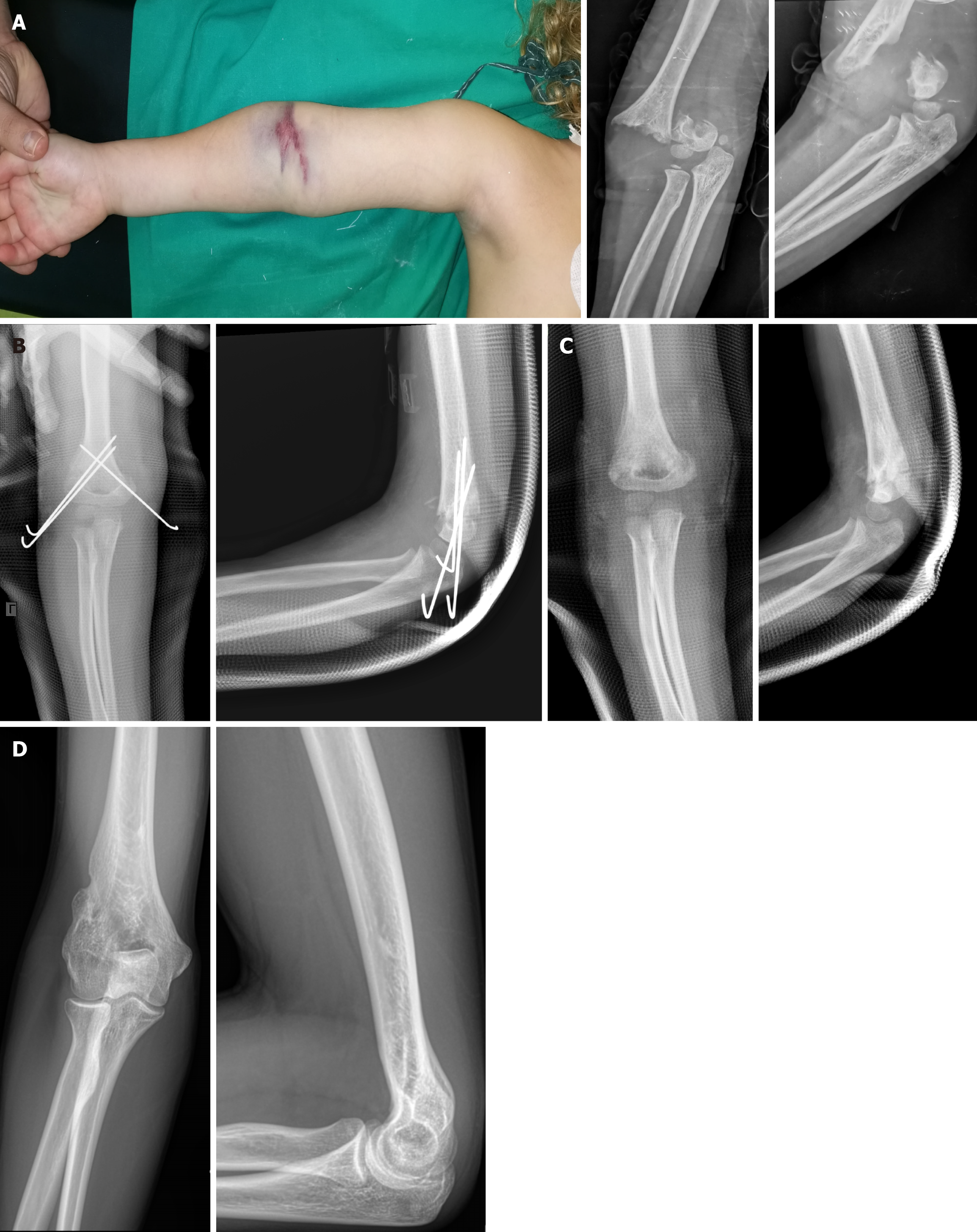
Figure 1 Typical ecchymosis in elbow fossa often suggesting vascular injury in a 4-year-old female with a right supracondylar humeral fracture of type IV according to modified Gartland classification.
A: Clinical and radiological presentation; B: The fracture was fixed after open reduction by lateral and medial approach; C: The K-wires were removed 1 month post-operatively; D: Right elbow X-rays at the age of 19 years.
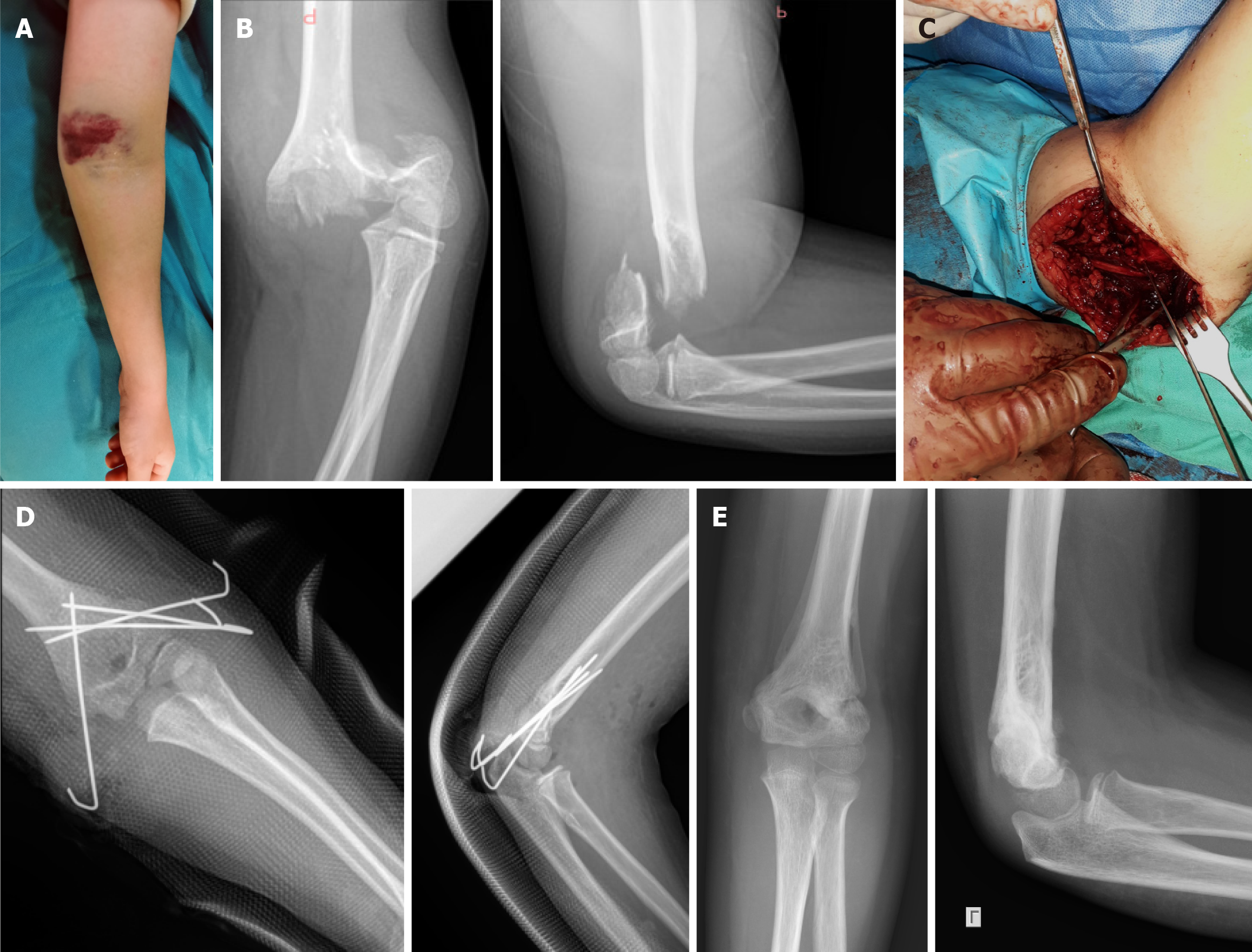
Figure 2 Significant anterior elbow haematoma, pink pulseless hand and anterior interosseous nerve palsy in an 8-year-old male with a Gartland type IV supracondylar humeral.
A: Clinical presentation; B: Radiological examination; C: Open reduction by lateral approach was unsuccessful due to periosteum and flexor muscles interposition on medial side requiring an enlarged medial approach and ulnar nerve exploration and protection; D: Four K-wires were applied (3 Lateral and 1 medial); E: K-wires were removed 5 weeks post-operatively. Normal pulses were palpable in hand 12 hours post-operatively and anterior interosseous nerve palsy recovered in 6 weeks.
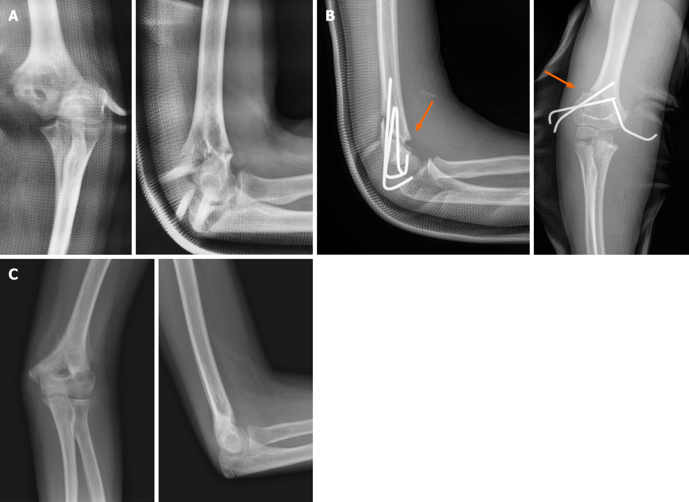
Figure 3 Malrotation can be easily missed, and it is poorly corrected by remodeling.
A: A 10-year-old male with a Gartland III supracondylar humeral fracture with marked internal rotation of the condyles; B: Despite open reduction and both column pinning, residual rotational deformity can be noticed by cortex mismatching in anteroposterior and lateral X-rays (orange arrows indicate cortex discontinuity); C: X-rays in 3 months follow-up with satisfactory functional outcome.
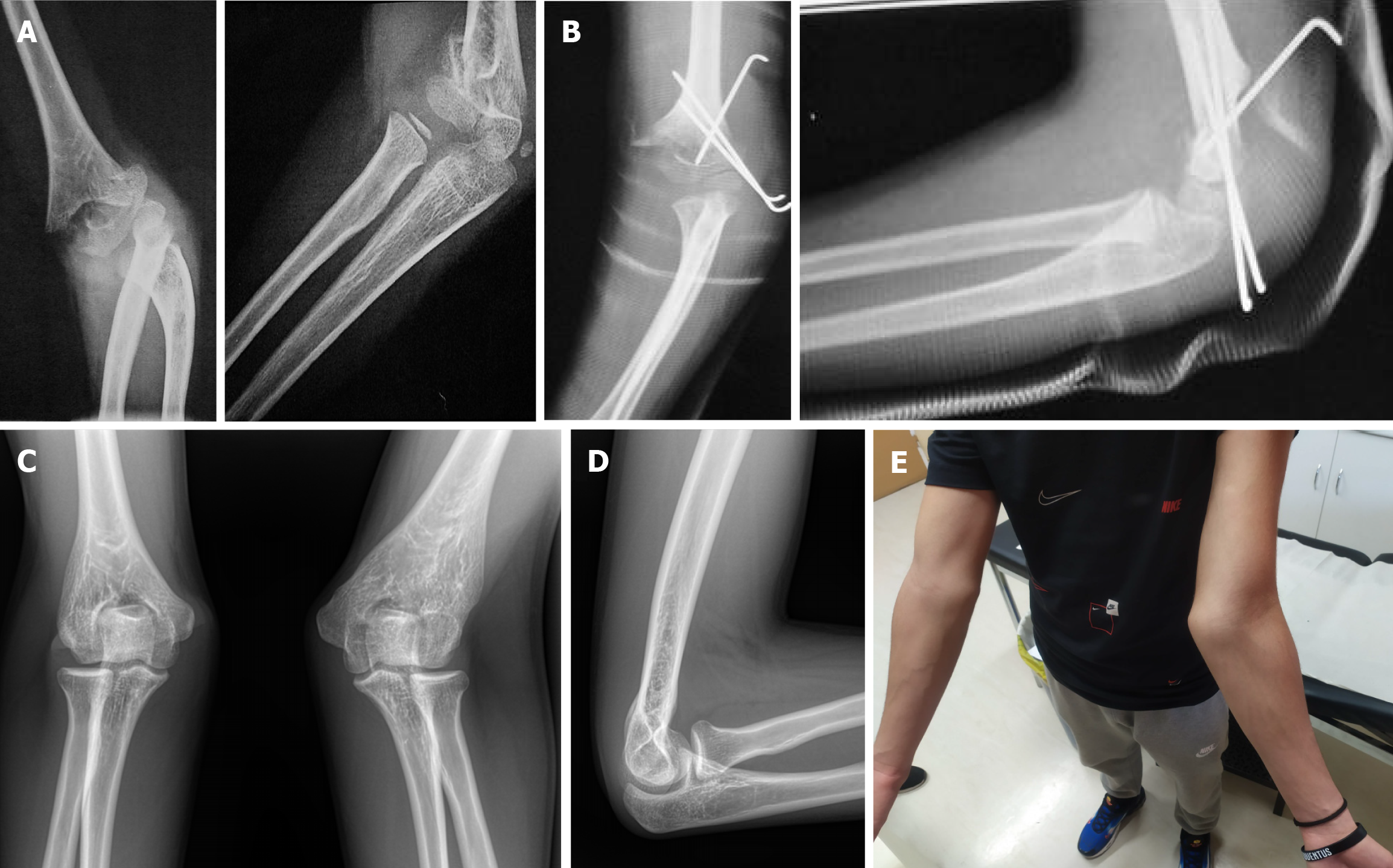
Figure 4 Deformity can be the result of improper reduction or physeal growth arrest (traumatic and iatrogenic).
A: A 7-year-old male with a Gartland type III supracondylar humeral fracture; B: Open reduction via lateral approach and stabilization by 3 Lateral K-wires with incomplete reduction on axial and sagittal level; C and D: In 9-year follow-up there is significant (28°) valgus deformity marked on X-rays (in comparison with the contralateral elbow); E: Elbow range of motion is normal during physical examination.
- Citation: Athanaselis ED, Metaxiotis N, Rigopoulos N, Hantes M, Dailiana ZH, Karachalios T, Varitimidis S. Open reduction can be a reasonable, safe and effective choice in complex paediatric supracondylar humeral fractures operative treatment. World J Orthop 2025; 16(10): 110461
- URL: https://www.wjgnet.com/2218-5836/full/v16/i10/110461.htm
- DOI: https://dx.doi.org/10.5312/wjo.v16.i10.110461


