©The Author(s) 2024.
World J Orthop. Aug 18, 2024; 15(8): 764-772
Published online Aug 18, 2024. doi: 10.5312/wjo.v15.i8.764
Published online Aug 18, 2024. doi: 10.5312/wjo.v15.i8.764
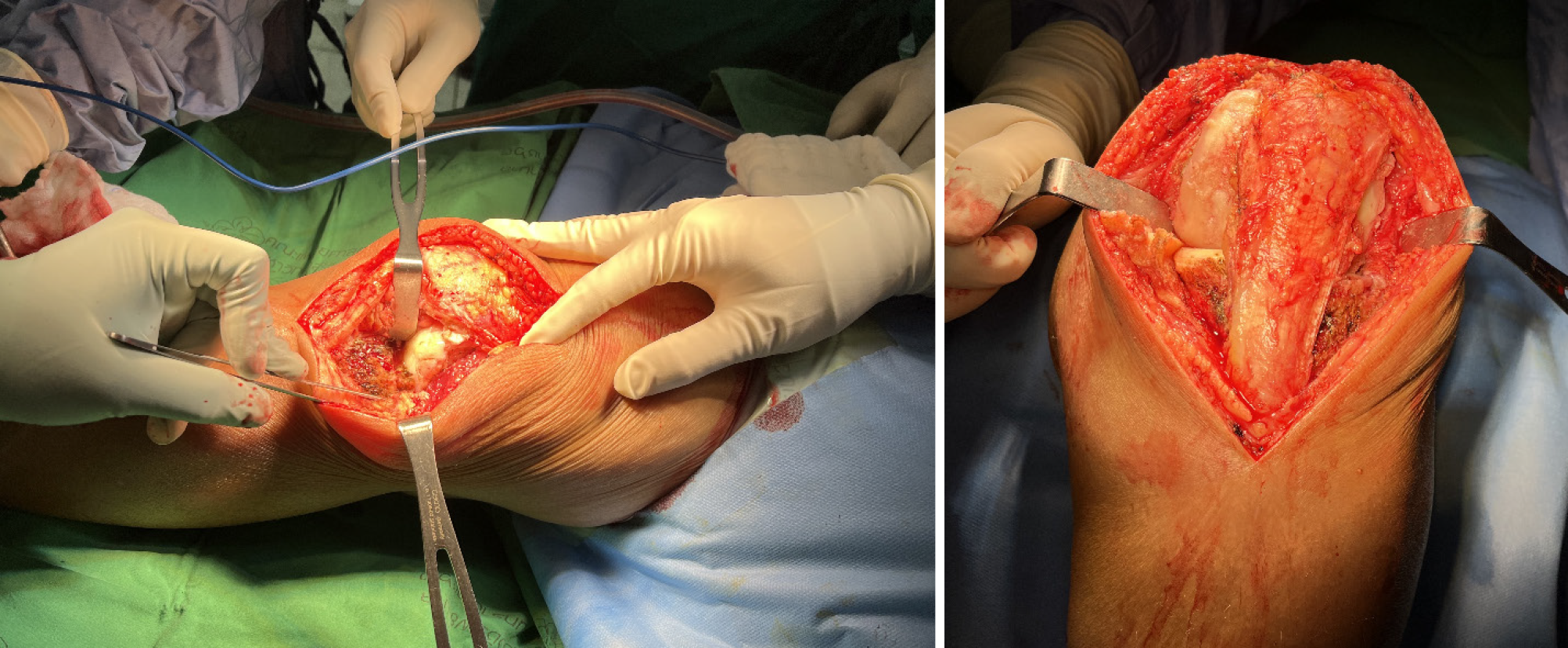
Figure 1 Midvastus arthrotomy combined with lateral parapatellar arthrotomy improved lateral side exposure and simultaneously assisted with the release of contracted lateral structures of the knee.
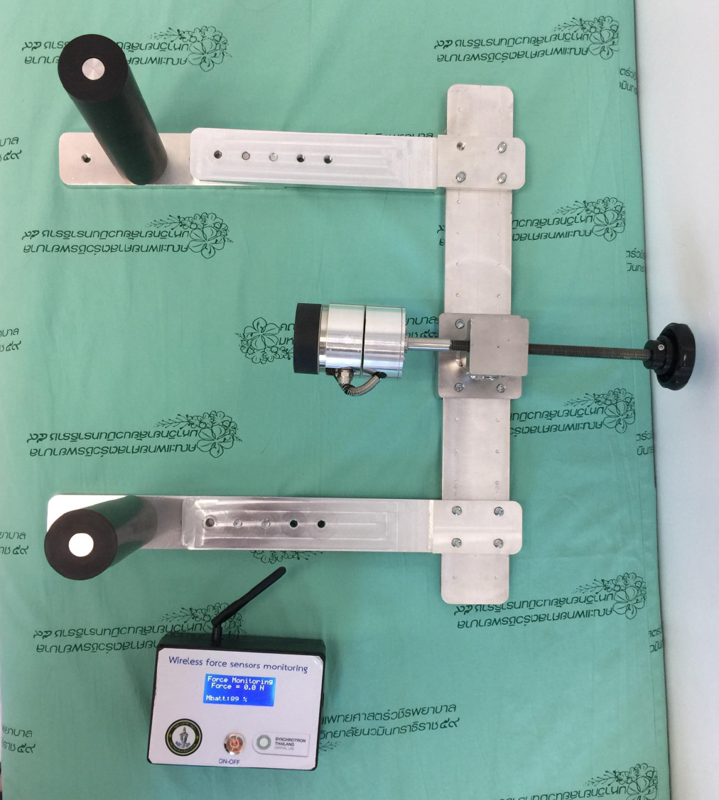
Figure 2 Digital ligament stress device (Patent number 2101002860, Navamindradhiraj University, Bangkok, Thailand) equipped with a sensor to measure the force applied during usage.
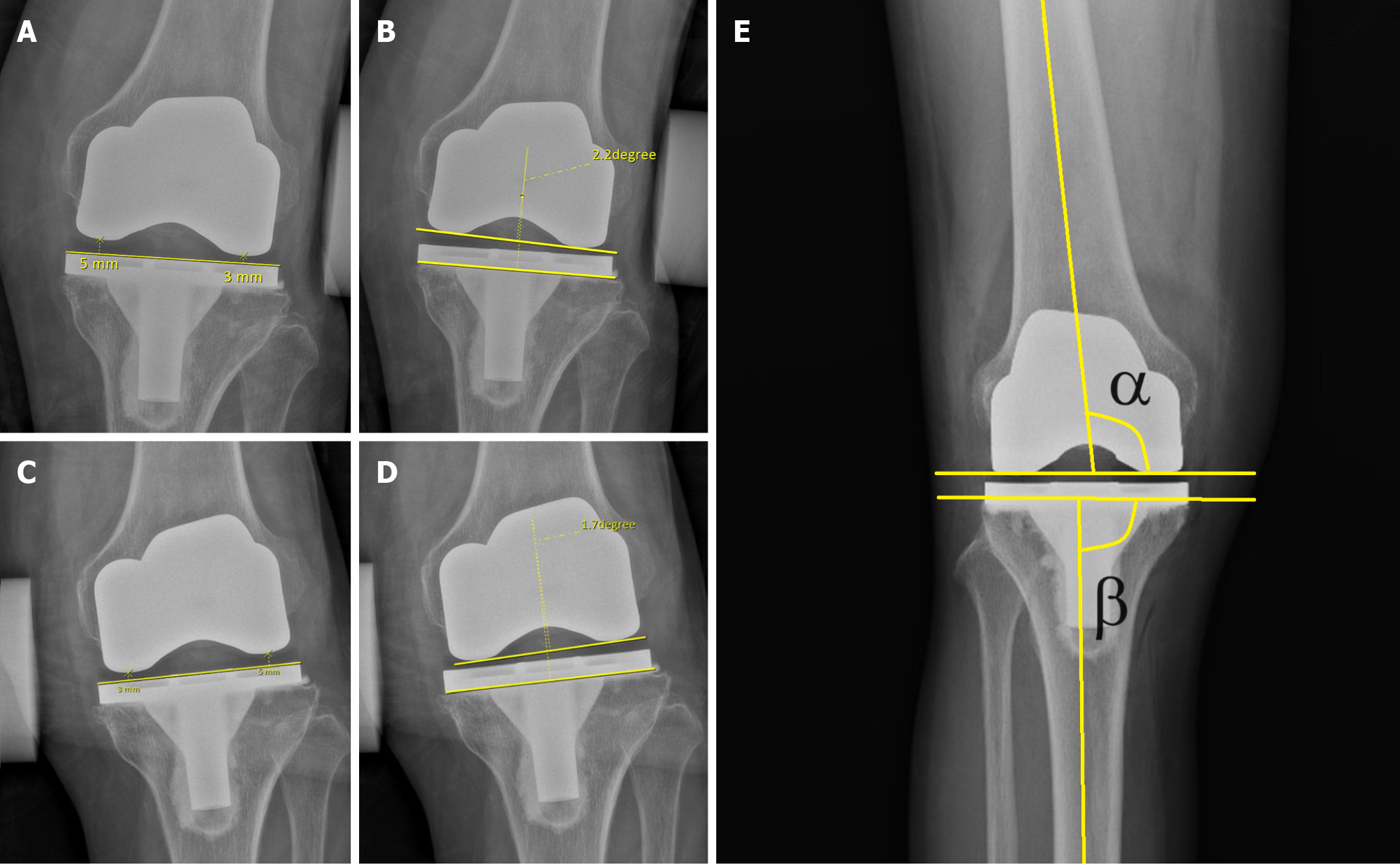
Figure 3 Measurement methods used in this study.
A: The valgus gap opening distance was measured by subtracting the medial gap from the lateral gap on the stress radiograph; B: The valgus gap opening angle was determined based on the angle between the line of the tibial tray and the line of the most distal part of the femoral component on the stress radiograph; C: The varus gap opening distance was measured by subtracting the lateral gap from the medial gap; D: The varus gap opening angle was determined based on the angle between the line of the tibial tray and the line of the most distal part of the femoral component; E: The alpha (α) and beta (β) angles of the femoral and tibia components on the antero-posterior radiograph.
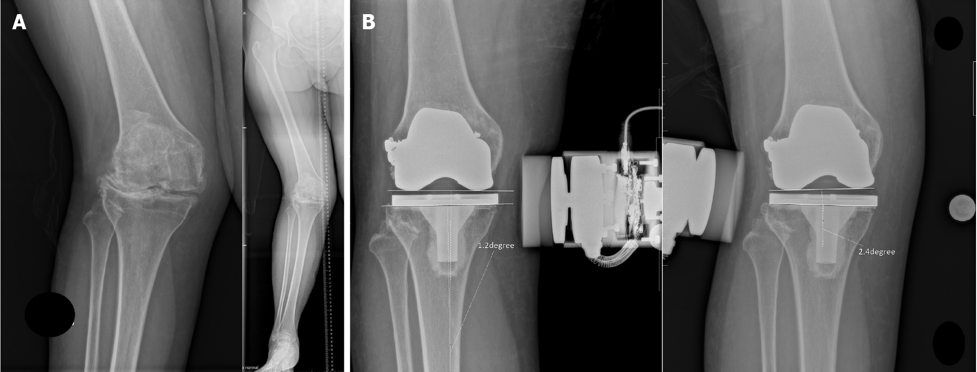
Figure 4 A case of a 65-year-old female with severe valgus osteoarthritis of the knee.
A: The preoperative radiograph shows a valgus deviation of the mechanical axis of 22 degrees. After initial soft tissue release, persistent tightness of the lateral structure was still noted, causing an unbalanced extension gap. Thus, a lateral condylar sliding osteotomy was performed intraoperatively; B: The knee was stable after stress radiography.
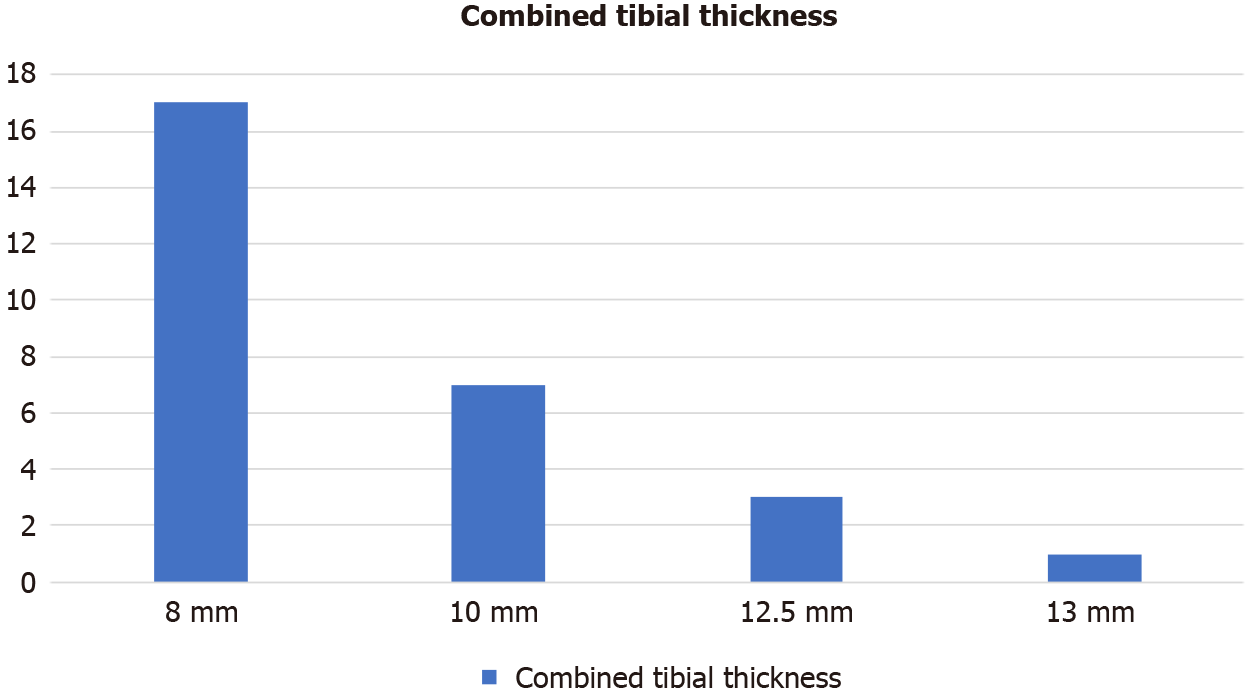
Figure 5 Distribution of the combined tibial thickness in this study.
A total of 17 knees (60.7%) were implanted with a combined tibial thickness of < 10 mm; 7 knees (25%) were implanted with a combined tibial thickness of 10 mm; 3 knees (10.7%) were implanted with a combined tibial thickness of 12.5 mm; and 1 knee (3.5%) was implanted with a combined tibial thickness of 13 mm.
- Citation: Chaiyakit P, Wattanapreechanon P. Coronal plane stability of cruciate-retaining total knee arthroplasty in valgus gonarthrosis patients: A mid-term evaluation using stress radiographs. World J Orthop 2024; 15(8): 764-772
- URL: https://www.wjgnet.com/2218-5836/full/v15/i8/764.htm
- DOI: https://dx.doi.org/10.5312/wjo.v15.i8.764


