©The Author(s) 2024.
World J Orthop. Jan 18, 2024; 15(1): 11-21
Published online Jan 18, 2024. doi: 10.5312/wjo.v15.i1.11
Published online Jan 18, 2024. doi: 10.5312/wjo.v15.i1.11
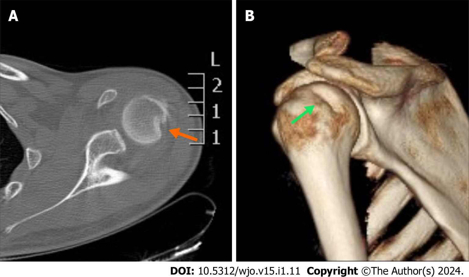
Figure 1 A 20-year-old patient after a forced abduction and hyperextension injury of his left shoulder with no support of his hand.
A: The computed tomography scan revealed an impaction fracture on the posterolateral part of the humeral head (orange arrow); B: A three-dimensional image in which the entire Hill-Sachs lesion was impressively displayed (green arrow).
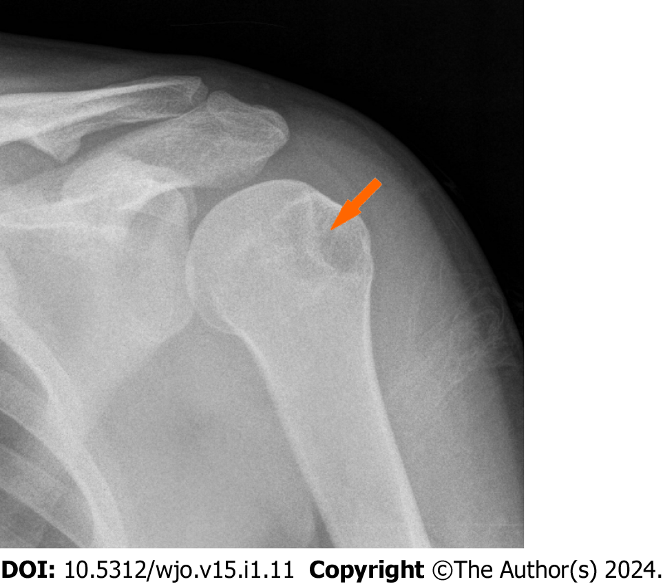
Figure 2 Left shoulder of a patient in internal rotation immediately after reduction of an anterior dislocation.
A large Hill-Sachs defect and a line of condensation was very obvious on this radiograph (orange arrow).
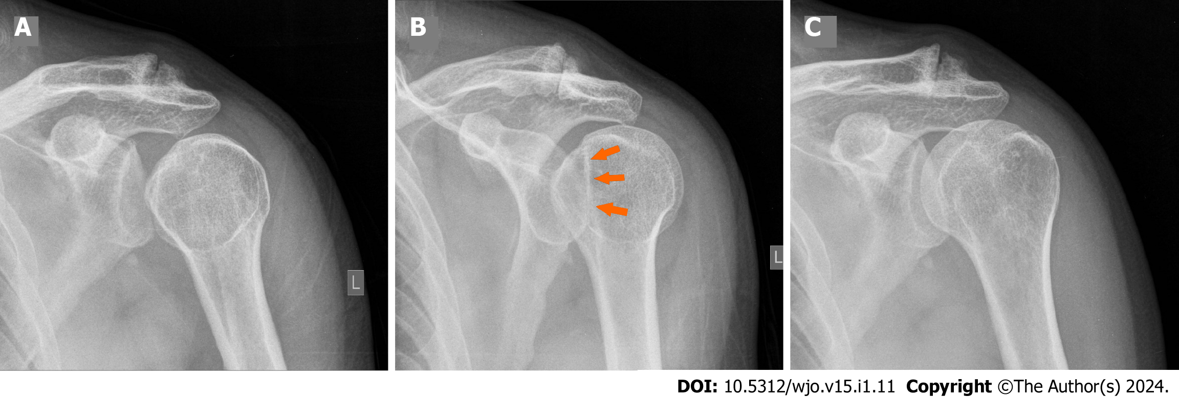
Figure 3 Posterior shoulder dislocation.
A: In the frontal view the distance of the medial cortical margin of the humeral head and the anterior glenoid rim was more than 6 mm, constituting a positive rim sign; B: A slightly oblique view with appreciable through sign (orange arrows). The humeral head was in internal rotation and hooked on the posterior rim of the glenoid fossa; C: The post reduction anteroposterior radiograph showed normal alignment and congruity of the glenohumeral articulation.
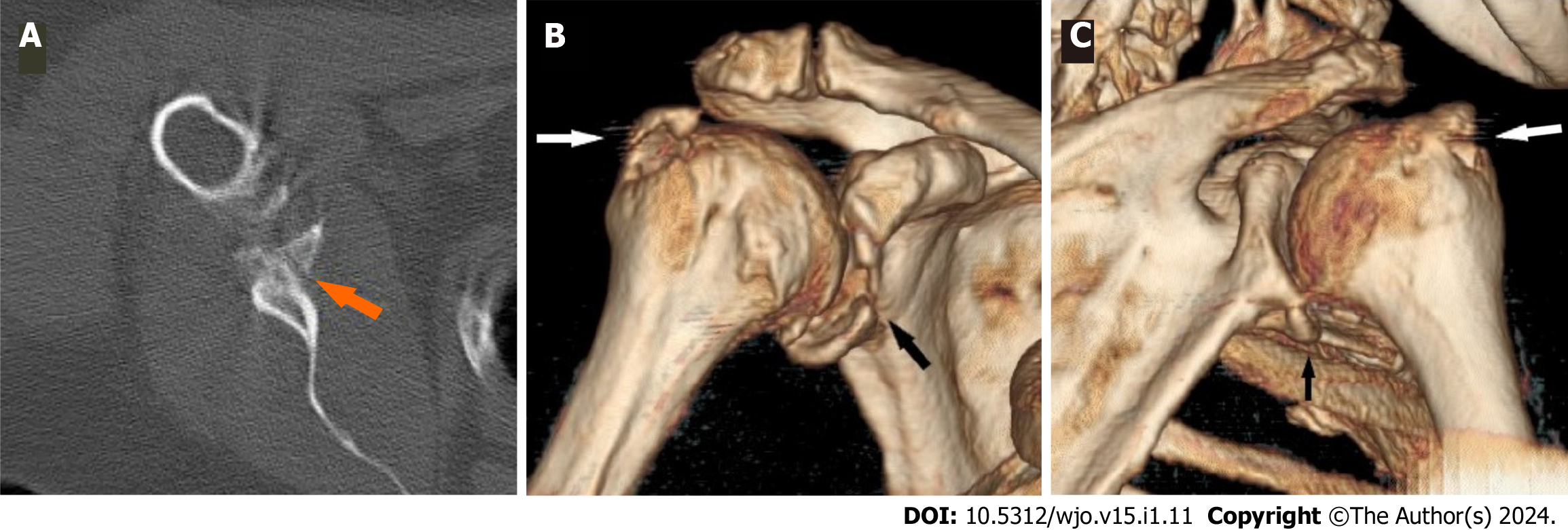
Figure 4 A 58-year-old female after a forward fall with the arm in flexion resulted in anterior dislocation with a bony Bankart lesion and greater tuberosity fracture.
A: The sagittal image illustrated a bony Bankart lesion (orange arrow); B: An anterior three-dimensional image on the same patient showed a large Bankart lesion (black arrow) and greater tuberosity fracture (white arrow); C: The absence of a Hill-Sachs lesion was evident on the posterior three-dimensional view.
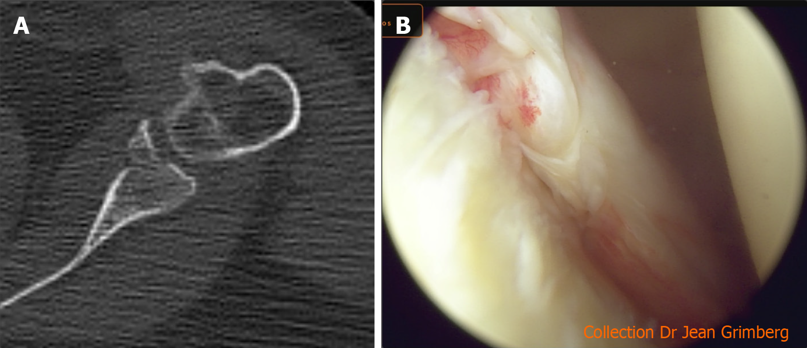
Figure 5 Bony Bankart lesion imaging.
A: Computed tomography scan of a bony Bankart lesion on the sagittal view; B: Image during arthroscopy of a Bankart lesion.
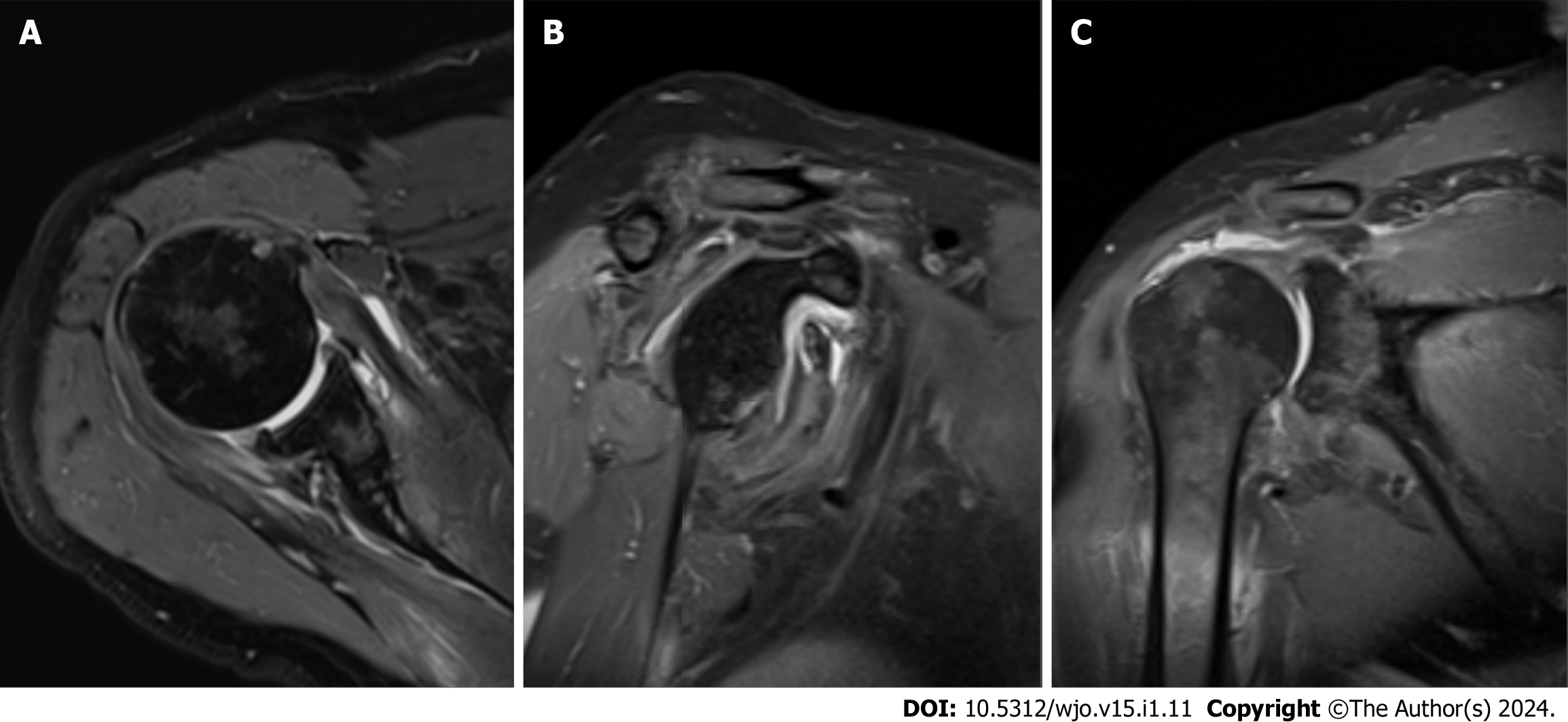
Figure 6 Magnetic resonance imaging of a 75-year-old male after a fall with forced abduction-hyperextension of the arm with support in the hand that resulted in anterior dislocation.
A: Anterior labral tears are best detected on the sagittal view; B: Bone edema on the anteroposterior margin of the glenoid; C: Magnetic resonance imaging is the modality of choice in cases of suspected rotator cuff tears.
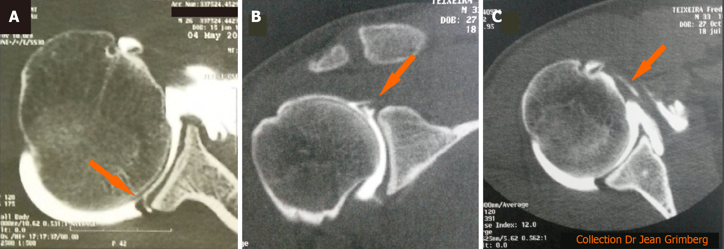
Figure 7 Computed tomography arthrography is useful in cases of false-negative magnetic resonance imaging results.
A: Posterior labral tear on the sagittal view; B: Superior labral anterior posterior lesions are best detected on the coronal view; C: Computed tomography arthrogram is the modality of choice in cases of undetected subscapular tears. The sagittal view showed widening of the bicipital canal, which is an indirect sign of subscapularis rupture and delamination with penetration of contrast fluid into the tendon (orange arrow).
- Citation: Mastrantonakis K, Karvountzis A, Yiannakopoulos CK, Kalinterakis G. Mechanisms of shoulder trauma: Current concepts. World J Orthop 2024; 15(1): 11-21
- URL: https://www.wjgnet.com/2218-5836/full/v15/i1/11.htm
- DOI: https://dx.doi.org/10.5312/wjo.v15.i1.11


