Copyright
©The Author(s) 2023.
World J Orthop. Apr 18, 2023; 14(4): 197-206
Published online Apr 18, 2023. doi: 10.5312/wjo.v14.i4.197
Published online Apr 18, 2023. doi: 10.5312/wjo.v14.i4.197
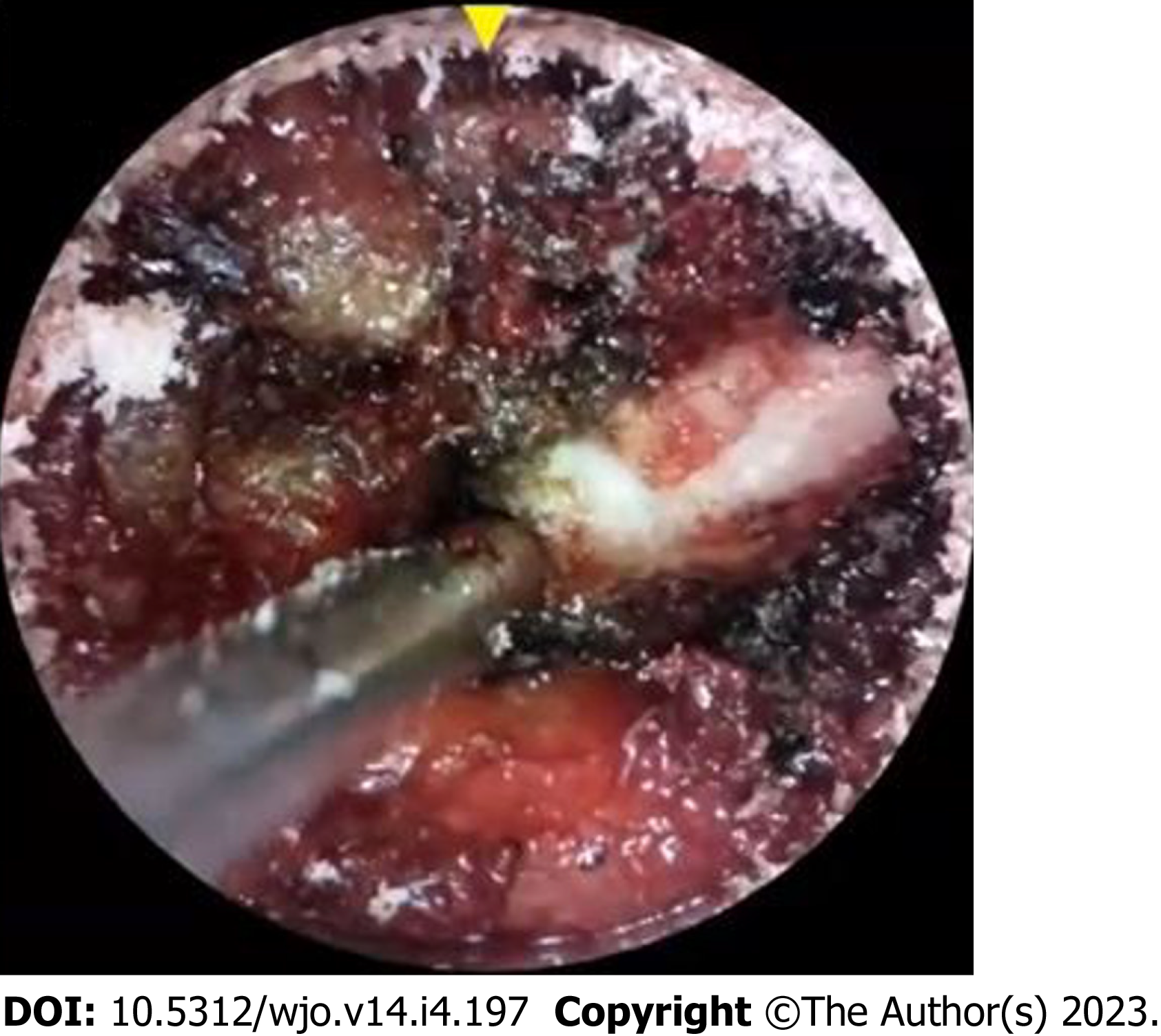
Figure 1 Direct endoscopic view from tubular/retractor-based camera that provides a two-dimensional image on a screen with digital zoom.
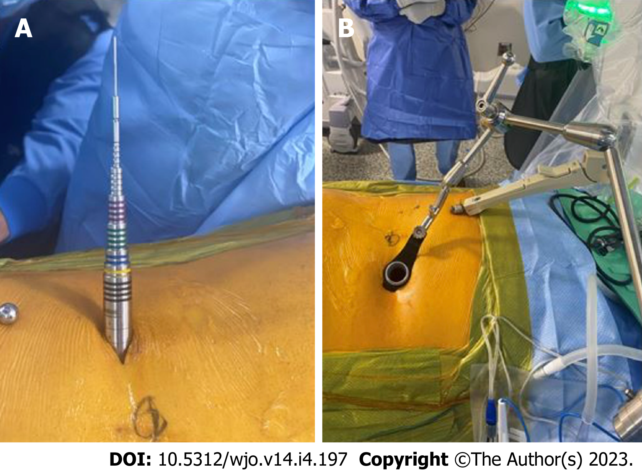
Figure 2 Minimally invasive surgery endoscopic technique.
A: Endoscopic cannula inserted in posterior lumbar region; B: Tubular/retractor-based setup where the camera can be inserted into the body through a channel called a working channel.
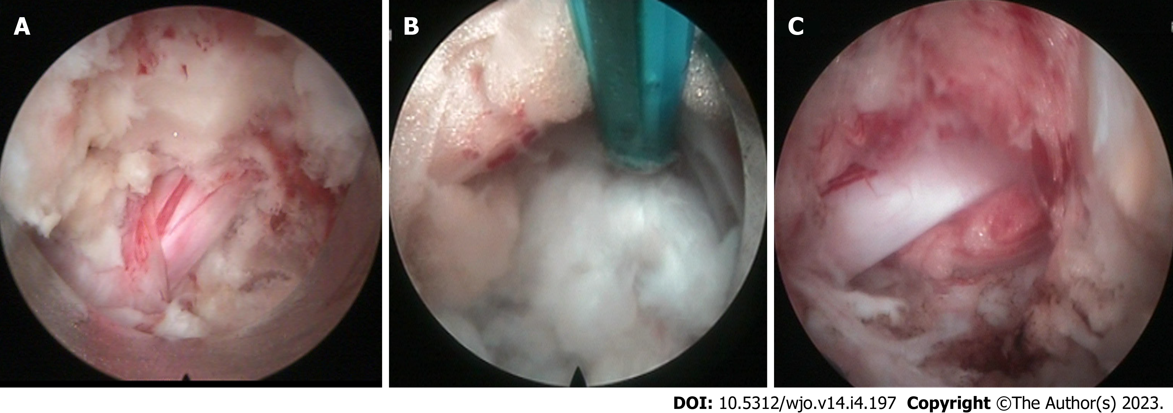
Figure 3 Direct two-dimensional endoscopic view (top of image as anatomically medial, bottom as lateral, left as cranial, and right as caudal) with dura mater exposed.
A: Disc has compressed nerve ventrally; B: Disc irrigated, exposed, and removed to alleviate nerve compression; and C: Nerve has been decompressed.
- Citation: Tang K, Goldman S, Avrumova F, Lebl DR. Background, techniques, applications, current trends, and future directions of minimally invasive endoscopic spine surgery: A review of literature. World J Orthop 2023; 14(4): 197-206
- URL: https://www.wjgnet.com/2218-5836/full/v14/i4/197.htm
- DOI: https://dx.doi.org/10.5312/wjo.v14.i4.197


