
















Copyright: ©Author(s) 2026.
World J Clin Oncol. Aug 24, 2026; 17(8): 124132
Published online Aug 24, 2026. doi: 10.5306/wjco.124132
Published online Aug 24, 2026. doi: 10.5306/wjco.124132
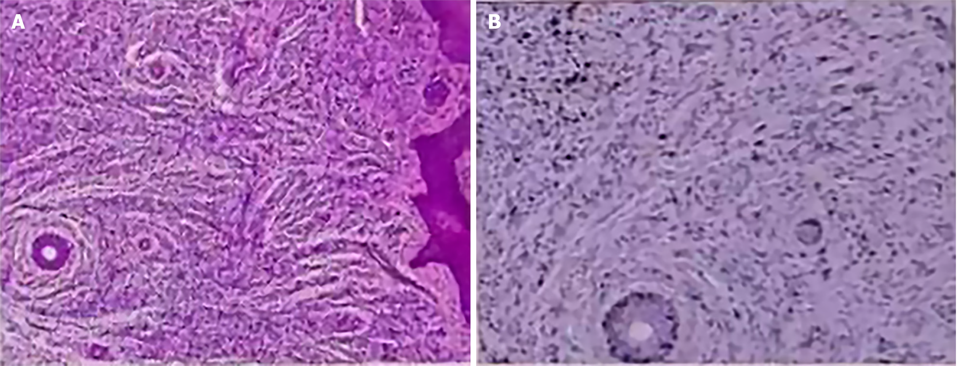
Figure 1 Initial incisional biopsy (misdiagnosed as infantile myofibroma).
A: Low power hematoxylin and eosin image showing a small biopsy specimen composed of spindle cell proliferation (× 40 magnification, scale bar = 500 μm); B: High power hematoxylin and eosin image demonstrating fascicular/nodular arrangement of bland spindle cells without the classic storiform pattern (× 200 magnification, scale bar = 100 μm). Immunohistochemistry showed CD34-, vimentin+, and S100-.
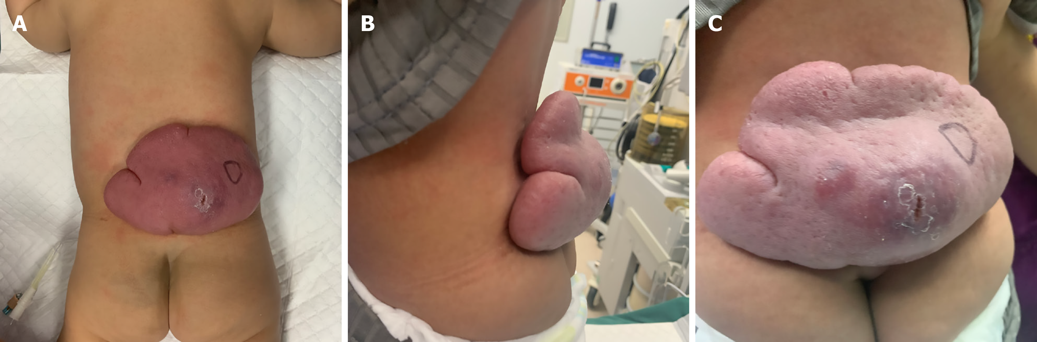
Figure 2 Preoperative clinical appearance of the dorsal mass.
A: Posterior view showing the overall extent and location of the multinodular, purplish lesion on the lower back; B: Lateral view demonstrating the degree of protrusion/elevation of the lesion; C: Close-up view showing the multinodular surface and purplish discoloration.
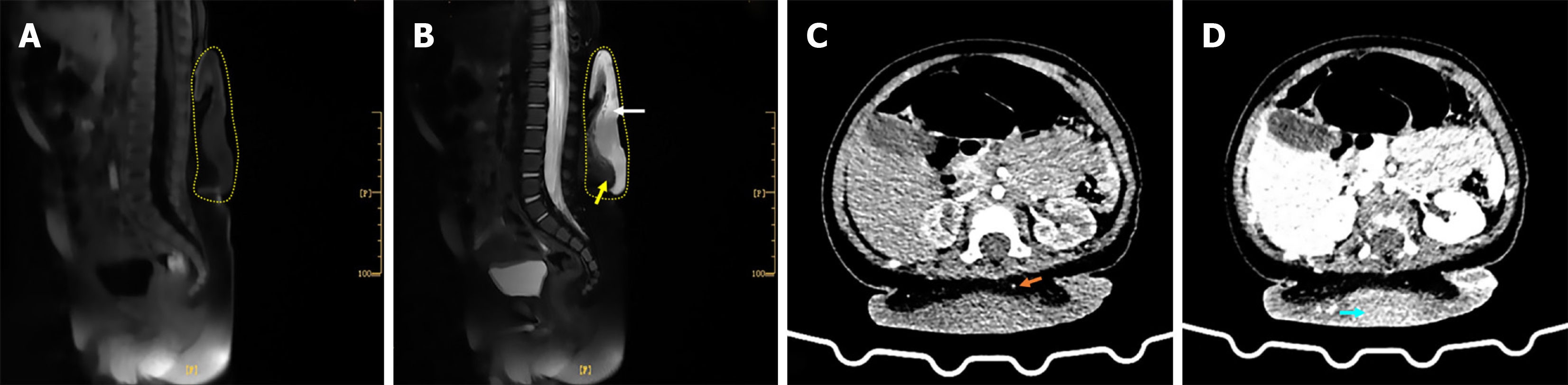
Figure 3 Preoperative imaging findings.
A: Sagittal T1 weighted magnetic resonance imaging showing the extent of the subcutaneous lesion (outlined by the yellow dashed line); B: Sagittal T2-weighted magnetic resonance imaging demonstrating a T2-hyperintense lesion (white arrow) measuring 20 cm × 13.0 cm, with ill-defined margins against the adjacent subcutaneous fat (yellow arrow); C and D: Contrast-enhanced computed tomography obtained during the arterial (C) and venous (D) phases demonstrating a 10.9 cm × 2.7 cm subcutaneous lesion with marked heterogeneous enhancement and no distinct plane separating the lesion from the underlying deep fascia. The orange arrow (C) indicates a tumor vessel, and the blue arrow (D) indicates an area of significant enhancement. Yellow dashed lines outline the lesion boundaries in (A and B).
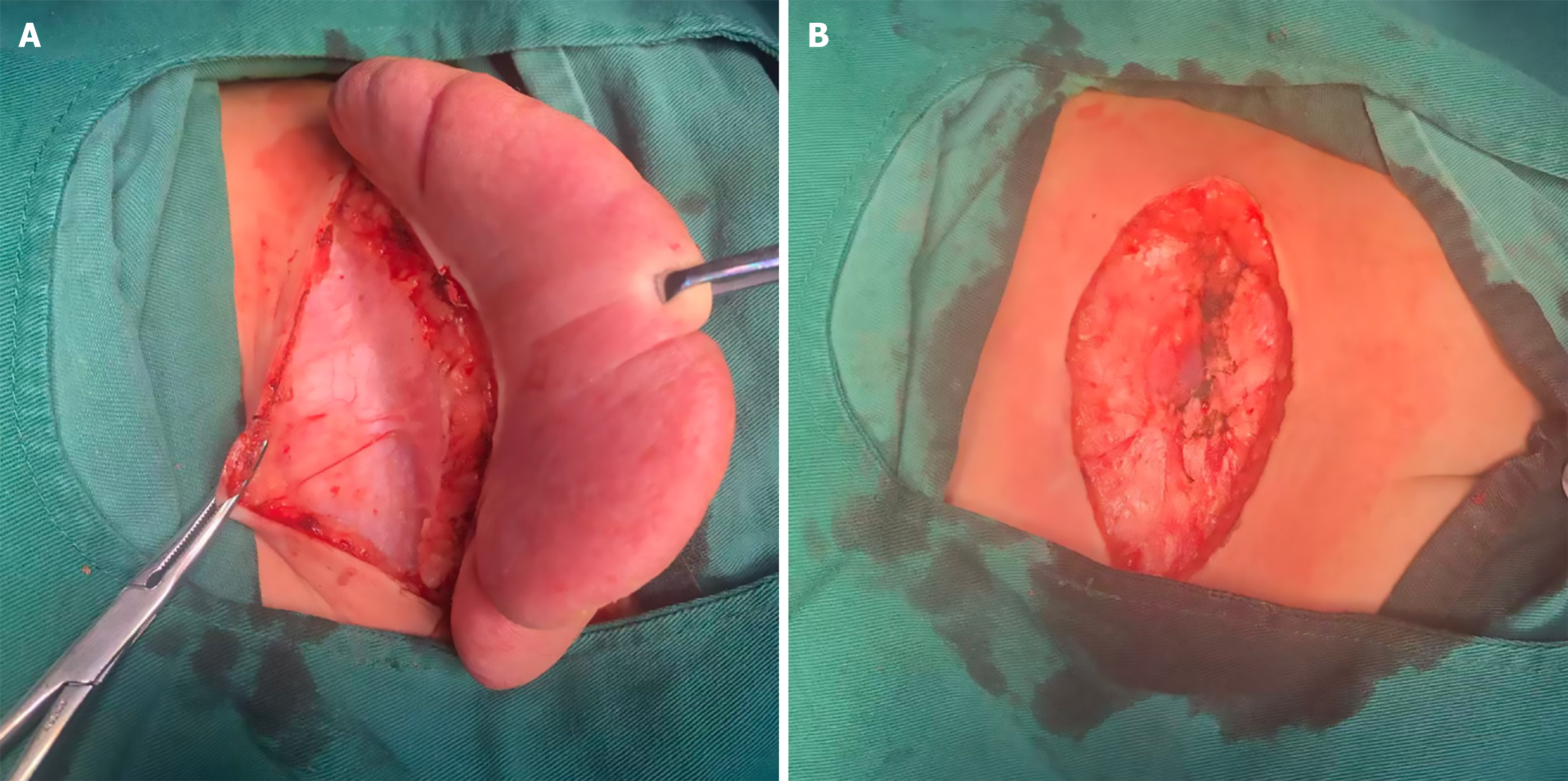
Figure 4 Intraoperative findings during tumor resection.
A: The tumor was primarily located in the dermis and appeared relatively well defined on gross inspection; B: Surgical defect following wide local excision with a 2-cm peripheral margin extending to the underlying muscular fascia, demonstrating complete tumor removal before primary wound closure.
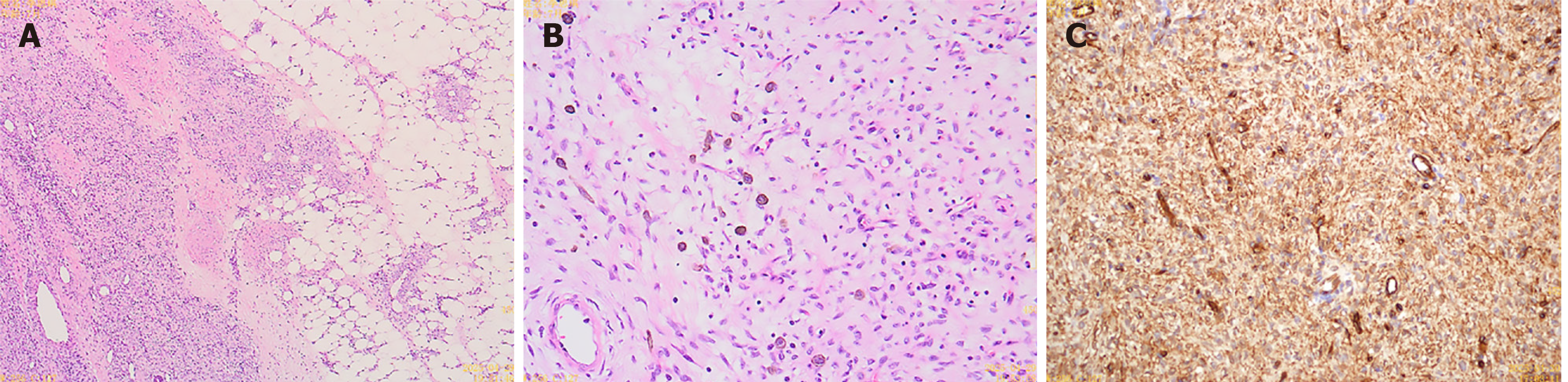
Figure 5 Final surgical specimen confirming classic dermatofibrosarcoma protuberans.
A: Low power hematoxylin and eosin image showing a dermal based spindle cell tumor with infiltrative extension into the subcutaneous adipose tissue in a characteristic honeycomb pattern (× 40 magnification, scale bar = 500 μm); B: Medium power hematoxylin and eosinimage demonstrating the classic storiform (cartwheel) arrangement of uniform spindle cells (× 100 magnification, scale bar = 200 μm); C: Immunohistochemical staining showing strong, diffuse positivity for CD34 antigen in the tumor cells (× 200 magnification, scale bar = 100 μm).
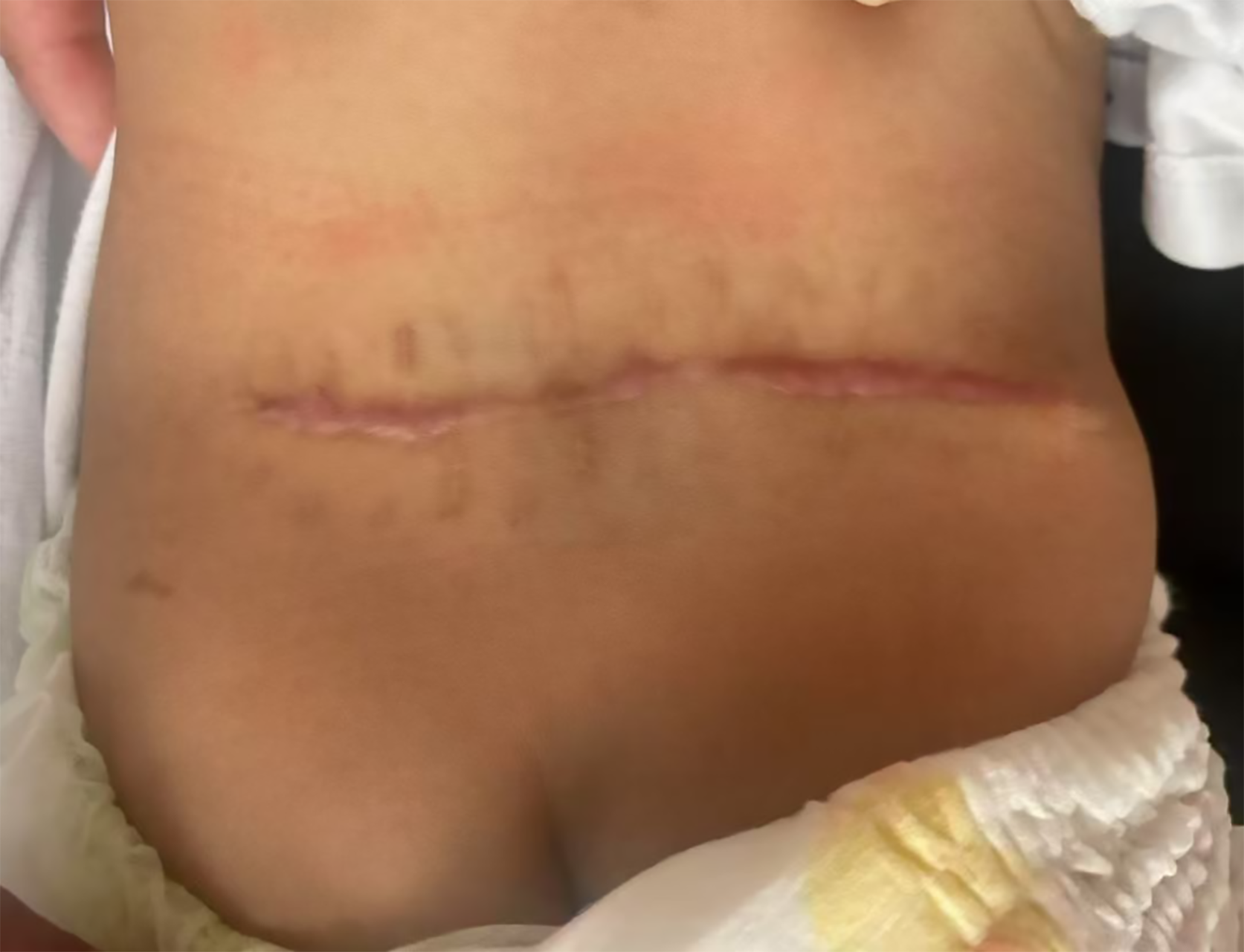
Figure 6
Postoperative appearance of the surgical incision at the 6-month follow-up, demonstrating a well-healed linear scar without evidence of wound complications.
- Citation: Zheng CJ, Wan CL, Zhang CB, Hai B, Huang CF. Congenital dermatofibrosarcoma protuberans in a seven-month-old infant initially misdiagnosed as infantile myofibroma: A case report. World J Clin Oncol 2026; 17(8): 124132
- URL: https://www.wjgnet.com/2218-4333/full/v17/i8/124132.htm
- DOI: https://dx.doi.org/10.5306/wjco.124132