©The Author(s) 2022.
World J Gastrointest Pharmacol Ther. Jul 5, 2022; 13(4): 33-46
Published online Jul 5, 2022. doi: 10.4292/wjgpt.v13.i4.33
Published online Jul 5, 2022. doi: 10.4292/wjgpt.v13.i4.33
Figure 1 Liver biopsy of a patient with biliary atresia.
A: Cholestasis (arrow) and ballooning degeneration (asterisks) (hematoxylin-eosin, magnification × 100); B: Portal fibrosis (hematoxylin-eosin, magnification × 100); C: Bile duct proliferation (Cytokeratin 19, magnification × 100).
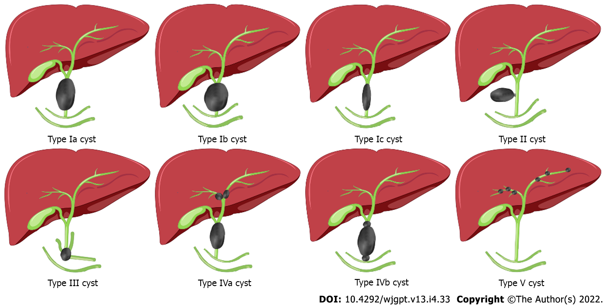
Figure 2 Classificiation of choledochal cysts[4].
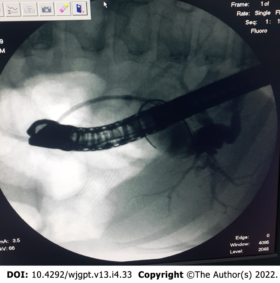
Figure 3 An endoscopic retrograde cholangiopancreatography image of type IVa choledochal cyst.
- Citation: Islek A, Tumgor G. Biliary atresia and congenital disorders of the extrahepatic bile ducts. World J Gastrointest Pharmacol Ther 2022; 13(4): 33-46
- URL: https://www.wjgnet.com/2150-5349/full/v13/i4/33.htm
- DOI: https://dx.doi.org/10.4292/wjgpt.v13.i4.33


