Copyright
©The Author(s) 2019.
World J Gastrointest Pharmacol Ther. Jan 21, 2019; 10(1): 1-21
Published online Jan 21, 2019. doi: 10.4292/wjgpt.v10.i1.1
Published online Jan 21, 2019. doi: 10.4292/wjgpt.v10.i1.1
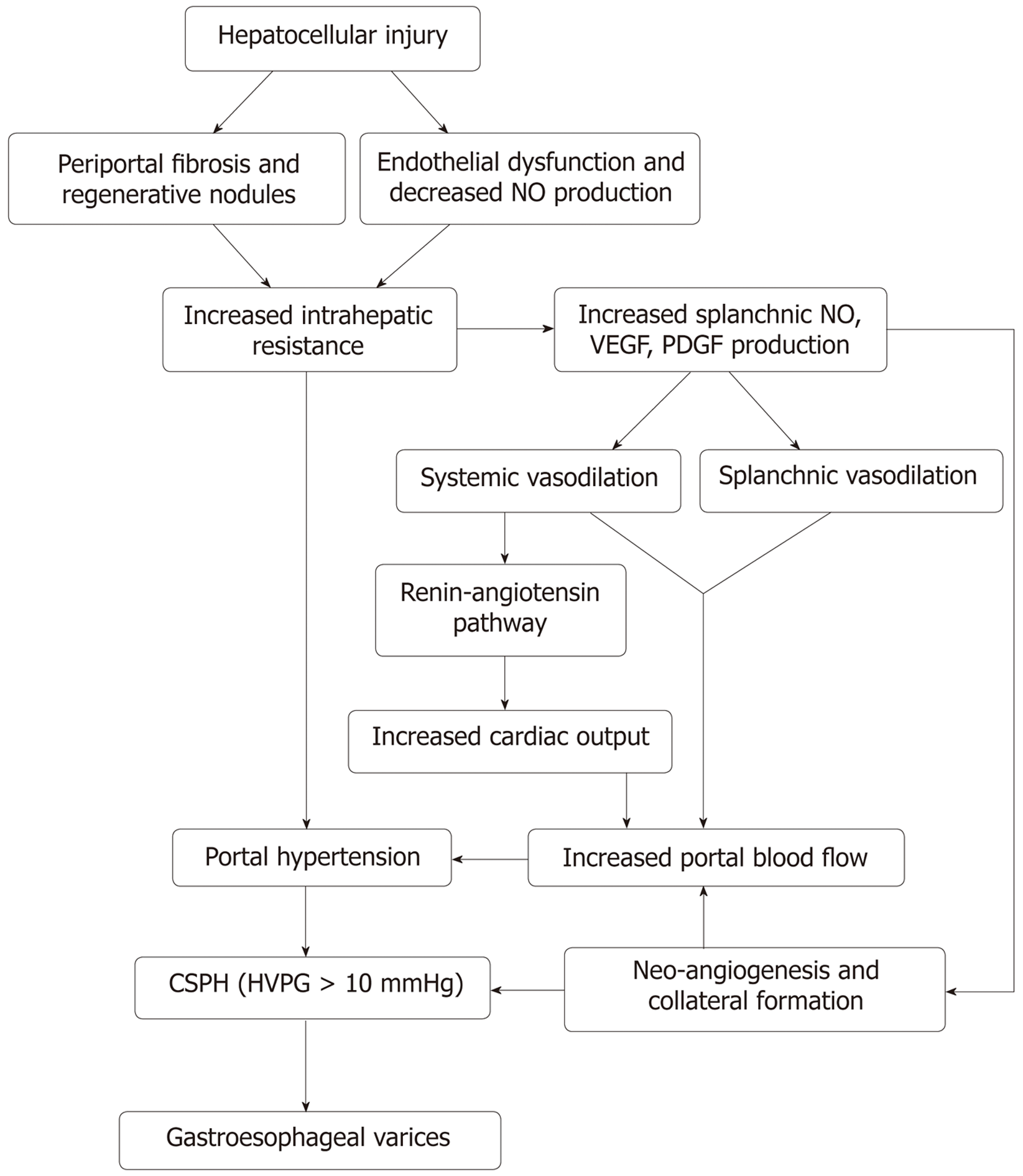
Figure 1 Mechanism of portal hypertension and the development of gastrointestinal varices.
VEGF: Vascular endothelial growth factor; PDGF: Platelet-derived growth factor; NO: Nitric oxide; HVPG: Hepatic venous pressure gradient.
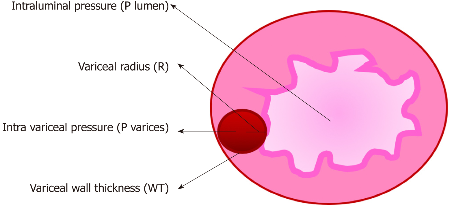
Figure 2 Mechanism of variceal bleeding.
P: Pressure; R: Radius; WT: Wall thickness.
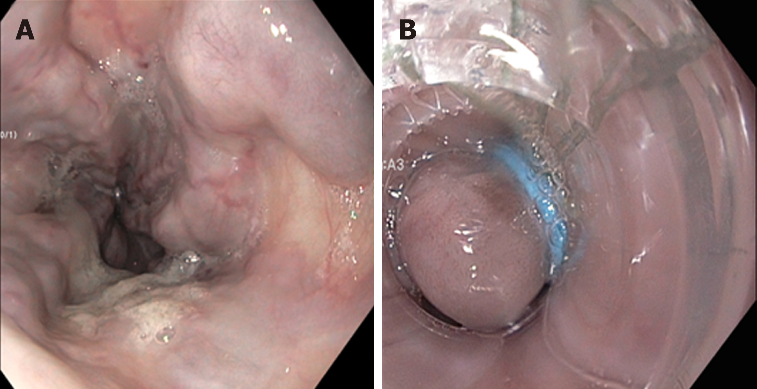
Figure 4 Endoscopic variceal ligation for primary prophylaxis.
A: Esophageal varices before banding; B: Esophageal varix post banding.
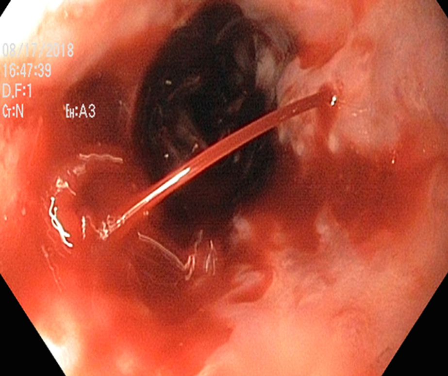
Figure 5 Bleeding esophageal varices.
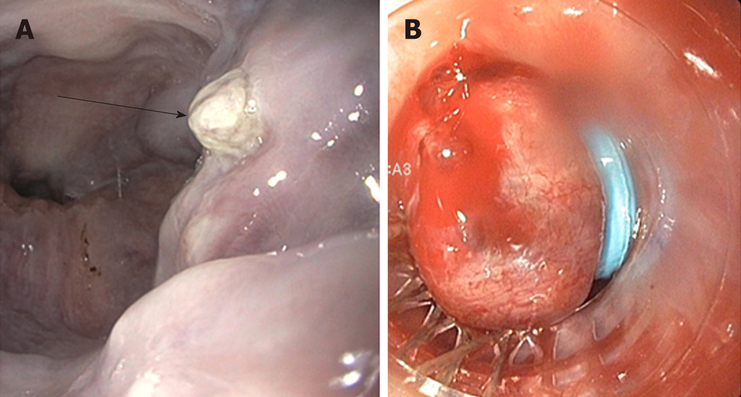
Figure 6 High-risk stigmata of bleeding from esophageal varices.
A: Platelet-fibrin plug on esophageal varix (white nipple sign); B: Bleeding esophageal varix post banding.
Figure 7 Endoscopic variceal band ligation.
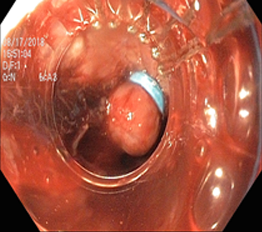
Figure 8 Metal stents for the treatment of bleeding esophageal varices.
A: Bleeding esophageal varix before stenting; B: Esophageal varix after metal stent.
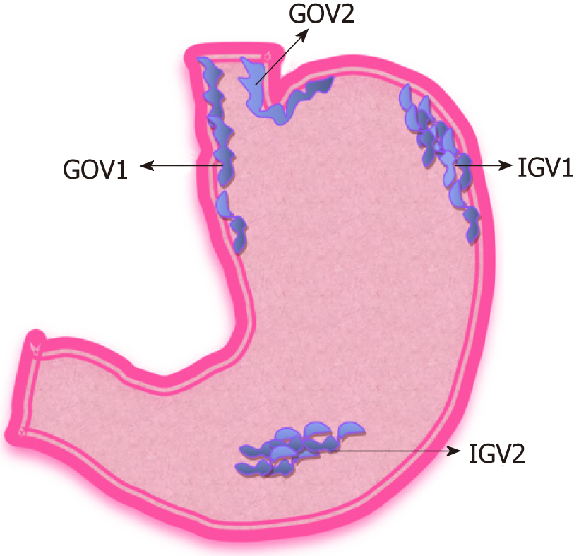
Figure 9 Sarin classification of gastric varices.
GOV1: Gastroesophageal varix type 1; GOV2: Gastroesophageal varix type 2; IGV1: Isolated gastric varix type 1; IGV2: Isolated gastric varix type 2.
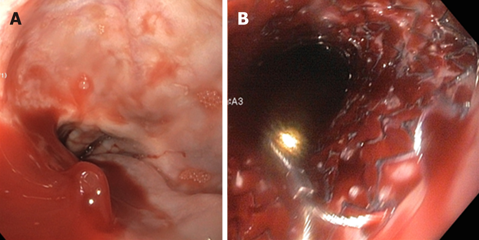
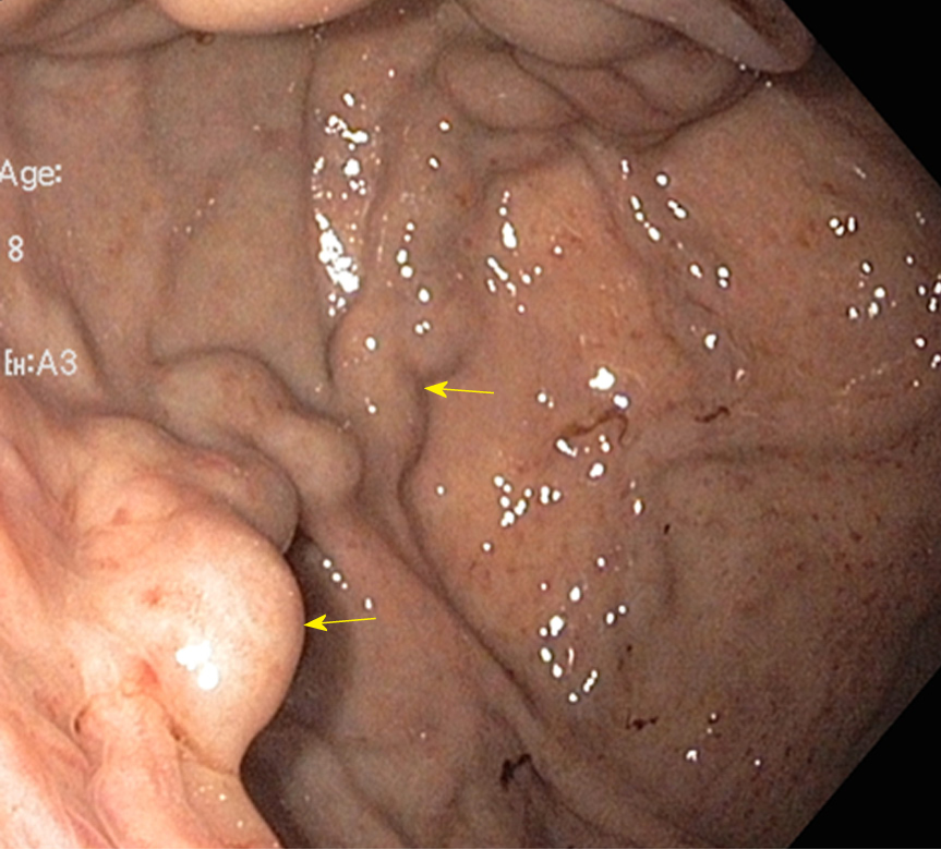
Figure 10 Gastric varices.
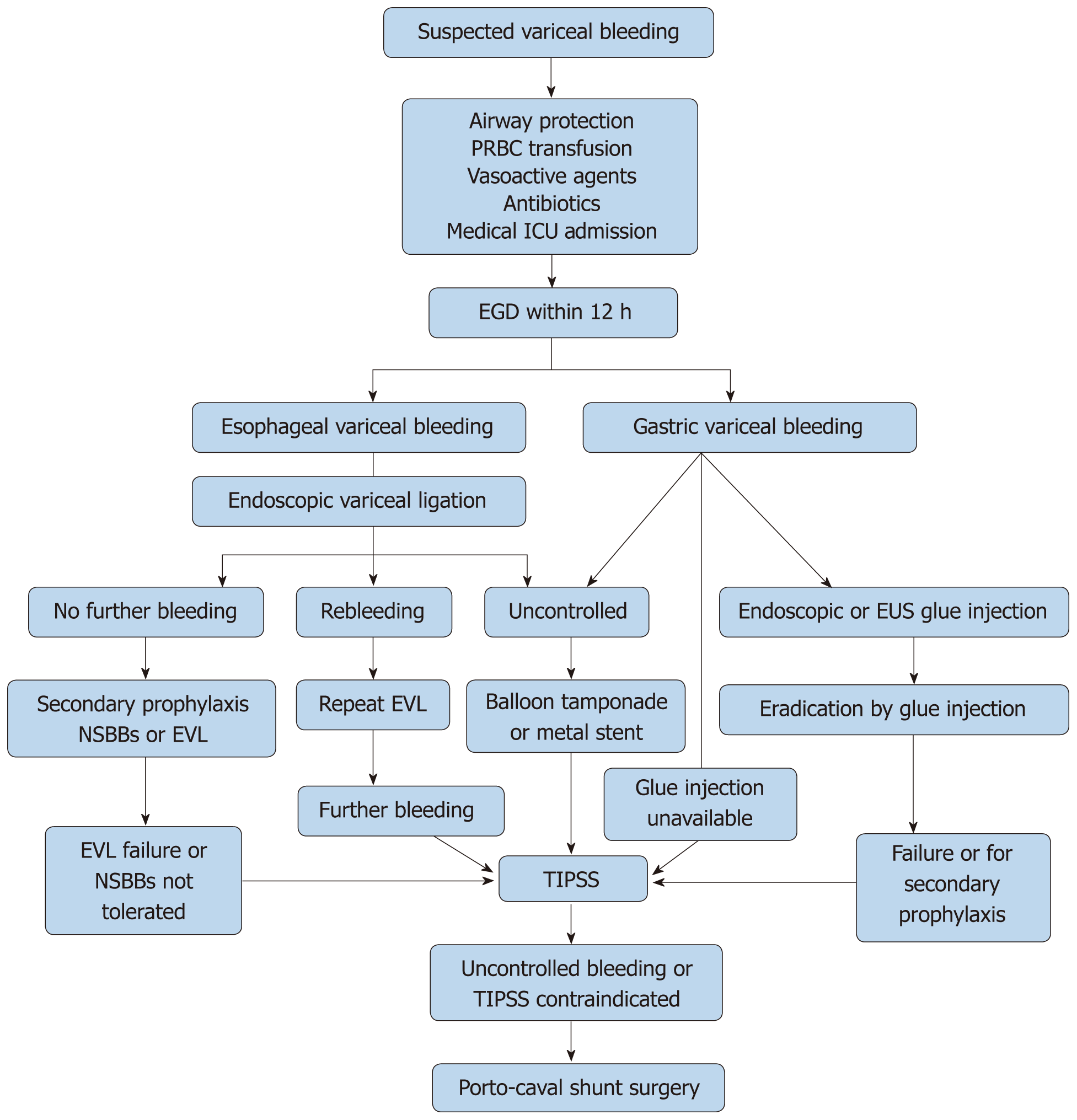
Figure 11 Algorithm for the management of acute variceal bleed.
ICU: Intensive care unit; EGD: Esophago-gastro duodenoscopy; NSBB: Nonselective beta blockers; EVL: Endoscopic variceal ligation; TIPS: Transjugular intrahepatic portosystemic shunt.
- Citation: Boregowda U, Umapathy C, Halim N, Desai M, Nanjappa A, Arekapudi S, Theethira T, Wong H, Roytman M, Saligram S. Update on the management of gastrointestinal varices. World J Gastrointest Pharmacol Ther 2019; 10(1): 1-21
- URL: https://www.wjgnet.com/2150-5349/full/v10/i1/1.htm
- DOI: https://dx.doi.org/10.4292/wjgpt.v10.i1.1


