Published online Feb 15, 2016. doi: 10.4291/wjgp.v7.i1.72
Peer-review started: July 12, 2015
First decision: September 22, 2015
Revised: December 2, 2015
Accepted: December 29, 2015
Article in press: January 4, 2016
Published online: February 15, 2016
Processing time: 211 Days and 14.8 Hours
Gastroesophageal reflux disease (GERD) is a common disorder of the gastrointestinal tract. In the last few decades, new technologies have evolved and have been applied to the functional study of the esophagus, allowing for the improvement of our knowledge of the pathophysiology of GERD. High-resolution manometry (HRM) permits greater understanding of the function of the esophagogastric junction and the risks associated with hiatal hernia. Moreover, HRM has been found to be more reproducible and sensitive than conventional water-perfused manometry to detect the presence of transient lower esophageal sphincter relaxation. Esophageal 24-h pH-metry with or without combined impedance is usually performed in patients with negative endoscopy and reflux symptoms who have a poor response to anti-reflux medical therapy to assess esophageal acid exposure and symptom-reflux correlations. In particular, esophageal 24-h impedance and pH monitoring can detect acid and non-acid reflux events. EndoFLIP is a recent technique poorly applied in clinical practice, although it provides a large amount of information about the esophagogastric junction. In the coming years, laryngopharyngeal symptoms could be evaluated with up and coming non-invasive or minimally invasive techniques, such as pepsin detection in saliva or pharyngeal pH-metry. Future studies are required of these techniques to evaluate their diagnostic accuracy and usefulness, although the available data are promising.
Core tip: In the last few decades, new technologies have evolved and have been applied to the functional study of the esophagus, allowing for the improvement of our knowledge of the pathophysiology of gastroesophageal reflux disease. High-resolution manometry permits a greater understanding of the function of the esophagogastric junction and the risks associated with hiatal hernia. The Chicago Classification V3.0 could define a hierarchic classification that accurately defines the major and minor disorders of esophageal motility. Esophageal 24-h pH-metry, especially when it is combined with impedance, is usually performed in patients with negative endoscopy and reflux symptoms who have a poor response to anti-reflux medical therapy to assess esophageal acid exposure and symptom-reflux correlations. In particular, esophageal 24-h impedance and pH monitoring are able to detect acid and non-acid reflux events. EndoFLIP is a recent technique poorly applied in clinical practice, although it provides a large amount of information about the esophagogastric junction. Recently, up and coming non-invasive or minimally invasive techniques, such as pepsin detection in saliva or pharyngeal pH-metry, have been suggested to detect laryngopharyngeal reflux disease. Future studies are required for these techniques to evaluate their accuracy and usefulness, although the available data are promising.
- Citation: de Bortoli N, Martinucci I, Bertani L, Russo S, Franchi R, Furnari M, Tolone S, Bodini G, Bolognesi V, Bellini M, Savarino V, Marchi S, Savarino EV. Esophageal testing: What we have so far. World J Gastrointest Pathophysiol 2016; 7(1): 72-85
- URL: https://www.wjgnet.com/2150-5330/full/v7/i1/72.htm
- DOI: https://dx.doi.org/10.4291/wjgp.v7.i1.72
Gastroesophageal reflux disease (GERD) is a highly prevalent disease in Western countries, affecting up to 20% of the general population, with important impacts on health care costs and the quality of life of patients[1]. According to the Montreal Definition, GERD develops when the reflux of gastric contents causes troublesome symptoms and/or complications[2,3].
In the past decade, it was realized that, in addition to the presence of esophageal mucosal lesions (i.e., erosions, intestinal metaplasia), the majority of GERD patients (approximately 70%) have typical reflux symptoms (i.e., heartburn, regurgitation) without any esophageal mucosal breaks on upper endoscopy; thus, they are considered to have non-erosive reflux disease (NERD)[2,4]. In keeping with this definition, a GERD diagnosis can be based on the presence of typical symptoms only. In contrast, several recent studies have emphasized that NERD represents a heterogeneous group of patients with several pathophysiological and clinical differences, and it should be better classified using appropriate techniques able to characterize gastro-esophageal refluxate because the management and therapeutic response can change on the basis of the main mechanisms of symptom generation[5-7]. Conventional pH monitoring was first considered a useful tool to identify GERD patients, by evaluating distal esophageal acid exposure time (AET), number of acid reflux episodes and the association between symptoms and acid reflux[8,9]. However, the growing acknowledgment that factors/stimuli different from acid were involved in symptom generation in GERD has paved the way toward the search for innovative diagnostic and therapeutic approaches to GERD[10]. Moreover, the more frequent request to evaluate patients refractory to therapy with proton pump inhibitors (PPIs) has provided further impetus in this direction[11-13]. Finally, it is relevant to bear in mind the increasing referral to our outpatient clinics of subjects with extra-esophageal symptoms suggestive of GERD, such as laryngeal and pulmonary symptoms, representing a true challenge in our clinical practice due to the difficulties in evaluating the potential relationship of their symptoms and GERD with appropriate management[14,15]. In this context, the advent of novel esophageal function testing, such as impedance-pH monitoring (MII-pH) and high resolution manometry (HRM), has allowed for relevant progress in the understanding of the pathophysiological mechanisms contributing to the development of GERD and, thereafter, its diagnosis and management. Moreover, the role of new technology to detect laryngopharyngeal reflux (LPR)[16-18], as well as the presence of pepsin in clinical samples[19], deserves careful consideration. The aim of the present review article is to report on the current literature about recent advances in diagnosing GERD.
GERD is primarily a motility disorder in which impairment of the esophago-gastric junction (EGJ) and ineffective esophageal motility (IEM) play an important roles[20-26].
Esophageal manometry, which assesses intraluminal esophageal pressures, peristalsis and bolus transit, is currently considered the gold standard to detect the esophageal motility abnormalities. Conventional manometry techniques record esophageal peristalsis using a catheter with 5 to 8 water-perfused channels, with or without a sleeve sensor to measure continuously the maximum lower esophageal sphincter (LES) pressure.
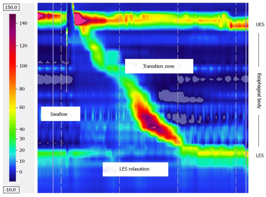
HRM was described for the first time in 1991, introducing an increased number of pressure sensors along the catheter and the use of spatio-temporal plots[27,28], leading to the subsequent development of the Chicago Classification for primary esophageal motility disorders[29,30]. In HRM systems, multiple sensors (up to 36) are distributed longitudinally and radially, closely spaced along the length of the manometric catheter. Two main types of manometric catheters are currently available, solid state and water perfused, each with different physical and performance characteristics and specific advantages and disadvantages concerning costs, preparation, the location of transducers, autoclave possibility and the rate of pressure increase. Nevertheless, HRM allows for simultaneous pressure readings within both the sphincters and the esophageal body, providing detailed esophageal pressure topography (EPT) (Figure 1). With HRM, pull-through techniques became unnecessary and several problems, such as artifacts attributable to swallow-induced sphincter movement[31] or EGJ conformational changes that can spontaneously occur, were overcome[32-34]. RM provides a dynamic representation of the pressure within and across the EGJ, and it also creates opportunities to quantify more precise measurement of EGJ relaxation and morphology[25,26,31], providing the opportunity to detect expiratory LES pressure and crural diaphragm (CD) contraction[35-39]. On this basis, the EGJ was recently reviewed and classified into three types: Type I (no LES-CD separation); type II (the LES and CD are spatially separated such that there is a double-peaked pressure profile, but the nadir pressure between LES and CD does not decrease to gastric pressure); an type III (the separation between peaks is > 2 cm, and the nadir pressure between LES-CD is equal to or less than the gastric pressure; in type IIIa, the pressure inversion point is located at the CD, while in type IIIb, it is placed at the LES level[30,36].
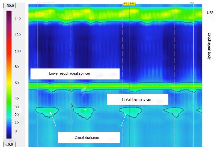
Pandolfino et al[36] compared the EPT attributes of the EGJ between 156 GERD patients and 75 asymptomatic controls. Although both lower LES pressure and greater LES-CD separation were associated with GERD, impaired CD function was most strongly associated factor and the only independent predictor of GERD. A study designed to analyze the relationship between obesity and the morphology of the EGJ pressure segment showed that obese subjects were more likely to have a spatial separation between the diaphragm and LES (Figure 2) and an augmented gastroesophageal pressure gradient[37]. These findings might partially explain why gastro-esophageal reflux is more frequent in obese subjects, as also evidenced in patients with NERD[40]. Bredenoord et al[32] showed that, in cases of small hiatal hernias in which intermittent reduction of the hernia frequently occurs, spatial separation of the CD and LES in the non-reduced state resulted in a 2-fold increase in acid and weakly acidic reflux.
Transient LES relaxations (TLESRs) are the most common mechanism of reflux. They occur independently from swallowing and are not accompanied by peristalsis, but they are accompanied by diaphragmatic inhibition, and they persist for longer periods than swallow-induced LES relaxations (> 10 s)[41,42]. In a recent study, Roman et al[43] demonstrated that HRM is reproducible and more sensitive than perfused-sleeve manometry to detect TLESRs, providing better inter-observer agreement. Notably, in GERD patients, there is not an increased frequency of TLESR compared with controls but only a greater frequency of acid reflux during TLESRs[44]. Bredenoord et al[45] investigated the factors associated with reflux during TLESRs but no differences were observed in TLESR duration, trans-sphincteric pressure gradient, the prevalence, duration and amplitude of esophageal pre-contractions or sphincteric post-contractions. Pandolfino et al[35] studied the largest number of TLESRs in the postprandial period. They observed that the key events associated with EGJ opening were CD inhibition, LES relaxation, distal esophageal muscular contraction and a positive gradient between the stomach and the esophagus, but in only a few cases was manometric signature of EGJ opening associated with evidence of reflux on pH-metry.
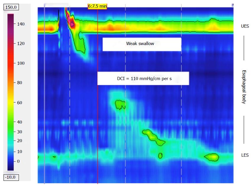
It has been shown that 21%-38% of patients with GERD present with severely impaired esophageal peristalsis (Figure 3), resulting in more severe reflux, slower acid clearance, worse mucosal injury, and more frequent respiratory symptoms[46]. The Chicago Classification V3.0 approved the term “IEM”, which is frequently used in conventional manometry[30]. In this recent Chicago Classification version, IEM was defined as 5 or more ineffective swallows of 10 with a DCI threshold of 450 mmHg-s-cm. No distinction need be made between failed swallows (DCI < 100 mmHg-s-cm) and weak swallows (450 mmHg-s-cm)[30].
Multiple rapid swallowing (MRS) as a provocative test has been suggested to diagnose IEM (in borderline conditions). MRS consists of administering 2 mL of water five times, for a total amount of 15 mL of water, in less than 10 s. MRS inhibits the esophageal body and LES during the first four swallows, and it is normally followed by an esophageal contraction of increased amplitude. It was suggested that many patients with suspected IEM had normalized esophageal contraction amplitude after MRS[47].
Manometric studies have described decreased pressure between striated and smooth esophageal muscle[48]. Pohl et al[49] correlated the size of the esophageal low-pressure zone and its possible relationship with esophageal symptoms (dysphagia, chest pain, and heartburn/regurgitation).
In conclusion, HRM is faster and easier to perform than conventional water-perfused manometry. Moreover, HRM does not require time-consuming pull-through maneuvers, and it allows for accurate evaluation of the intrinsic and extrinsic components of EGJ, thus improving the identification of TLESRs. However, the clinical application of this technique in GERD remains very limited; TLESRs cannot be used in the diagnosis of GERD because their prevalence is similar between GERD patients and normal subjects. Furthermore, minor esophageal motility abnormalities, observed in GERD patients, are not specific and can either be primarily or secondarily related to GERD[50]. However, HRM represents an important advance in the assessment of esophageal motor function and a promising technique for the evaluation of the mechanical abnormalities involved in GERD. Further studies are needed to evaluate additional possible benefits of this technique in clinical practice.
Multichannel intraluminal impedance (MII) has been promoted to detect the movement of fluid, solid, and air in the esophagus regardless of its pH[51].
This new device, which combines MII-pH analysis, provides a sophisticated characterization of reflux episodes over a 24-h period. The most common catheter allows for 6 channels for intraluminal impedance (at 3, 5, 7, 9, 15, and 17 cm) and a pH sensor (at 5 cm above the upper border of the LES)[52].
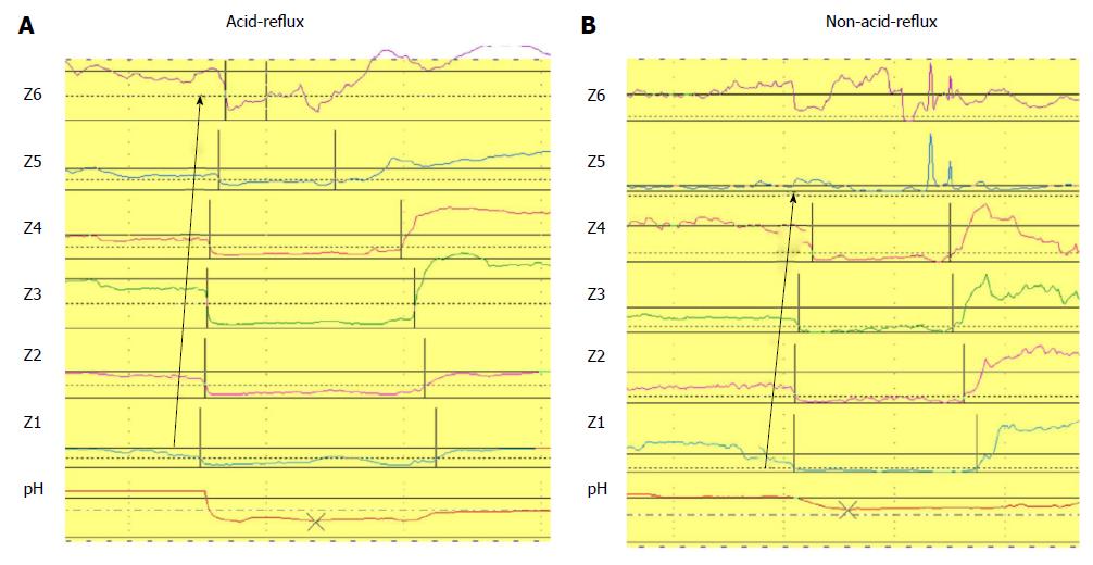
MII-pH is an innovative technique that provides a detailed characterization of each reflux event including chemical (acid and non-acid reflux) and physical properties (liquid, mixed, gas) (Figure 4)[53]. To date, non-acid reflux represents the majority of reflux episodes in patients with GERD on PPI therapy[54,55]. Indeed, the total number of reflux episodes is not affected by acid suppressive therapy, and weakly acidic reflux accounts for approximately 90% of all reflux episodes in patients on PPIs, thus representing a potential mechanism underlying the failure of PPI treatment in patients with reflux-related symptoms[56,57]. Moreover, MII-pH monitoring, as well as pH-metry alone, provides the opportunity to assess the temporal relationship between the occurrence of refluxes and the onset of symptoms[58,59]. The relationship between symptoms and reflux events can be evaluated with the symptom index (SI) and symptom association probability (SAP), which are the most commonly used symptom indices[59].
Based on esophageal pH monitoring, NERD patients with physiological esophageal AET and a close temporal relationship between symptoms and reflux events have been defined as hypersensitive to acid stimuli. In contrast, in agreement with the Rome III criteria, patients with heartburn, normal upper endoscopy, physiological AET, and negative correspondence between symptoms and reflux and who fail to respond to PPIs are defined as having functional heartburn (FH)[8,9,60]. In this regard, the advent of MII-pH monitoring has improved the diagnostic yield of GERD patients, mainly by identifying a positive SAP or SI with weakly acidic or non-acid reflux[61-68]. In particular, if pH-metry and the patient’s response to PPI therapy are compared with MII-pH, we can observe an underestimation of GERD patients[69,70].
In contrast, all of the available diagnostic tests for GERD have some limitations. The drawbacks of MII-pH are mainly due to the day-to-day variability of the test[71-73]. Additionally, the reflux-symptom correlation in patients with GERD who do not respond to PPI therapy is actually also calculated by the SI or SAP if its validity is still uncertain[74,75]. Recently Zerbib et al[76] reported that MII-pH findings were not always able to predict the response to PPIs in patients with typical reflux-related symptoms when the test is performed off PPI therapy.
Regarding the clinical utility of pathophysiological investigations in patients with heartburn, we described a group of patients (more than 19% of the whole population enrolled) with heartburn who completely responded to PPI, in whom GERD was not diagnosed with the MII-pH test[5]. Thus, our data suggested that PPI response alone should not be always considered a good predictor of a GERD diagnosis[5]. Overall, it is notable that NERD patients vary greatly from a pathophysiological point of view and should be accurately studied by means of MII-pH to undertake the best therapeutic approach[6]. Indeed, a meta-analysis found that the once defined low response rate in NERD was likely the result of the inclusion of patients with heartburn who did not have reflux disease[77].
Recently, the ability to perform MII-pH testing to understand GERD pathophysiology better has improved through the introduction of up and coming parameters, such as the post-reflux swallow-induced peristaltic wave (PSPW) index, which indicates the efficacy of esophageal clearance[78], and baseline impedance values, which indicate the presence of a lack of integrity in the esophageal mucosa[79]. Constant changes in esophageal chemical clearance could represent a specific mechanism involved in GERD pathophysiology. PSPW has been shown to be lower in patients with abnormal AET, compared to healthy volunteers (HVs) or FH patients. Moreover, this parameter was not altered after medical or surgical therapy[78]. Further, Kessing et al[80] described lower values of baseline impedance levels in the distal esophagus of patients with abnormal esophageal AET, compared to HVs. The authors described a negative correlation between baseline impedance levels and esophageal AET[80]. We recently described a large group of patients with typical GERD symptoms, negative endoscopy and any pathophysiological characteristics of GERD (normal AET, number of refluxes and negative SI and SAP). We observed that patients with good symptom relief after PPI therapy had lower baseline impedance values than FH patients (non-responders). FH patients showed similar baseline values to HVs. Moreover, we observed almost the same results when analyzing the PSPW index, which was lower in responders than in non-responders and HV groups. A direct linear correlation between PSPW and baseline impedance values has been described. Overall, these data suggest that baseline impedance values and PSPW could be considered up-and-coming parameters that could be helpful in better investigating patients with GERD-related symptoms, particularly when symptom-reflux association indexes fail to do so[81].
MII-pH testing showed that acid reflux events and their clearance were determinant factors that provoked esophageal mucosal breaks. Non-acid reflux does not appear to be directly related to the development of esophageal mucosa lesions; however, it is definitely involved in the genesis of symptoms in both NERD and erosive esophagitis (EE) patients. Ambulatory MII-pH studies have suggested that patients with moderate or severe esophagitis have rates of weakly acidic reflux similar to or slightly greater than healthy controls[69]. In this regard, it is important to emphasize that weakly acidic reflux is not synonymous with bile reflux. A simultaneous Bilitech and MII-pH study showed no relationship between the percentage of time for bilirubin absorbance and weakly acidic or weakly alkaline reflux. Indeed, this study showed that the greatest number of bile refluxes occurred concomitantly with acid refluxes[82].
In patients with Barrett’s esophagus (BE), MII-pH testing showed overall more severe reflux disease with a greater number of acid and weakly acidic reflux events and higher proximal extension[71]. By means of MII-pH, Savarino et al[71] showed that patients with BE and EE had greater numbers of acid and weakly acidic reflux episodes, higher percentages of proximal migration of the refluxate and higher total acid and volume clearance. Notably, it has been emphasized that a significantly increased amount of total reflux occurs in both the long and short segments of BE, compared with EE. In BE patients, the MII-pH tracks are not easy to analyze, especially in the long segment of BE. Inflammation and histologic modifications are supposed to reduce baseline impedance values thus impairing the ability to detect the real number of reflux episodes. This phenomenon has led some investigators to be more accurate during the manual analysis of MII-pH in patients with BE[62]. However, some authors have shown the viability of MII-pH even in BE patients[71,83]. In clinical practice, BE patients should be evaluated with MII-pH “on” PPI therapy to evaluate the effectiveness of acid suppression.
The presence of EE, which represents clear endoscopic evidence of esophageal mucosal injury (30% of all endoscopic series in patients with GERD-related symptoms), can obviously be responsible for symptoms provoked by the refluxate. In contrast, the absence of macroscopic damage makes it difficult to clarify the occurrence of the same symptoms in patients with NERD, and it is reasonable to hypothesize that microscopic damage is responsible for the GERD-related symptoms in them[84]. According to this assumption, recent investigations have focused their attention on the evaluation of the presence of dilated intercellular spaces, considered the most important microscopic alteration involved in symptom perception in patients with GERD[85]. Recently Savarino et al[86] showed that the frequency of microscopic esophagitis did not differ between patients with FH and control subjects and was significantly lower in FH patients than in the EE and well-defined NERD patients evaluated with MII-pH.
The overall MII-pH assessment “on” or “off” PPI therapy is actually a matter of discussion. In a recent seminar, Bredenoord et al[52] affirmed that combined MII-pH is better performed off PPIs when the diagnosis of GERD has not yet been established and on-PPIs when the diagnosis has been already made (i.e., positive endoscopy for EE, MII-pH off-therapy already performed, BE surveillance). In particular, MII-pH off PPIs is useful to investigate the causes of ineffective PPI treatment. To confirm the presence of GERD, Hemmink et al[87] reported that PPI-resistant patients should preferably be evaluated with MII-pH monitoring after cessation of PPI treatment. This approach could increase the likelihood of observing a positive relationship between symptoms and reflux.
GERD is considered an important cause of laryngeal inflammation. Laryngoscopic examinations are very important to exclude abnormalities in the laryngeal mucosa or vocal cords (nodules or neoplastic lesions), but it is not sufficiently specific/sensitive to detect LPR[88]. We recently described that MII-pH analysis detected GERD at least in 40% of patients who were diagnosed with LPRD. In particular, MII-pH analysis was able to identify patients with NERD, those with HE and those without GERD, whereas laryngeal symptoms and laryngoscopic findings were not able to do so[89].
Finally, some interesting studies, performed by means of MII-pH analysis, evaluated the effectiveness of raft-forming gel formulation in “add-on” treatment for GERD symptoms. These studies have clearly demonstrated the efficacy of raft-forming gel preparation in reducing the total number of refluxes and their proximal migration[90-92].
To conclude, MII-pH is able to improve the ability to detect GERD compared with pH-metry alone in patients with GERD-related symptoms. MII-pH is able to provide more information and to exclude GERD diagnosis definitively in PPI non-responder patients (FH). The outcomes studies are unmet clinical needs for determining whether MII-pH truly leads to a change in management.
Combined multichannel intraluminal impedance and manometry has been considered a very helpful device because it provides information about esophageal contraction and bolus transit simultaneously. Tutuian et al[93] described results from a large cohort of patients (350), showing a very strong correlation between dysphagia and incomplete bolus transit. Moreover, the largest defects of bolus transit were correlated with diagnoses of achalasia, scleroderma, distal esophageal spasm and IEM. Outcome studies are needed to clarify better the role of this technique in clinical diagnosis.
The wireless pH capsule (BRAVO) is a novel technology able to assess esophageal exposure to acid. This device is placed transorally 6 cm above the EGJ during an upper endoscopy, usually under sedation, or 5 cm above the LES, previously detected by means of esophageal manometry[94]. The capsule transmits recording data via telemetry to an external receiver. This method is usually performed after the failure of a PPI test, alternatively at MII-pH or during an upper endoscopy before the PPI test. Capsule detachment occurs spontaneously in most cases between 2-3 d and 2 wk[95]. Adverse events or complications (severe or persistent chest pain) are very rare (1% to 2%)[94]. The technique is usually well tolerated, and the principal side effect reported is mild to moderate chest pain. The recording time of the wireless capsule is usually 48 h, showing overall good reproducibility between the 2 d of recording[96]. However, several studies have shown a relevant day-to-day variability responsible for discrepancies in AET between the first and the second days of recording[97,98]. The reasons for these findings remain controversial and are the subject of debate.
A great day-to-day variability in AET might be frequent during 24-h pH-metry testing. pH monitoring prolonged to 48 h is routinely performed with the BRAVO technique, and it should provide a better overview of esophageal acid exposure and symptom-reflux correlation.
The prolonged pH-monitoring period increases the likelihood of detecting reflux events (12.5% increase), potentially improving symptom association (5.2% increase)[99]. These data were confirmed in patients on anti-reflux therapy[99], in patients with endoscopy negative heartburn[100] and in patients with non-cardiac chest pain[101].
Overall, BRAVO is a good method for reflux monitoring, especially in patients with less frequent symptoms (i.e., non-cardiac chest pain) and in those who refuse catheter-based techniques, but it has some limitations, including higher cost than MII-pH and the possible risk of misplacement; more importantly, non-acid reflux events cannot be detected.
An important property of the reflux barrier that is not assessed by manometry is distensibility (i.e., the ease with which the EGJ is opened)[102]. If pressure and radius can be measured simultaneously in the LES, the circumferential tension in the wall can be estimated, as it has been firstly demonstrated by McMahon et al[103].
The EndoFLIP (Endo Functional Luminal Imaging Probe system; Crospon Ltd., Galway, Ireland) consists of a polyurethane balloon (maximum volume of 60 mL) mounted on the distal 14 cm of a probe (length 240 cm, diameter 25 mm) attached to the EndoFLIP unit. This balloon assumes a 10-cm long cylindrical shape with maximum diameter of 2.5 cm when filled. Along a 7.5-cm segment within the balloon, 17 ring electrodes are spaced 5 mm apart to obtain 16 CSA measurements using an impedance planimetry technique. The probe also contains a solid-state pressure transducer to measure intra-balloon pressure.
The EndoFLIP system provides real-time and dynamic information on EGJ distention that is visualized as cylinders of different diameters, based on instantaneous CSA measurements (included instant pressure measurements).
Using this technique, Kwiatek et al[104] compared GERD patients with healthy controls, with the following major findings: (1) the EGJ was 2 to 3 times more distensible in GERD patients than controls; (2) 20- to 30-mL distention volumes provided, in patients with GERD, a two- to three-fold increased EGJ distensibility, compared with controls; and (3) the endoscopic estimation of the flap valve grade seemed poorly correlated with EndoFLIP measurements.
Recently, Regan et al[105] used the EndoFLIP system to measure upper esophageal sphincter (UES) distensibility. EndoFLIP provided definitive information regarding UES compliance without the need for fluoroscopy. The results regarding the anatomic parameters and physiology of UES were directly matched with current knowledge. Few data are yet available, but EndoFLIP could be able to provide determinant information about UES in patients with dysphagia before and after surgery or rehabilitation.
As mentioned above, the diagnosis and treatment of GERD are particularly difficult in cases of LPR[106].
Some authors have described LPR by means of dual-channel impedance and pH monitoring[107,108]. These studies have described the frequency, location, and direction of any gas or liquid refluxate along the esophagus, as well as in the hypopharynx[107]. Despite initial enthusiasm[108,109], outcome studies with impedance monitoring have been lacking, and their clinical significance with regard to medically recalcitrant LPR patients remains unclear[110].
The Dx-pH measurement system, called Restech (Respiratory Technology Corp., San Diego, CA, United States), was designed to detect aerosolized acid, thus identifying patients whose symptoms are due to acidic mist or liquid refluxing into the pharynx. Because the distal part containing the pH sensor does not traverse the UES, it is more comfortable than a conventional pH catheter, and it does not require esophageal manometry[17].
Restech has a higher frequency of measurement than older catheter-based pH probes and wireless pH probes, with a pH measurement obtained every 0.5 s (sampling rate, 2 Hz), compared with traditional catheter-based pH probes, which sample every 4 s to 5 s (at a rate of 0.2-0.25 Hz), and wireless pH probes, with a pH measurement every 6 s (0.17 Hz). This increased sampling rate theoretically allows Restech to detect more reflux events, which is a characteristic that could prove useful in patients with LPR[111].
Recently, Worrell et al[112] demonstrated, that in patients with extraesophageal reflux symptoms who underwent antireflux surgery, esophageal pH monitoring in the proximal esophagus failed to recognize 50% of the patients who recorded good outcomes post-antireflux surgery. Vailati et al[113] showed the high specificity and reasonable sensitivity of the Restech technique, which could be considered an interesting tool that can be used before therapy in patients with pharyngoesophageal reflux. In contrast, Ummarino et al[114] did not show any correlation, although chronic coughing was the only symptom reported by patients, and when they compared MII-pH and Restech, the superiority of the first technique seemed clear. Similarly, Becker et al[115] evaluated, in a prospective, single-center trial, the differences between MII-pH and Dx-pH. They demonstrated that acid pharyngeal pH levels detected with Dx-pH were not correlated with GERD, and acid esophageal reflux episodes did not result in pharyngeal pH alterations. Mazzoleni et al[116], observed a very poor correlation between Dx-pH probe oropharyngeal monitoring and MII-pH in a group of 68 patients.
Until now, the use of Dx-pH recordings could not be recommended in clinical practice, given the discrepancies between traditional MII-pH monitoring and the current technologies used to measure oro-pharyngeal pH events.
Recent studies have shown that patients with LPR do not reap great benefit from PPI therapy because, at this level, the damage is not mediated by acid but rather by pepsin[117]. For this reason, it was recently decided to search for pepsin as a marker of reflux because the enzyme is secreted only by gastric main cells[118]; therefore, its presence above UES is a certain sign of GERD.
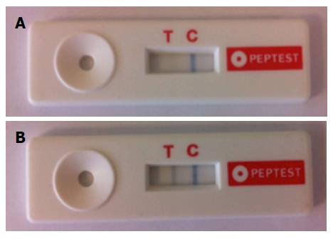
The pepsin lateral flow device (LFD), also called the PEP-Test (RDBiomed, Hull, United Kingdom), is a rapid, non-invasive test to detect salivary pepsin as a surrogate marker for GERD. The PEP-Test is an immunological in-vitro diagnostic medical device that contains two pepsin monoclonal antibodies; this test is able to detect pepsin in a clinical sample of saliva/sputum quickly and easily; the limit of detection is 16 ng/mL of pepsin. The patient must collect 30 mL of saliva, and this sample is centrifuged at 4000 rpm for 5 min; then, 80 μL from the surface layer are drawn up and mixed with a buffer solution using a vortex mixer. The sample obtained is applied in the circular well of the PEP-Test device, and after a few minutes, it is possible to check for the presence of pepsin: If it occurs on antigen-antibody binding, two blue lines appear on the display, while if pepsin is not present, it will be possible to see only the control line. The blue lines are visible on the display because the monoclonal antibody is directly labelled with blue latex beads, which detach at the time of Ag-Ab binding. Saritas Yuksel et al[19] evaluated the PEP-Test (defined as ELISA LFD) in 47 patients using pH-metry as a reference standard. The results were compared with a control population, which underwent only the PEP-Test, which has sensitivity and a specificity of 87% (Figure 5).
More recently Hayat et al[119] compared the PEP-test with MII-pH and showed a higher prevalence and concentration of salivary pepsin in patients with GERD or HE, compared with patients with FH and healthy controls. The authors suggested that salivary pepsin might complement questionnaires in the office-based diagnosis of patients with GERD-related symptoms.
The most important advantages of the PEP-Test are its low cost, its non-invasiveness, and, not to be underestimated, its feasibility while the patient is undergoing PPI therapy. However, given the very limited data available in the medical literature, further studies are needed to understand and assess the clinical utility of this test.
The reflux of gastric contents into the esophagus results in a broad and varied GERD spectrum. Thus, advances in the diagnosis of GERD represent the necessary progress in clinical investigation and therapeutic management as well as progress for more-in-depth assessment of the underlying pathophysiological mechanisms.
The pathogenesis of GERD is multifactorial, including esophageal motility abnormalities of which the most important are TLESRs and hypotensive LES and IEM. Recently, the role of esophageal dysmotility has gained relevant interest, showing an increased prevalence with increasing severity of GERD, from NERD to EE and BE[120]. To date, HRM is the gold standard for characterizing esophageal motility disorders. Indeed, HRM improves characterization of LES zones and esophageal body motility, increasing diagnostic yield and accuracy[121]. Moreover, HRM must be regarded as the new gold standard for detecting TLESRs[43]. However, the value of HRM in clinical practice has yet to be established fully Exclusion of severe esophageal motility disorders (i.e., achalasia) is important because such diseases can present with heartburn and regurgitation, which could lead to an erroneous diagnosis of GERD[122].
Ambulatory MII-pH has become the gold standard for investigating GERD, owing mainly to its ability to detect both acid and non-acid reflux[68,123]. Of particular importance, MII-pH has enabled the subdivision of patients with NERD into different subsets, including patients with an excess of acid and those with symptomatic non-acid reflux, and MII-pH has the ability to identify patients with FH whose symptoms are not GERD related and who must be excluded from the realm of GERD[6,56,109].
Moreover, diagnosing patients with HE to non-acid reflux, MII-pH has contributed to narrowing down the population of patients with FH[70]. Recently, it has been demonstrated that a more-in-depth pathophysiological evaluation of MII-pH tracings, including baseline impedance levels and PSPW index evaluation, could be helpful in better investigating patients with heartburn and appropriately identifying those with reflux disease and particularly those with HE, when symptom-reflux association analysis fails to do so[5,78,81].
According to the Montreal Classification, extra-esophageal symptoms of GERD can occur, such as hoarseness, coughing, and asthma[2]. However, establishing that extra-esophageal symptoms caused by GERD can be difficult, and in this context, the advent of new technologies deserves careful consideration. The Dx-pH measurement system is a sensitive and minimally invasive device for detecting acidic refluxate in the oropharynx. The PEP-Test is a sensitive and specific in vitro diagnostic test that allows for the rapid detection of pepsin, a marker of reflux, in a clinical sample. Overall, further studies are warranted to substantiate the clinical roles of these new technologies in diagnosing GERD.
| 1. | Dent J, El-Serag HB, Wallander MA, Johansson S. Epidemiology of gastro-oesophageal reflux disease: a systematic review. Gut. 2005;54:710-717. [RCA] [PubMed] [DOI] [Full Text] [Cited by in Crossref: 1256] [Cited by in RCA: 1271] [Article Influence: 60.5] [Reference Citation Analysis (1)] |
| 2. | Vakil N, van Zanten SV, Kahrilas P, Dent J, Jones R. The Montreal definition and classification of gastroesophageal reflux disease: a global evidence-based consensus. Am J Gastroenterol. 2006;101:1900-1920; quiz 1943. [PubMed] |
| 3. | Fuchs KH, Babic B, Breithaupt W, Dallemagne B, Fingerhut A, Furnee E, Granderath F, Horvath P, Kardos P, Pointner R. EAES recommendations for the management of gastroesophageal reflux disease. Surg Endosc. 2014;28:1753-1773. [RCA] [PubMed] [DOI] [Full Text] [Cited by in Crossref: 125] [Cited by in RCA: 137] [Article Influence: 11.4] [Reference Citation Analysis (0)] |
| 4. | Modlin IM, Hunt RH, Malfertheiner P, Moayyedi P, Quigley EM, Tytgat GN, Tack J, Heading RC, Holtman G, Moss SF. Diagnosis and management of non-erosive reflux disease--the Vevey NERD Consensus Group. Digestion. 2009;80:74-88. [PubMed] |
| 5. | de Bortoli N, Martinucci I, Savarino E, Bellini M, Bredenoord AJ, Franchi R, Bertani L, Furnari M, Savarino V, Blandizzi C. Proton pump inhibitor responders who are not confirmed as GERD patients with impedance and pH monitoring: who are they? Neurogastroenterol Motil. 2014;26:28-35. [RCA] [PubMed] [DOI] [Full Text] [Cited by in Crossref: 62] [Cited by in RCA: 64] [Article Influence: 5.3] [Reference Citation Analysis (0)] |
| 6. | Savarino E, Zentilin P, Savarino V. NERD: an umbrella term including heterogeneous subpopulations. Nat Rev Gastroenterol Hepatol. 2013;10:371-380. [RCA] [PubMed] [DOI] [Full Text] [Cited by in Crossref: 142] [Cited by in RCA: 181] [Article Influence: 13.9] [Reference Citation Analysis (0)] |
| 7. | Giacchino M, Savarino V, Savarino E. Distinction between patients with non-erosive reflux disease and functional heartburn. Ann Gastroenterol. 2013;26:283-289. [PubMed] |
| 8. | Martinez SD, Malagon IB, Garewal HS, Cui H, Fass R. Non-erosive reflux disease (NERD)--acid reflux and symptom patterns. Aliment Pharmacol Ther. 2003;17:537-545. [PubMed] |
| 9. | Fass R, Fennerty MB, Vakil N. Nonerosive reflux disease--current concepts and dilemmas. Am J Gastroenterol. 2001;96:303-314. [RCA] [PubMed] [DOI] [Full Text] [Cited by in Crossref: 169] [Cited by in RCA: 185] [Article Influence: 7.4] [Reference Citation Analysis (0)] |
| 10. | Woodland P, Sifrim D. The refluxate: The impact of its magnitude, composition and distribution. Best Pract Res Clin Gastroenterol. 2010;24:861-871. [RCA] [PubMed] [DOI] [Full Text] [Cited by in Crossref: 25] [Cited by in RCA: 22] [Article Influence: 1.4] [Reference Citation Analysis (0)] |
| 11. | Sifrim D, Zerbib F. Diagnosis and management of patients with reflux symptoms refractory to proton pump inhibitors. Gut. 2012;61:1340-1354. [RCA] [PubMed] [DOI] [Full Text] [Cited by in Crossref: 224] [Cited by in RCA: 252] [Article Influence: 18.0] [Reference Citation Analysis (0)] |
| 12. | Smout AJ. The patient with GORD and chronically recurrent problems. Best Pract Res Clin Gastroenterol. 2007;21:365-378. [RCA] [PubMed] [DOI] [Full Text] [Cited by in Crossref: 12] [Cited by in RCA: 12] [Article Influence: 0.6] [Reference Citation Analysis (0)] |
| 13. | Furnari M, de Bortoli N, Savarino V, Marchi S, Savarino E. Not all anti-reflux treatment failures are due to persistence of abnormal esophageal acid exposure. Surg Endosc. 2014;28:1382-1383. [RCA] [PubMed] [DOI] [Full Text] [Cited by in Crossref: 1] [Cited by in RCA: 1] [Article Influence: 0.1] [Reference Citation Analysis (0)] |
| 14. | Savarino E, Carbone R, Marabotto E, Furnari M, Sconfienza L, Ghio M, Zentilin P, Savarino V. Gastro-oesophageal reflux and gastric aspiration in idiopathic pulmonary fibrosis patients. Eur Respir J. 2013;42:1322-1331. [RCA] [PubMed] [DOI] [Full Text] [Cited by in Crossref: 187] [Cited by in RCA: 174] [Article Influence: 13.4] [Reference Citation Analysis (0)] |
| 15. | Savarino E, Bazzica M, Zentilin P, Pohl D, Parodi A, Cittadini G, Negrini S, Indiveri F, Tutuian R, Savarino V. Gastroesophageal reflux and pulmonary fibrosis in scleroderma: a study using pH-impedance monitoring. Am J Respir Crit Care Med. 2009;179:408-413. [RCA] [PubMed] [DOI] [Full Text] [Cited by in Crossref: 188] [Cited by in RCA: 192] [Article Influence: 10.7] [Reference Citation Analysis (0)] |
| 16. | Martinucci I, de Bortoli N, Savarino E, Nacci A, Romeo SO, Bellini M, Savarino V, Fattori B, Marchi S. Optimal treatment of laryngopharyngeal reflux disease. Ther Adv Chronic Dis. 2013;4:287-301. [RCA] [PubMed] [DOI] [Full Text] [Cited by in Crossref: 55] [Cited by in RCA: 66] [Article Influence: 5.1] [Reference Citation Analysis (1)] |
| 17. | Wiener GJ, Tsukashima R, Kelly C, Wolf E, Schmeltzer M, Bankert C, Fisk L, Vaezi M. Oropharyngeal pH monitoring for the detection of liquid and aerosolized supraesophageal gastric reflux. J Voice. 2009;23:498-504. [RCA] [PubMed] [DOI] [Full Text] [Cited by in Crossref: 94] [Cited by in RCA: 82] [Article Influence: 4.6] [Reference Citation Analysis (0)] |
| 18. | Savarino E, Zentilin P, Savarino V, Tenca A, Penagini R, Clarke JO, Bravi I, Zerbib F, Yüksel ES. Functional testing: pharyngeal pH monitoring and high-resolution manometry. Ann N Y Acad Sci. 2013;1300:226-235. [RCA] [PubMed] [DOI] [Full Text] [Cited by in Crossref: 12] [Cited by in RCA: 13] [Article Influence: 1.0] [Reference Citation Analysis (0)] |
| 19. | Saritas Yuksel E, Hong SK, Strugala V, Slaughter JC, Goutte M, Garrett CG, Dettmar PW, Vaezi MF. Rapid salivary pepsin test: blinded assessment of test performance in gastroesophageal reflux disease. Laryngoscope. 2012;122:1312-1316. [RCA] [PubMed] [DOI] [Full Text] [Cited by in Crossref: 84] [Cited by in RCA: 94] [Article Influence: 6.7] [Reference Citation Analysis (0)] |
| 20. | Savarino E, Giacchino M, Savarino V. Dysmotility and reflux disease. Curr Opin Otolaryngol Head Neck Surg. 2013;21:548-556. [RCA] [PubMed] [DOI] [Full Text] [Cited by in Crossref: 2] [Cited by in RCA: 7] [Article Influence: 0.6] [Reference Citation Analysis (0)] |
| 21. | Martinucci I, de Bortoli N, Giacchino M, Bodini G, Marabotto E, Marchi S, Savarino V, Savarino E. Esophageal motility abnormalities in gastroesophageal reflux disease. World J Gastrointest Pharmacol Ther. 2014;5:86-96. [RCA] [PubMed] [DOI] [Full Text] [Full Text (PDF)] [Cited by in CrossRef: 51] [Cited by in RCA: 55] [Article Influence: 4.6] [Reference Citation Analysis (0)] |
| 22. | Dodds WJ, Dent J, Hogan WJ, Helm JF, Hauser R, Patel GK, Egide MS. Mechanisms of gastroesophageal reflux in patients with reflux esophagitis. N Engl J Med. 1982;307:1547-1552. [RCA] [PubMed] [DOI] [Full Text] [Cited by in Crossref: 741] [Cited by in RCA: 652] [Article Influence: 14.8] [Reference Citation Analysis (0)] |
| 23. | Orlando RC. Overview of the mechanisms of gastroesophageal reflux. Am J Med. 2001;111 Suppl 8A:174S-177S. [PubMed] |
| 24. | Dent J, Holloway RH, Toouli J, Dodds WJ. Mechanisms of lower oesophageal sphincter incompetence in patients with symptomatic gastrooesophageal reflux. Gut. 1988;29:1020-1028. [PubMed] |
| 25. | Tolone S, de Cassan C, de Bortoli N, Roman S, Galeazzi F, Salvador R, Marabotto E, Furnari M, Zentilin P, Marchi S. Esophagogastric junction morphology is associated with a positive impedance-pH monitoring in patients with GERD. Neurogastroenterol Motil. 2015;27:1175-1182. [RCA] [PubMed] [DOI] [Full Text] [Cited by in Crossref: 78] [Cited by in RCA: 95] [Article Influence: 8.6] [Reference Citation Analysis (0)] |
| 26. | Tolone S, De Bortoli N, Marabotto E, de Cassan C, Bodini G, Roman S, Furnari M, Savarino V, Docimo L, Savarino E. Esophagogastric junction contractility for clinical assessment in patients with GERD: a real added value? Neurogastroenterol Motil. 2015;27:1423-1431. [RCA] [PubMed] [DOI] [Full Text] [Cited by in Crossref: 70] [Cited by in RCA: 88] [Article Influence: 8.0] [Reference Citation Analysis (0)] |
| 27. | Clouse RE, Staiano A. Topography of the esophageal peristaltic pressure wave. Am J Physiol. 1991;261:G677-G684. [PubMed] |
| 28. | Clouse RE, Staiano A, Alrakawi A. Development of a topographic analysis system for manometric studies in the gastrointestinal tract. Gastrointest Endosc. 1998;48:395-401. [PubMed] |
| 29. | Kahrilas PJ, Ghosh SK, Pandolfino JE. Esophageal motility disorders in terms of pressure topography: the Chicago Classification. J Clin Gastroenterol. 2008;42:627-635. [RCA] [PubMed] [DOI] [Full Text] [Full Text (PDF)] [Cited by in Crossref: 249] [Cited by in RCA: 217] [Article Influence: 12.1] [Reference Citation Analysis (0)] |
| 30. | Kahrilas PJ, Bredenoord AJ, Fox M, Gyawali CP, Roman S, Smout AJ, Pandolfino JE. The Chicago Classification of esophageal motility disorders, v3.0. Neurogastroenterol Motil. 2015;27:160-174. [RCA] [PubMed] [DOI] [Full Text] [Cited by in Crossref: 1373] [Cited by in RCA: 1485] [Article Influence: 135.0] [Reference Citation Analysis (0)] |
| 31. | Pandolfino JE, Ghosh SK, Zhang Q, Jarosz A, Shah N, Kahrilas PJ. Quantifying EGJ morphology and relaxation with high-resolution manometry: a study of 75 asymptomatic volunteers. Am J Physiol Gastrointest Liver Physiol. 2006;290:G1033-G1040. [RCA] [PubMed] [DOI] [Full Text] [Cited by in Crossref: 172] [Cited by in RCA: 163] [Article Influence: 8.2] [Reference Citation Analysis (0)] |
| 32. | Bredenoord AJ, Weusten BL, Timmer R, Smout AJ. Intermittent spatial separation of diaphragm and lower esophageal sphincter favors acidic and weakly acidic reflux. Gastroenterology. 2006;130:334-340. [RCA] [PubMed] [DOI] [Full Text] [Cited by in Crossref: 114] [Cited by in RCA: 104] [Article Influence: 5.2] [Reference Citation Analysis (0)] |
| 33. | Edmundowicz SA, Clouse RE. Shortening of the esophagus in response to swallowing. Am J Physiol. 1991;260:G512-G516. [PubMed] |
| 34. | Staiano A, Clouse RE. Detection of incomplete lower esophageal sphincter relaxation with conventional point-pressure sensors. Am J Gastroenterol. 2001;96:3258-3267. [RCA] [PubMed] [DOI] [Full Text] [Cited by in Crossref: 34] [Cited by in RCA: 34] [Article Influence: 1.4] [Reference Citation Analysis (0)] |
| 35. | Pandolfino JE, Zhang QG, Ghosh SK, Han A, Boniquit C, Kahrilas PJ. Transient lower esophageal sphincter relaxations and reflux: mechanistic analysis using concurrent fluoroscopy and high-resolution manometry. Gastroenterology. 2006;131:1725-1733. [RCA] [PubMed] [DOI] [Full Text] [Cited by in Crossref: 152] [Cited by in RCA: 141] [Article Influence: 7.1] [Reference Citation Analysis (0)] |
| 36. | Pandolfino JE, Kim H, Ghosh SK, Clarke JO, Zhang Q, Kahrilas PJ. High-resolution manometry of the EGJ: an analysis of crural diaphragm function in GERD. Am J Gastroenterol. 2007;102:1056-1063. [RCA] [PubMed] [DOI] [Full Text] [Cited by in Crossref: 197] [Cited by in RCA: 198] [Article Influence: 10.4] [Reference Citation Analysis (0)] |
| 37. | Pandolfino JE, El-Serag HB, Zhang Q, Shah N, Ghosh SK, Kahrilas PJ. Obesity: a challenge to esophagogastric junction integrity. Gastroenterology. 2006;130:639-649. [RCA] [PubMed] [DOI] [Full Text] [Cited by in Crossref: 379] [Cited by in RCA: 366] [Article Influence: 18.3] [Reference Citation Analysis (0)] |
| 38. | Bredenoord AJ, Weusten BL, Roelofs JM, Smout AJ. The gastro-oesophageal pressure inversion point revisited. Scand J Gastroenterol. 2003;38:812-818. [PubMed] |
| 39. | Bredenoord AJ, Weusten BL, Carmagnola S, Smout AJ. Double-peaked high-pressure zone at the esophagogastric junction in controls and in patients with a hiatal hernia: a study using high-resolution manometry. Dig Dis Sci. 2004;49:1128-1135. [PubMed] |
| 40. | Savarino E, Zentilin P, Marabotto E, Bonfanti D, Inferrera S, Assandri L, Sammito G, Gemignani L, Furnari M, Dulbecco P. Overweight is a risk factor for both erosive and non-erosive reflux disease. Dig Liver Dis. 2011;43:940-945. [RCA] [PubMed] [DOI] [Full Text] [Cited by in Crossref: 42] [Cited by in RCA: 52] [Article Influence: 3.5] [Reference Citation Analysis (0)] |
| 41. | Mittal RK, Holloway RH, Penagini R, Blackshaw LA, Dent J. Transient lower esophageal sphincter relaxation. Gastroenterology. 1995;109:601-610. [PubMed] |
| 42. | Holloway RH, Penagini R, Ireland AC. Criteria for objective definition of transient lower esophageal sphincter relaxation. Am J Physiol. 1995;268:G128-G133. [PubMed] |
| 43. | Roman S, Zerbib F, Belhocine K, des Varannes SB, Mion F. High resolution manometry to detect transient lower oesophageal sphincter relaxations: diagnostic accuracy compared with perfused-sleeve manometry, and the definition of new detection criteria. Aliment Pharmacol Ther. 2011;34:384-393. [RCA] [PubMed] [DOI] [Full Text] [Cited by in Crossref: 39] [Cited by in RCA: 37] [Article Influence: 2.5] [Reference Citation Analysis (0)] |
| 44. | Sifrim D, Holloway R. Transient lower esophageal sphincter relaxations: how many or how harmful? Am J Gastroenterol. 2001;96:2529-2532. [RCA] [PubMed] [DOI] [Full Text] [Cited by in Crossref: 92] [Cited by in RCA: 72] [Article Influence: 2.9] [Reference Citation Analysis (0)] |
| 45. | Bredenoord AJ, Weusten BL, Timmer R, Smout AJ. Gastro-oesophageal reflux of liquids and gas during transient lower oesophageal sphincter relaxations. Neurogastroenterol Motil. 2006;18:888-893. [RCA] [PubMed] [DOI] [Full Text] [Cited by in Crossref: 25] [Cited by in RCA: 24] [Article Influence: 1.2] [Reference Citation Analysis (0)] |
| 46. | Diener U, Patti MG, Molena D, Fisichella PM, Way LW. Esophageal dysmotility and gastroesophageal reflux disease. J Gastrointest Surg. 2001;5:260-265. [PubMed] |
| 47. | Fornari F, Bravi I, Penagini R, Tack J, Sifrim D. Multiple rapid swallowing: a complementary test during standard oesophageal manometry. Neurogastroenterol Motil. 2009;21:718-e41. [RCA] [PubMed] [DOI] [Full Text] [Cited by in Crossref: 142] [Cited by in RCA: 130] [Article Influence: 7.6] [Reference Citation Analysis (0)] |
| 48. | Ghosh SK, Janiak P, Schwizer W, Hebbard GS, Brasseur JG. Physiology of the esophageal pressure transition zone: separate contraction waves above and below. Am J Physiol Gastrointest Liver Physiol. 2006;290:G568-G576. [RCA] [PubMed] [DOI] [Full Text] [Cited by in Crossref: 70] [Cited by in RCA: 60] [Article Influence: 3.0] [Reference Citation Analysis (0)] |
| 49. | Pohl D, Ribolsi M, Savarino E, Frühauf H, Fried M, Castell DO, Tutuian R. Characteristics of the esophageal low-pressure zone in healthy volunteers and patients with esophageal symptoms: assessment by high-resolution manometry. Am J Gastroenterol. 2008;103:2544-2549. [RCA] [PubMed] [DOI] [Full Text] [Cited by in Crossref: 36] [Cited by in RCA: 35] [Article Influence: 1.9] [Reference Citation Analysis (0)] |
| 50. | Vinjirayer E, Gonzalez B, Brensinger C, Bracy N, Obelmejias R, Katzka DA, Metz DC. Ineffective motility is not a marker for gastroesophageal reflux disease. Am J Gastroenterol. 2003;98:771-776. [RCA] [PubMed] [DOI] [Full Text] [Cited by in Crossref: 33] [Cited by in RCA: 36] [Article Influence: 1.6] [Reference Citation Analysis (0)] |
| 51. | Dolder M, Tutuian R. Laboratory based investigations for diagnosing gastroesophageal reflux disease. Best Pract Res Clin Gastroenterol. 2010;24:787-798. [RCA] [PubMed] [DOI] [Full Text] [Cited by in Crossref: 9] [Cited by in RCA: 5] [Article Influence: 0.3] [Reference Citation Analysis (0)] |
| 52. | Bredenoord AJ, Pandolfino JE, Smout AJ. Gastro-oesophageal reflux disease. Lancet. 2013;381:1933-1942. [RCA] [PubMed] [DOI] [Full Text] [Cited by in Crossref: 107] [Cited by in RCA: 101] [Article Influence: 7.8] [Reference Citation Analysis (0)] |
| 53. | Zentilin P, Iiritano E, Dulbecco P, Bilardi C, Savarino E, De Conca S, Parodi A, Reglioni S, Vigneri S, Savarino V. Normal values of 24-h ambulatory intraluminal impedance combined with pH-metry in subjects eating a Mediterranean diet. Dig Liver Dis. 2006;38:226-232. [PubMed] |
| 54. | Frazzoni M, Savarino E, Manno M, Melotti G, Mirante VG, Mussetto A, Bertani H, Manta R, Conigliaro R. Reflux patterns in patients with short-segment Barrett’s oesophagus: a study using impedance-pH monitoring off and on proton pump inhibitor therapy. Aliment Pharmacol Ther. 2009;30:508-515. [PubMed] |
| 55. | Boeckxstaens GE, Smout A. Systematic review: role of acid, weakly acidic and weakly alkaline reflux in gastro-oesophageal reflux disease. Aliment Pharmacol Ther. 2010;32:334-343. [RCA] [PubMed] [DOI] [Full Text] [Cited by in Crossref: 104] [Cited by in RCA: 98] [Article Influence: 6.1] [Reference Citation Analysis (0)] |
| 56. | Mainie I, Tutuian R, Shay S, Vela M, Zhang X, Sifrim D, Castell DO. Acid and non-acid reflux in patients with persistent symptoms despite acid suppressive therapy: a multicentre study using combined ambulatory impedance-pH monitoring. Gut. 2006;55:1398-1402. [PubMed] |
| 57. | Zerbib F, Roman S, Ropert A, des Varannes SB, Pouderoux P, Chaput U, Mion F, Vérin E, Galmiche JP, Sifrim D. Esophageal pH-impedance monitoring and symptom analysis in GERD: a study in patients off and on therapy. Am J Gastroenterol. 2006;101:1956-1963. [PubMed] |
| 58. | Savarino E, Zentilin P, Tutuian R, Pohl D, Gemignani L, Malesci A, Savarino V. Impedance-pH reflux patterns can differentiate non-erosive reflux disease from functional heartburn patients. J Gastroenterol. 2012;47:159-168. [RCA] [PubMed] [DOI] [Full Text] [Cited by in Crossref: 80] [Cited by in RCA: 94] [Article Influence: 6.7] [Reference Citation Analysis (0)] |
| 59. | Bredenoord AJ, Weusten BL, Smout AJ. Symptom association analysis in ambulatory gastro-oesophageal reflux monitoring. Gut. 2005;54:1810-1817. [PubMed] |
| 60. | de Bortoli N, Martinucci I, Bellini M, Savarino E, Savarino V, Blandizzi C, Marchi S. Overlap of functional heartburn and gastroesophageal reflux disease with irritable bowel syndrome. World J Gastroenterol. 2013;19:5787-5797. [RCA] [PubMed] [DOI] [Full Text] [Full Text (PDF)] [Cited by in CrossRef: 38] [Cited by in RCA: 41] [Article Influence: 3.2] [Reference Citation Analysis (0)] |
| 61. | Savarino E, Zentilin P, Tutuian R, Pohl D, Casa DD, Frazzoni M, Cestari R, Savarino V. The role of nonacid reflux in NERD: lessons learned from impedance-pH monitoring in 150 patients off therapy. Am J Gastroenterol. 2008;103:2685-2693. [RCA] [PubMed] [DOI] [Full Text] [Cited by in Crossref: 178] [Cited by in RCA: 202] [Article Influence: 11.2] [Reference Citation Analysis (0)] |
| 62. | Sifrim D, Castell D, Dent J, Kahrilas PJ. Gastro-oesophageal reflux monitoring: review and consensus report on detection and definitions of acid, non-acid, and gas reflux. Gut. 2004;53:1024-1031. [PubMed] |
| 63. | Shay S, Tutuian R, Sifrim D, Vela M, Wise J, Balaji N, Zhang X, Adhami T, Murray J, Peters J. Twenty-four hour ambulatory simultaneous impedance and pH monitoring: a multicenter report of normal values from 60 healthy volunteers. Am J Gastroenterol. 2004;99:1037-1043. [RCA] [PubMed] [DOI] [Full Text] [Cited by in Crossref: 413] [Cited by in RCA: 386] [Article Influence: 17.5] [Reference Citation Analysis (0)] |
| 64. | Blondeau K, Tack J. Pro: Impedance testing is useful in the management of GERD. Am J Gastroenterol. 2009;104:2664-2666. [RCA] [PubMed] [DOI] [Full Text] [Cited by in Crossref: 21] [Cited by in RCA: 17] [Article Influence: 1.0] [Reference Citation Analysis (0)] |
| 65. | Chen CL, Hsu PI. Current advances in the diagnosis and treatment of nonerosive reflux disease. Gastroenterol Res Pract. 2013;2013:653989. [RCA] [PubMed] [DOI] [Full Text] [Full Text (PDF)] [Cited by in Crossref: 20] [Cited by in RCA: 21] [Article Influence: 1.6] [Reference Citation Analysis (0)] |
| 66. | Bredenoord AJ, Weusten BL, Timmer R, Conchillo JM, Smout AJ. Addition of esophageal impedance monitoring to pH monitoring increases the yield of symptom association analysis in patients off PPI therapy. Am J Gastroenterol. 2006;101:453-459. [RCA] [PubMed] [DOI] [Full Text] [Cited by in Crossref: 122] [Cited by in RCA: 108] [Article Influence: 5.4] [Reference Citation Analysis (0)] |
| 67. | Savarino E, Pohl D, Zentilin P, Dulbecco P, Sammito G, Sconfienza L, Vigneri S, Camerini G, Tutuian R, Savarino V. Functional heartburn has more in common with functional dyspepsia than with non-erosive reflux disease. Gut. 2009;58:1185-1191. [PubMed] |
| 68. | de Bortoli N, Martinucci I, Savarino E, Franchi R, Bertani L, Russo S, Ceccarelli L, Costa F, Bellini M, Blandizzi C. Lower pH values of weakly acidic refluxes as determinants of heartburn perception in gastroesophageal reflux disease patients with normal esophageal acid exposure. Dis Esophagus. 2014;Epub ahead of print. [RCA] [PubMed] [DOI] [Full Text] [Cited by in Crossref: 12] [Cited by in RCA: 12] [Article Influence: 1.2] [Reference Citation Analysis (0)] |
| 69. | Savarino E, Tutuian R, Zentilin P, Dulbecco P, Pohl D, Marabotto E, Parodi A, Sammito G, Gemignani L, Bodini G. Characteristics of reflux episodes and symptom association in patients with erosive esophagitis and nonerosive reflux disease: study using combined impedance-pH off therapy. Am J Gastroenterol. 2010;105:1053-1061. [RCA] [PubMed] [DOI] [Full Text] [Cited by in Crossref: 155] [Cited by in RCA: 179] [Article Influence: 11.2] [Reference Citation Analysis (0)] |
| 70. | Savarino E, Marabotto E, Zentilin P, Frazzoni M, Sammito G, Bonfanti D, Sconfienza L, Assandri L, Gemignani L, Malesci A. The added value of impedance-pH monitoring to Rome III criteria in distinguishing functional heartburn from non-erosive reflux disease. Dig Liver Dis. 2011;43:542-547. [RCA] [PubMed] [DOI] [Full Text] [Cited by in Crossref: 111] [Cited by in RCA: 129] [Article Influence: 8.6] [Reference Citation Analysis (0)] |
| 71. | Savarino E, Zentilin P, Frazzoni M, Cuoco DL, Pohl D, Dulbecco P, Marabotto E, Sammito G, Gemignani L, Tutuian R. Characteristics of gastro-esophageal reflux episodes in Barrett’s esophagus, erosive esophagitis and healthy volunteers. Neurogastroenterol Motil. 2010;22:1061-e1280. [RCA] [PubMed] [DOI] [Full Text] [Cited by in Crossref: 64] [Cited by in RCA: 71] [Article Influence: 4.4] [Reference Citation Analysis (0)] |
| 72. | Zentilin P, Dulbecco P, Savarino E, Giannini E, Savarino V. Combined multichannel intraluminal impedance and pH-metry: a novel technique to improve detection of gastro-oesophageal reflux literature review. Dig Liver Dis. 2004;36:565-569. [RCA] [PubMed] [DOI] [Full Text] [Cited by in Crossref: 62] [Cited by in RCA: 70] [Article Influence: 3.2] [Reference Citation Analysis (0)] |
| 73. | Pandolfino JE, Vela MF. Esophageal-reflux monitoring. Gastrointest Endosc. 2009;69:917-30, 930.e1. [PubMed] |
| 74. | Hershcovici T, Wendel CS, Fass R. Symptom indexes in refractory gastroesophageal reflux disease: overrated or misunderstood? Clin Gastroenterol Hepatol. 2011;9:816-817. [RCA] [PubMed] [DOI] [Full Text] [Cited by in Crossref: 8] [Cited by in RCA: 8] [Article Influence: 0.5] [Reference Citation Analysis (0)] |
| 75. | Slaughter JC, Goutte M, Rymer JA, Oranu AC, Schneider JA, Garrett CG, Hagaman D, Vaezi MF. Caution about overinterpretation of symptom indexes in reflux monitoring for refractory gastroesophageal reflux disease. Clin Gastroenterol Hepatol. 2011;9:868-874. [RCA] [PubMed] [DOI] [Full Text] [Cited by in Crossref: 107] [Cited by in RCA: 118] [Article Influence: 7.9] [Reference Citation Analysis (0)] |
| 76. | Zerbib F, Belhocine K, Simon M, Capdepont M, Mion F, Bruley des Varannes S, Galmiche JP. Clinical, but not oesophageal pH-impedance, profiles predict response to proton pump inhibitors in gastro-oesophageal reflux disease. Gut. 2012;61:501-506. [RCA] [PubMed] [DOI] [Full Text] [Cited by in Crossref: 96] [Cited by in RCA: 96] [Article Influence: 6.9] [Reference Citation Analysis (0)] |
| 77. | Weijenborg PW, Cremonini F, Smout AJ, Bredenoord AJ. PPI therapy is equally effective in well-defined non-erosive reflux disease and in reflux esophagitis: a meta-analysis. Neurogastroenterol Motil. 2012;24:747-757, e350. [RCA] [PubMed] [DOI] [Full Text] [Cited by in Crossref: 94] [Cited by in RCA: 115] [Article Influence: 8.2] [Reference Citation Analysis (0)] |
| 78. | Frazzoni M, Manta R, Mirante VG, Conigliaro R, Frazzoni L, Melotti G. Esophageal chemical clearance is impaired in gastro-esophageal reflux disease--a 24-h impedance-pH monitoring assessment. Neurogastroenterol Motil. 2013;25:399-406, e295. [RCA] [PubMed] [DOI] [Full Text] [Cited by in Crossref: 136] [Cited by in RCA: 135] [Article Influence: 10.4] [Reference Citation Analysis (0)] |
| 79. | Farré R, Blondeau K, Clement D, Vicario M, Cardozo L, Vieth M, Mertens V, Pauwels A, Silny J, Jimenez M. Evaluation of oesophageal mucosa integrity by the intraluminal impedance technique. Gut. 2011;60:885-892. [RCA] [PubMed] [DOI] [Full Text] [Cited by in Crossref: 236] [Cited by in RCA: 220] [Article Influence: 14.7] [Reference Citation Analysis (0)] |
| 80. | Kessing BF, Bredenoord AJ, Weijenborg PW, Hemmink GJ, Loots CM, Smout AJ. Esophageal acid exposure decreases intraluminal baseline impedance levels. Am J Gastroenterol. 2011;106:2093-2097. [RCA] [PubMed] [DOI] [Full Text] [Cited by in Crossref: 175] [Cited by in RCA: 189] [Article Influence: 12.6] [Reference Citation Analysis (0)] |
| 81. | Martinucci I, de Bortoli N, Savarino E, Piaggi P, Bellini M, Antonelli A, Savarino V, Frazzoni M, Marchi S. Esophageal baseline impedance levels in patients with pathophysiological characteristics of functional heartburn. Neurogastroenterol Motil. 2014;26:546-555. [RCA] [PubMed] [DOI] [Full Text] [Cited by in Crossref: 155] [Cited by in RCA: 195] [Article Influence: 16.3] [Reference Citation Analysis (0)] |
| 82. | Pace F, Sangaletti O, Pallotta S, Molteni P, Porro GB. Biliary reflux and non-acid reflux are two distinct phenomena: a comparison between 24-hour multichannel intraesophageal impedance and bilirubin monitoring. Scand J Gastroenterol. 2007;42:1031-1039. [RCA] [PubMed] [DOI] [Full Text] [Cited by in Crossref: 36] [Cited by in RCA: 30] [Article Influence: 1.6] [Reference Citation Analysis (0)] |
| 83. | Eisen GM, Sandler RS, Murray S, Gottfried M. The relationship between gastroesophageal reflux disease and its complications with Barrett’s esophagus. Am J Gastroenterol. 1997;92:27-31. [PubMed] |
| 84. | Dent J. Microscopic esophageal mucosal injury in nonerosive reflux disease. Clin Gastroenterol Hepatol. 2007;5:4-16. [RCA] [PubMed] [DOI] [Full Text] [Cited by in Crossref: 92] [Cited by in RCA: 83] [Article Influence: 4.4] [Reference Citation Analysis (0)] |
| 85. | Zentilin P, Savarino V, Mastracci L, Spaggiari P, Dulbecco P, Ceppa P, Savarino E, Parodi A, Mansi C, Fiocca R. Reassessment of the diagnostic value of histology in patients with GERD, using multiple biopsy sites and an appropriate control group. Am J Gastroenterol. 2005;100:2299-2306. [RCA] [PubMed] [DOI] [Full Text] [Cited by in Crossref: 161] [Cited by in RCA: 162] [Article Influence: 7.7] [Reference Citation Analysis (2)] |
| 86. | Savarino E, Zentilin P, Mastracci L, Dulbecco P, Marabotto E, Gemignani L, Bruzzone L, de Bortoli N, Frigo AC, Fiocca R. Microscopic esophagitis distinguishes patients with non-erosive reflux disease from those with functional heartburn. J Gastroenterol. 2013;48:473-482. [RCA] [PubMed] [DOI] [Full Text] [Cited by in Crossref: 149] [Cited by in RCA: 145] [Article Influence: 11.2] [Reference Citation Analysis (0)] |
| 87. | Hemmink GJ, Bredenoord AJ, Weusten BL, Monkelbaan JF, Timmer R, Smout AJ. Esophageal pH-impedance monitoring in patients with therapy-resistant reflux symptoms: ‘on’ or ‘off’ proton pump inhibitor? Am J Gastroenterol. 2008;103:2446-2453. [RCA] [PubMed] [DOI] [Full Text] [Cited by in Crossref: 156] [Cited by in RCA: 160] [Article Influence: 8.9] [Reference Citation Analysis (0)] |
| 88. | Vaezi MF. Gastroesophageal reflux-related chronic laryngitis: con. Arch Otolaryngol Head Neck Surg. 2010;136:908-909. [RCA] [PubMed] [DOI] [Full Text] [Cited by in Crossref: 7] [Cited by in RCA: 8] [Article Influence: 0.5] [Reference Citation Analysis (0)] |
| 89. | de Bortoli N, Nacci A, Savarino E, Martinucci I, Bellini M, Fattori B, Ceccarelli L, Costa F, Mumolo MG, Ricchiuti A. How many cases of laryngopharyngeal reflux suspected by laryngoscopy are gastroesophageal reflux disease-related? World J Gastroenterol. 2012;18:4363-4370. [RCA] [PubMed] [DOI] [Full Text] [Full Text (PDF)] [Cited by in CrossRef: 102] [Cited by in RCA: 131] [Article Influence: 9.4] [Reference Citation Analysis (0)] |
| 90. | Zentilin P, Dulbecco P, Savarino E, Parodi A, Iiritano E, Bilardi C, Reglioni S, Vigneri S, Savarino V. An evaluation of the antireflux properties of sodium alginate by means of combined multichannel intraluminal impedance and pH-metry. Aliment Pharmacol Ther. 2005;21:29-34. [RCA] [PubMed] [DOI] [Full Text] [Cited by in Crossref: 54] [Cited by in RCA: 65] [Article Influence: 3.1] [Reference Citation Analysis (1)] |
| 91. | Giannini EG, Zentilin P, Dulbecco P, Iiritano E, Bilardi C, Savarino E, Mansi C, Savarino V. A comparison between sodium alginate and magaldrate anhydrous in the treatment of patients with gastroesophageal reflux symptoms. Dig Dis Sci. 2006;51:1904-1909. [RCA] [PubMed] [DOI] [Full Text] [Cited by in Crossref: 23] [Cited by in RCA: 25] [Article Influence: 1.3] [Reference Citation Analysis (0)] |
| 92. | Savarino E, de Bortoli N, Zentilin P, Martinucci I, Bruzzone L, Furnari M, Marchi S, Savarino V. Alginate controls heartburn in patients with erosive and nonerosive reflux disease. World J Gastroenterol. 2012;18:4371-4378. [RCA] [PubMed] [DOI] [Full Text] [Full Text (PDF)] [Cited by in CrossRef: 48] [Cited by in RCA: 59] [Article Influence: 4.2] [Reference Citation Analysis (0)] |
| 93. | Tutuian R, Castell DO. Combined multichannel intraluminal impedance and manometry clarifies esophageal function abnormalities: study in 350 patients. Am J Gastroenterol. 2004;99:1011-1019. [RCA] [PubMed] [DOI] [Full Text] [Cited by in Crossref: 212] [Cited by in RCA: 199] [Article Influence: 9.0] [Reference Citation Analysis (0)] |
| 94. | Pandolfino JE, Richter JE, Ours T, Guardino JM, Chapman J, Kahrilas PJ. Ambulatory esophageal pH monitoring using a wireless system. Am J Gastroenterol. 2003;98:740-749. [RCA] [PubMed] [DOI] [Full Text] [Cited by in Crossref: 390] [Cited by in RCA: 354] [Article Influence: 15.4] [Reference Citation Analysis (1)] |
| 95. | Marchese M, Spada C, Iacopini F, Familiari P, Shah SG, Tringali A, Costamagna G. Nonendoscopic transnasal placement of a wireless capsule for esophageal pH monitoring: feasibility, safety, and efficacy of a manometry-guided procedure. Endoscopy. 2006;38:813-818. [RCA] [PubMed] [DOI] [Full Text] [Cited by in Crossref: 13] [Cited by in RCA: 13] [Article Influence: 0.7] [Reference Citation Analysis (0)] |
| 96. | Bhat YM, McGrath KM, Bielefeldt K. Wireless esophageal pH monitoring: new technique means new questions. J Clin Gastroenterol. 2006;40:116-121. [PubMed] |
| 97. | Lacy BE, O’Shana T, Hynes M, Kelley ML, Weiss JE, Paquette L, Rothstein RI. Safety and tolerability of transoral Bravo capsule placement after transnasal manometry using a validated conversion factor. Am J Gastroenterol. 2007;102:24-32. [RCA] [PubMed] [DOI] [Full Text] [Cited by in Crossref: 38] [Cited by in RCA: 29] [Article Influence: 1.5] [Reference Citation Analysis (0)] |
| 98. | Ayazi S, Lipham JC, Portale G, Peyre CG, Streets CG, Leers JM, Demeester SR, Banki F, Chan LS, Hagen JA. Bravo catheter-free pH monitoring: normal values, concordance, optimal diagnostic thresholds, and accuracy. Clin Gastroenterol Hepatol. 2009;7:60-67. [RCA] [PubMed] [DOI] [Full Text] [Cited by in Crossref: 85] [Cited by in RCA: 73] [Article Influence: 4.3] [Reference Citation Analysis (0)] |
| 99. | Prakash C, Clouse RE. Value of extended recording time with wireless pH monitoring in evaluating gastroesophageal reflux disease. Clin Gastroenterol Hepatol. 2005;3:329-334. [PubMed] |
| 100. | Ang D, Teo EK, Ang TL, Ong J, Poh CH, Tan J, Fock KM. To Bravo or not? A comparison of wireless esophageal pH monitoring and conventional pH catheter to evaluate non-erosive gastroesophageal reflux disease in a multiracial Asian cohort. J Dig Dis. 2010;11:19-27. [RCA] [PubMed] [DOI] [Full Text] [Cited by in Crossref: 18] [Cited by in RCA: 17] [Article Influence: 1.1] [Reference Citation Analysis (0)] |
| 101. | Prakash C, Clouse RE. Wireless pH monitoring in patients with non-cardiac chest pain. Am J Gastroenterol. 2006;101:446-452. [RCA] [PubMed] [DOI] [Full Text] [Cited by in Crossref: 52] [Cited by in RCA: 47] [Article Influence: 2.4] [Reference Citation Analysis (0)] |
| 102. | Fox M. Review article: identifying the causes of reflux events and symptoms - new approaches. Aliment Pharmacol Ther. 2011;33:36-42. |
| 103. | McMahon BP, Frøkjaer JB, Drewes AM, Gregersen H. A new measurement of oesophago-gastric junction competence. Neurogastroenterol Motil. 2004;16:543-546. [RCA] [PubMed] [DOI] [Full Text] [Cited by in Crossref: 46] [Cited by in RCA: 49] [Article Influence: 2.2] [Reference Citation Analysis (0)] |
| 104. | Kwiatek MA, Pandolfino JE, Hirano I, Kahrilas PJ. Esophagogastric junction distensibility assessed with an endoscopic functional luminal imaging probe (EndoFLIP). Gastrointest Endosc. 2010;72:272-278. [RCA] [PubMed] [DOI] [Full Text] [Full Text (PDF)] [Cited by in Crossref: 215] [Cited by in RCA: 194] [Article Influence: 12.1] [Reference Citation Analysis (0)] |
| 105. | Regan J, Walshe M, Rommel N, Tack J, McMahon BP. New measures of upper esophageal sphincter distensibility and opening patterns during swallowing in healthy subjects using EndoFLIP®. Neurogastroenterol Motil. 2013;25:e25-e34. [RCA] [PubMed] [DOI] [Full Text] [Cited by in Crossref: 33] [Cited by in RCA: 34] [Article Influence: 2.6] [Reference Citation Analysis (0)] |
| 106. | Vaezi MF, Hicks DM, Abelson TI, Richter JE. Laryngeal signs and symptoms and gastroesophageal reflux disease (GERD): a critical assessment of cause and effect association. Clin Gastroenterol Hepatol. 2003;1:333-344. [PubMed] |
| 107. | Carroll TL, Fedore LW, Aldahlawi MM. pH Impedance and high-resolution manometry in laryngopharyngeal reflux disease high-dose proton pump inhibitor failures. Laryngoscope. 2012;122:2473-2481. [RCA] [PubMed] [DOI] [Full Text] [Cited by in Crossref: 37] [Cited by in RCA: 39] [Article Influence: 2.8] [Reference Citation Analysis (0)] |
| 108. | Richter JE. Typical and atypical presentations of gastroesophageal reflux disease. The role of esophageal testing in diagnosis and management. Gastroenterol Clin North Am. 1996;25:75-102. [PubMed] |
| 109. | Mainie I, Tutuian R, Agrawal A, Adams D, Castell DO. Combined multichannel intraluminal impedance-pH monitoring to select patients with persistent gastro-oesophageal reflux for laparoscopic Nissen fundoplication. Br J Surg. 2006;93:1483-1487. [RCA] [PubMed] [DOI] [Full Text] [Cited by in Crossref: 225] [Cited by in RCA: 196] [Article Influence: 10.3] [Reference Citation Analysis (0)] |
| 110. | Vaezi MF. Laryngitis and gastroesophageal reflux disease: increasing prevalence or poor diagnostic tests? Am J Gastroenterol. 2004;99:786-788. [RCA] [PubMed] [DOI] [Full Text] [Cited by in Crossref: 32] [Cited by in RCA: 29] [Article Influence: 1.3] [Reference Citation Analysis (0)] |
| 111. | Sun G, Muddana S, Slaughter JC, Casey S, Hill E, Farrokhi F, Garrett CG, Vaezi MF. A new pH catheter for laryngopharyngeal reflux: Normal values. Laryngoscope. 2009;119:1639-1643. [RCA] [PubMed] [DOI] [Full Text] [Cited by in Crossref: 83] [Cited by in RCA: 70] [Article Influence: 4.1] [Reference Citation Analysis (0)] |
| 112. | Worrell SG, DeMeester SR, Greene CL, Oh DS, Hagen JA. Pharyngeal pH monitoring better predicts a successful outcome for extraesophageal reflux symptoms after antireflux surgery. Surg Endosc. 2013;27:4113-4118. [RCA] [PubMed] [DOI] [Full Text] [Cited by in Crossref: 38] [Cited by in RCA: 39] [Article Influence: 3.0] [Reference Citation Analysis (0)] |
| 113. | Vailati C, Mazzoleni G, Bondi S, Bussi M, Testoni PA, Passaretti S. Oropharyngeal pH monitoring for laryngopharyngeal reflux: is it a reliable test before therapy? J Voice. 2013;27:84-89. [RCA] [PubMed] [DOI] [Full Text] [Cited by in Crossref: 39] [Cited by in RCA: 42] [Article Influence: 3.0] [Reference Citation Analysis (0)] |
| 114. | Ummarino D, Vandermeulen L, Roosens B, Urbain D, Hauser B, Vandenplas Y. Gastroesophageal reflux evaluation in patients affected by chronic cough: Restech versus multichannel intraluminal impedance/pH metry. Laryngoscope. 2013;123:980-984. [RCA] [PubMed] [DOI] [Full Text] [Cited by in Crossref: 44] [Cited by in RCA: 45] [Article Influence: 3.2] [Reference Citation Analysis (0)] |
| 115. | Becker V, Graf S, Schlag C, Schuster T, Feussner H, Schmid RM, Bajbouj M. First agreement analysis and day-to-day comparison of pharyngeal pH monitoring with pH/impedance monitoring in patients with suspected laryngopharyngeal reflux. J Gastrointest Surg. 2012;16:1096-1101. [RCA] [PubMed] [DOI] [Full Text] [Cited by in Crossref: 47] [Cited by in RCA: 50] [Article Influence: 3.6] [Reference Citation Analysis (0)] |
| 116. | Mazzoleni G, Vailati C, Lisma DG, Testoni PA, Passaretti S. Correlation between oropharyngeal pH-monitoring and esophageal pH-impedance monitoring in patients with suspected GERD-related extra-esophageal symptoms. Neurogastroenterol Motil. 2014;26:1557-1564. [RCA] [PubMed] [DOI] [Full Text] [Cited by in Crossref: 28] [Cited by in RCA: 33] [Article Influence: 2.8] [Reference Citation Analysis (0)] |
| 117. | Bardhan KD, Strugala V, Dettmar PW. Reflux revisited: advancing the role of pepsin. Int J Otolaryngol. 2012;2012:646901. [RCA] [PubMed] [DOI] [Full Text] [Full Text (PDF)] [Cited by in Crossref: 85] [Cited by in RCA: 98] [Article Influence: 6.5] [Reference Citation Analysis (0)] |
| 118. | Knight J, Lively MO, Johnston N, Dettmar PW, Koufman JA. Sensitive pepsin immunoassay for detection of laryngopharyngeal reflux. Laryngoscope. 2005;115:1473-1478. [RCA] [PubMed] [DOI] [Full Text] [Cited by in Crossref: 90] [Cited by in RCA: 104] [Article Influence: 5.0] [Reference Citation Analysis (0)] |
| 119. | Hayat JO, Gabieta-Somnez S, Yazaki E, Kang JY, Woodcock A, Dettmar P, Mabary J, Knowles CH, Sifrim D. Pepsin in saliva for the diagnosis of gastro-oesophageal reflux disease. Gut. 2015;64:373-380. [RCA] [PubMed] [DOI] [Full Text] [Cited by in Crossref: 119] [Cited by in RCA: 128] [Article Influence: 11.6] [Reference Citation Analysis (0)] |
| 120. | Savarino E, Gemignani L, Pohl D, Zentilin P, Dulbecco P, Assandri L, Marabotto E, Bonfanti D, Inferrera S, Fazio V. Oesophageal motility and bolus transit abnormalities increase in parallel with the severity of gastro-oesophageal reflux disease. Aliment Pharmacol Ther. 2011;34:476-486. [RCA] [PubMed] [DOI] [Full Text] [Cited by in Crossref: 143] [Cited by in RCA: 157] [Article Influence: 10.5] [Reference Citation Analysis (0)] |
| 121. | Fox MR, Bredenoord AJ. Oesophageal high-resolution manometry: moving from research into clinical practice. Gut. 2008;57:405-423. [RCA] [PubMed] [DOI] [Full Text] [Cited by in Crossref: 274] [Cited by in RCA: 261] [Article Influence: 14.5] [Reference Citation Analysis (0)] |
| 122. | Kessing BF, Bredenoord AJ, Smout AJ. Erroneous diagnosis of gastroesophageal reflux disease in achalasia. Clin Gastroenterol Hepatol. 2011;9:1020-1024. [RCA] [PubMed] [DOI] [Full Text] [Cited by in Crossref: 53] [Cited by in RCA: 57] [Article Influence: 3.8] [Reference Citation Analysis (0)] |
| 123. | Shay SS, Bomeli S, Richter J. Multichannel intraluminal impedance accurately detects fasting, recumbent reflux events and their clearing. Am J Physiol Gastrointest Liver Physiol. 2002;283:G376-G383. [RCA] [PubMed] [DOI] [Full Text] [Cited by in Crossref: 52] [Cited by in RCA: 50] [Article Influence: 2.1] [Reference Citation Analysis (0)] |
Open-Access: This article is an open-access article which was selected by an in-house editor and fully peer-reviewed by external reviewers. It is distributed in accordance with the Creative Commons Attribution Non Commercial (CC BY-NC 4.0) license, which permits others to distribute, remix, adapt, build upon this work non-commercially, and license their derivative works on different terms, provided the original work is properly cited and the use is non-commercial. See: http://creativecommons.org/licenses/by-nc/4.0/
P- Reviewer: Dumitrascu DL, Hillman LC, Pehl C, Solazzo A S- Editor: Ji FF L- Editor: A E- Editor: Jiao XK


