Copyright
©2011 Baishideng Publishing Group Co.
World J Gastrointest Pathophysiol. Dec 15, 2011; 2(6): 88-92
Published online Dec 15, 2011. doi: 10.4291/wjgp.v2.i6.88
Published online Dec 15, 2011. doi: 10.4291/wjgp.v2.i6.88
Figure 1 Gross appearance of adenomyoma of the periampullary region.
The lesions are observed as intramural nodules covered by mucosa and they protrude into the lumen (A, B, arrows).
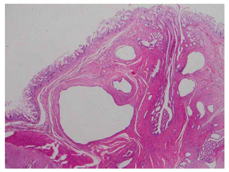
Figure 2 Low-power view of adenomyoma of the small intestine.
A nodular lesion mainly occupies the submucosa (hematoxylin and eosin stain, × 10).
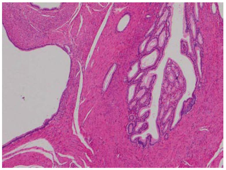
Figure 3 High-power view of adenomyoma of the small intestine.
The lesion consists of glandular structures of various sizes and interlacing smooth muscle bundles (hematoxylin and eosin stain, × 40).
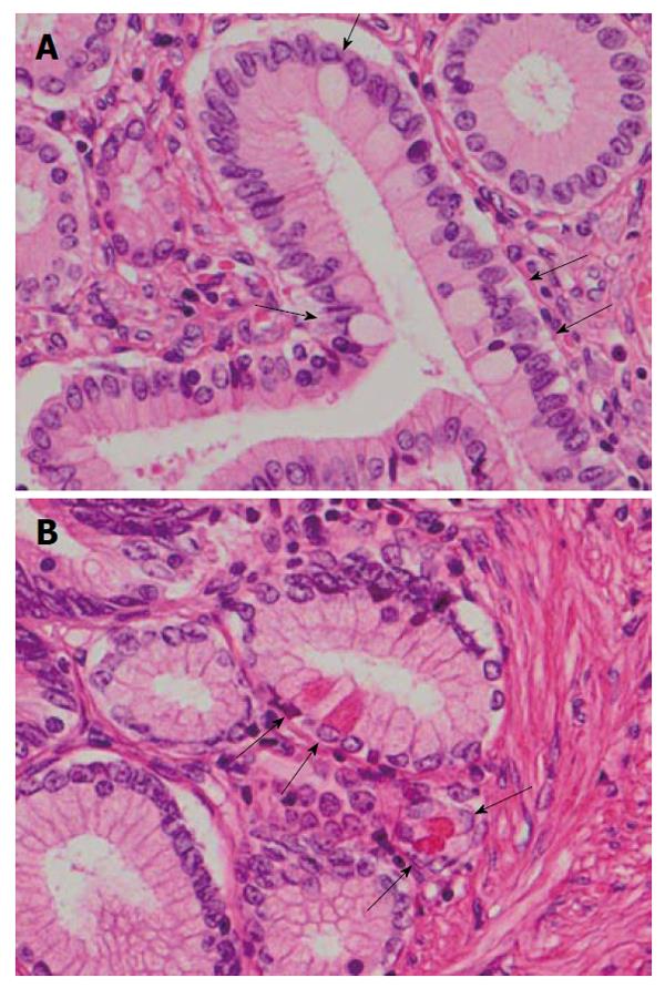
Figure 4 Appearance of goblet cells (A, arrows) and Paneth cells (B, arrows) in the intestinal adenomyoma (hematoxylin and eosin stain, × 400).
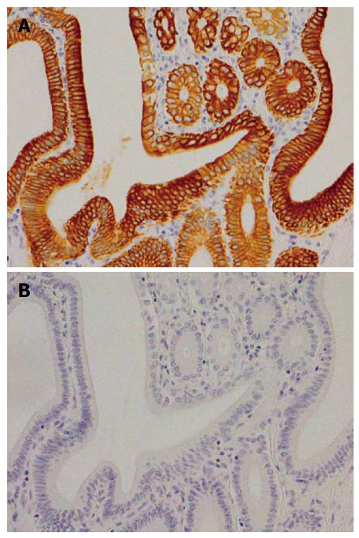
Figure 5 Results of immunohistochemical staining for cytokeratin 7 and cytokeratin 20.
The glandular element of the lesion is positive for cytokeratin 7 (A) and negative for cytokeratin 20 (B) (× 200).
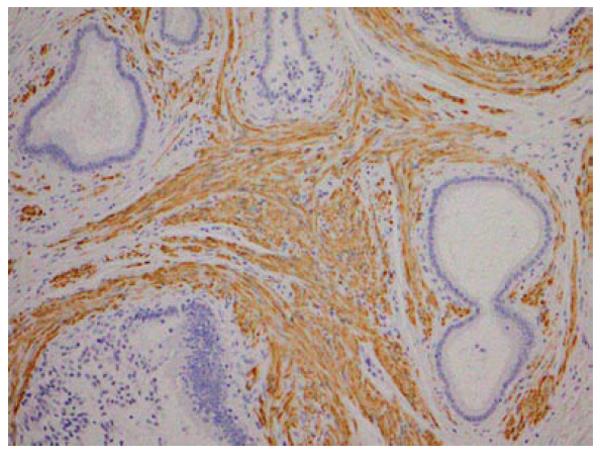
Figure 6 Results of immunohistochemical staining for desmin.
Smooth muscle cells surrounding the glandular elements are positive for desmin (× 100).
- Citation: Takahashi Y, Fukusato T. Adenomyoma of the small intestine. World J Gastrointest Pathophysiol 2011; 2(6): 88-92
- URL: https://www.wjgnet.com/2150-5330/full/v2/i6/88.htm
- DOI: https://dx.doi.org/10.4291/wjgp.v2.i6.88


