©The Author(s) 2025.
World J Gastrointest Pathophysiol. Jun 22, 2025; 16(2): 107265
Published online Jun 22, 2025. doi: 10.4291/wjgp.v16.i2.107265
Published online Jun 22, 2025. doi: 10.4291/wjgp.v16.i2.107265
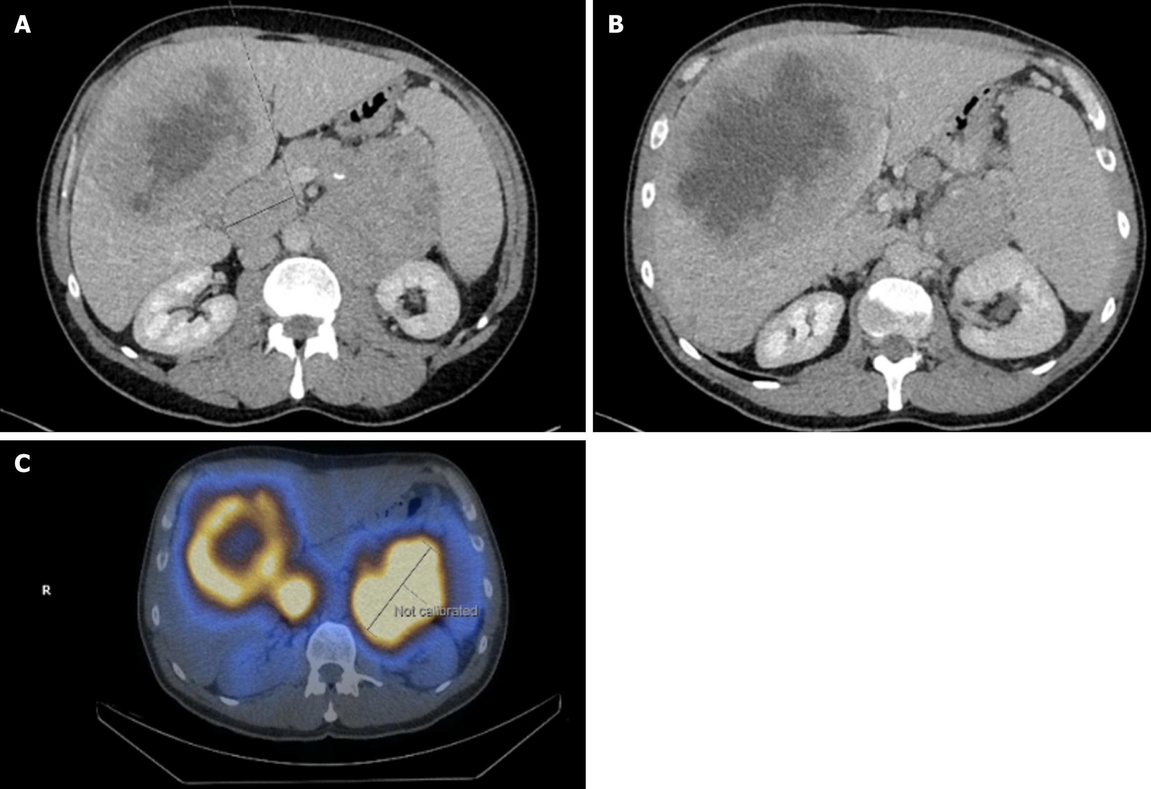
Figure 2 Initial CT-TAP confirmed 10 cm × 9.
5 cm 1° mass in the pancreatic tail, extending diffusely into the pancreatic body. A and B: Evidence of metastasis – 15 cm necrotic mass in segment 8 of the liver (B); C: Increased Octreotide uptake is shown on the OctreoScan, confirming the presence of somatostatin receptor positivity.
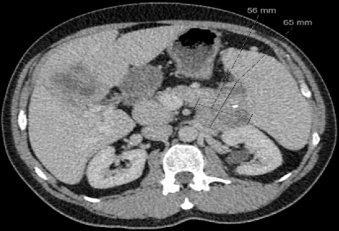
Figure 3 The smallest 1° tumor size 20 months post-initial treatment with Everolimus and somatostatin analogue therapy.
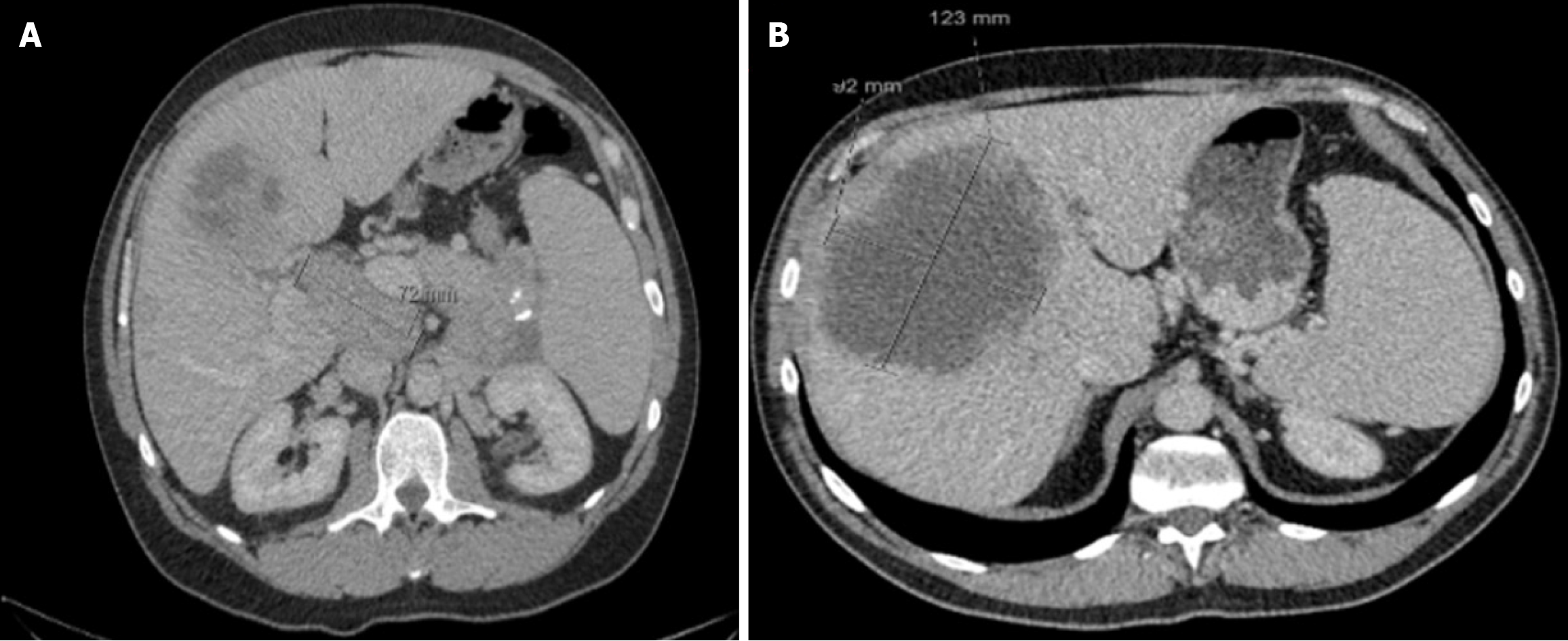
Figure 4 48 months post-treatment showing disease progression (increase in 1° tumor size and secondary hepatic metastasis and nodal disease).
A and B: Increased size in secondary hepatic mass (12.3 cm × 9.2 cm) at 49 months.
- Citation: Rikhraj N, Fernandez CJ, Ganakumar V, Pappachan JM. Pancreatic neuroendocrine tumors: A case-based evidence review. World J Gastrointest Pathophysiol 2025; 16(2): 107265
- URL: https://www.wjgnet.com/2150-5330/full/v16/i2/107265.htm
- DOI: https://dx.doi.org/10.4291/wjgp.v16.i2.107265


