©The Author(s) 2022.
World J Gastrointest Pathophysiol. May 22, 2022; 13(3): 107-113
Published online May 22, 2022. doi: 10.4291/wjgp.v13.i3.107
Published online May 22, 2022. doi: 10.4291/wjgp.v13.i3.107
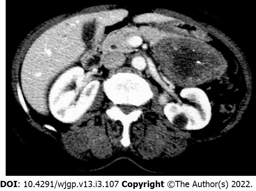
Figure 1
Computed tomography scan showing solid and cystic tumor in the body and tail of the pancreas (pancreatic schwannoma).
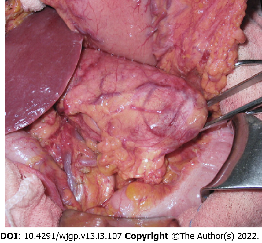
Figure 2
Laparotomy view of pancreatic body mass.
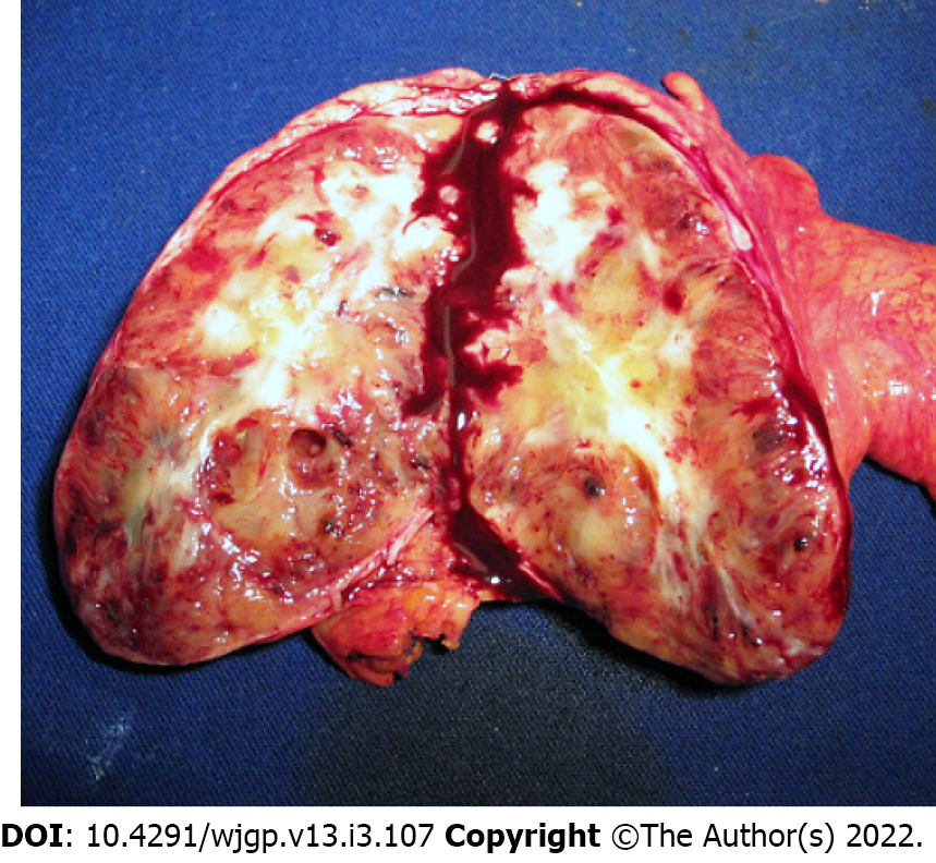
Figure 3
Macroscopic examination showed a well-encapsulated, pale yellow solid pancreatic tumor with areas of hemorrhage.
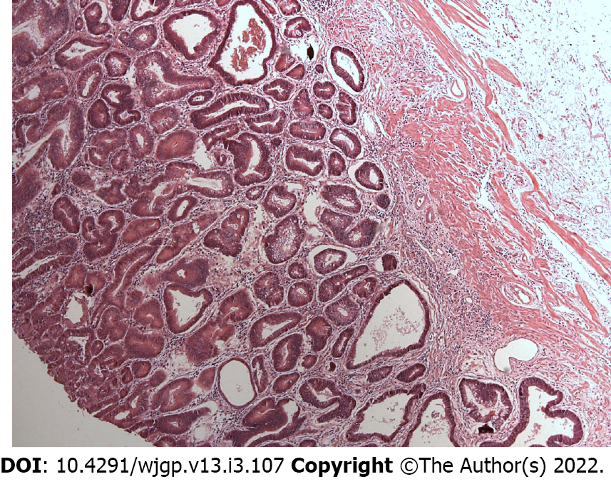
Figure 4 Representative area of moderately differentiated gastric adenocarcinoma.
Hematoxylin and eosin; Magnification × 50.
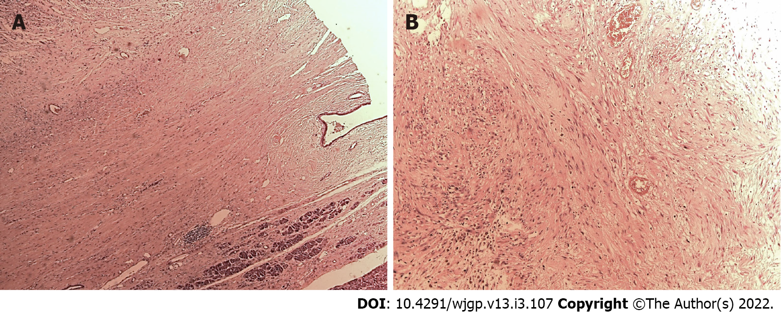
Figure 5 Microscopic examination.
A and B: Representative areas of pancreatic schwannoma; Hematoxylin and eosin; Magnification × 20).
- Citation: Ribeiro MB, Abe ES, Kondo A, Safatle-Ribeiro AV, Pereira MA, Zilberstein B, Ribeiro Jr U. Gastric cancer with concurrent pancreatic schwannoma: A case report. World J Gastrointest Pathophysiol 2022; 13(3): 107-113
- URL: https://www.wjgnet.com/2150-5330/full/v13/i3/107.htm
- DOI: https://dx.doi.org/10.4291/wjgp.v13.i3.107


