Copyright
©The Author(s) 2020.
World J Gastrointest Pathophysiol. May 12, 2020; 11(3): 43-56
Published online May 12, 2020. doi: 10.4291/wjgp.v11.i3.43
Published online May 12, 2020. doi: 10.4291/wjgp.v11.i3.43
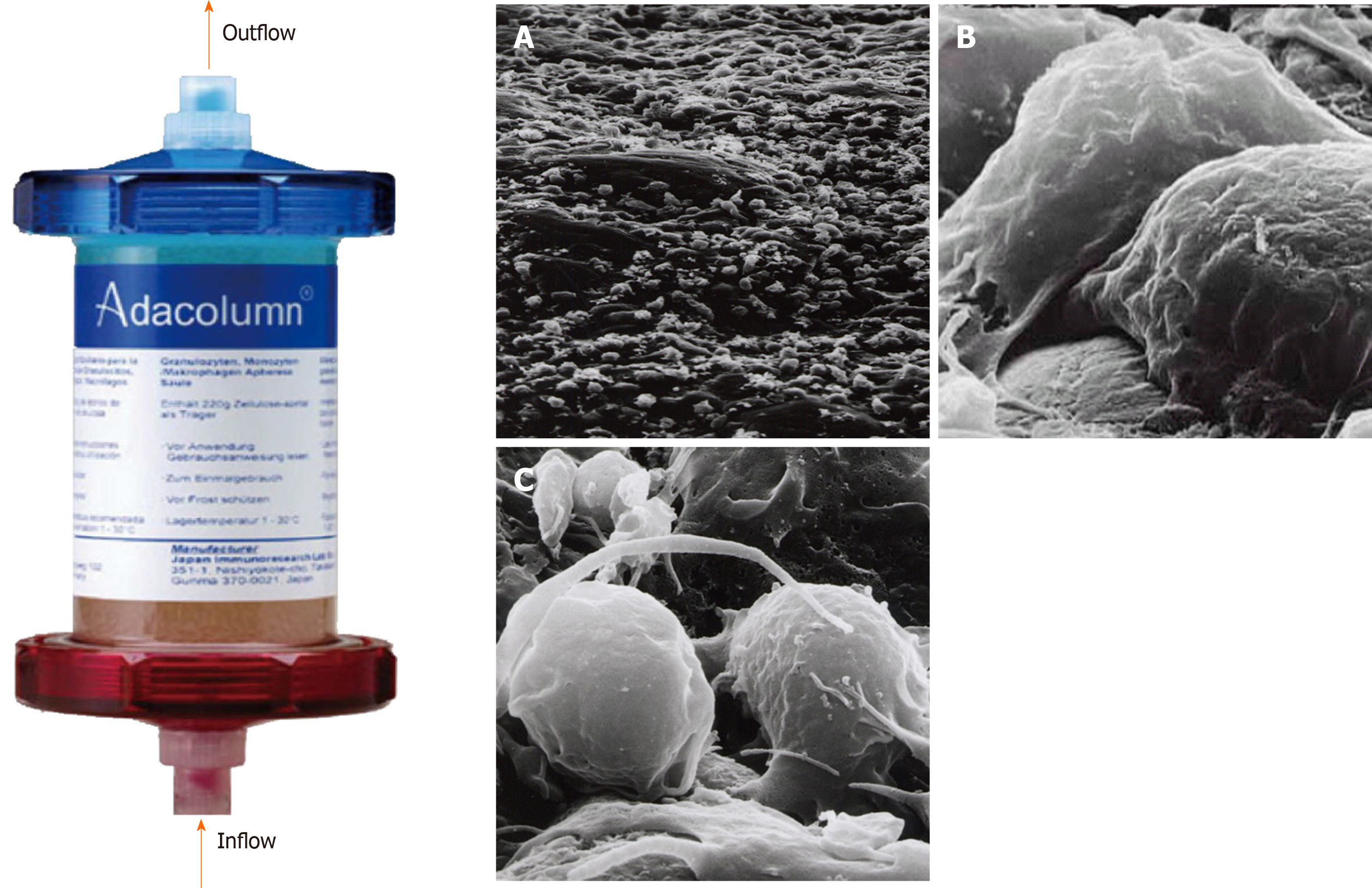
Figure 1 Photograph of Adacolumn and scanning electron photomicrograph of the acetate beads after treatment.
Adacolumn is filled with cellulose acetate beads of 2 mm in diameter (adsorptive carriers) bathed in sterile saline. The blood from the antecubital vein of one arm flows into the column and returns to the antecubital vein in the contralateral arm. A: A low power view (400 ×) of the acetate beads in a column after treatment with cells covering the surface of the carrier; B: Viewed at 10000 ×. Neutrophils were adsorbed onto the beads; C: Viewed at 12000 ×. Activated monocyte/macrophages are seen (taken by Dr. A. Saniabadi of Japan Immunoresearch Laboratories). Modified from reference[31].
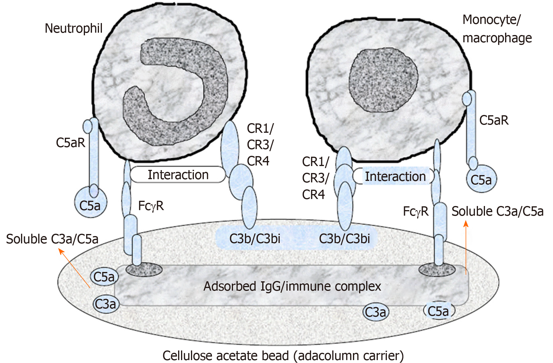
Figure 2 A schematic diagram of the selective adhesion of myeloid granulocyte and monocyte to cellulose acetate carriers.
Cellulose acetate beads inside the Adacolumn are capable of selectively adsorbing circulating neutrophils and monocytes by binding to IgG fragments (Fcγ) and immune complements complexes. Lymphocytes are not absorbed as they rarely express complement receptor. Modified from reference[31].
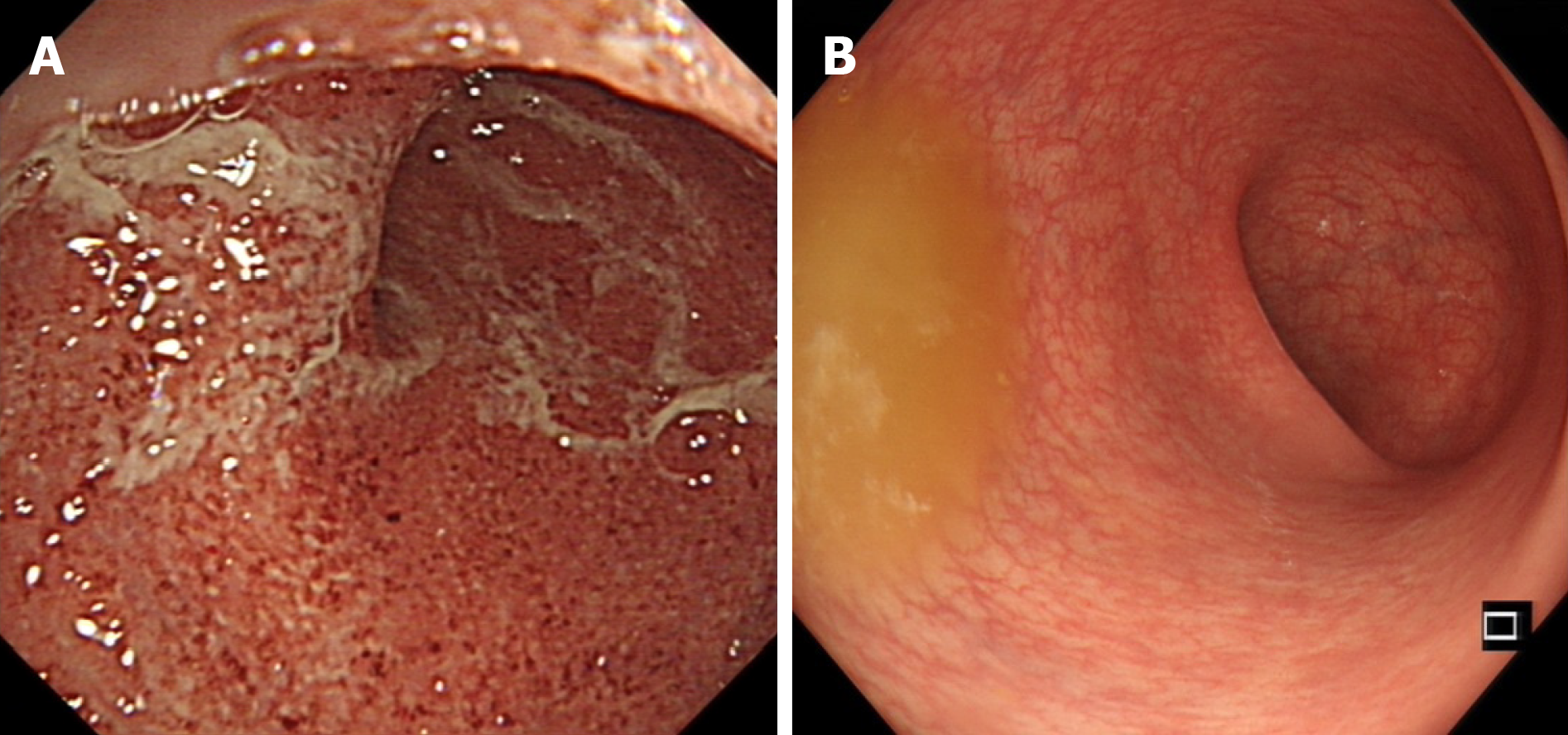
Figure 3 Endoscopic photographs of an ulcerative colitis patient who responded well to selective granulocyte and monocyte apheresis.
A: Endoscopic photograph before granulocytes and monocytes apheresis therapy; B: Endoscopic photograph after ten sessions of granulocytes and monocytes apheresis therapy.
Figure 4 Changes of Mayo scores in 30 ulcerative colitis patients at entry and after ten granulocytes and monocytes apheresis sessions.
Mayo scores were significantly decreased after ten granulocytes and monocytes apheresis sessions compared with that at entry[29]. GMA: Granulocytes and monocytes apheresis.
- Citation: Chen XL, Mao JW, Wang YD. Selective granulocyte and monocyte apheresis in inflammatory bowel disease: Its past, present and future. World J Gastrointest Pathophysiol 2020; 11(3): 43-56
- URL: https://www.wjgnet.com/2150-5330/full/v11/i3/43.htm
- DOI: https://dx.doi.org/10.4291/wjgp.v11.i3.43


