©2010 Baishideng.
World J Gastrointest Pathophysiol. Aug 15, 2010; 1(3): 109-111
Published online Aug 15, 2010. doi: 10.4291/wjgp.v1.i3.109
Published online Aug 15, 2010. doi: 10.4291/wjgp.v1.i3.109
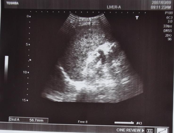
Figure 1 Ultrasound showing splenomegaly with multiple hypoechoic lesions in the spleen.
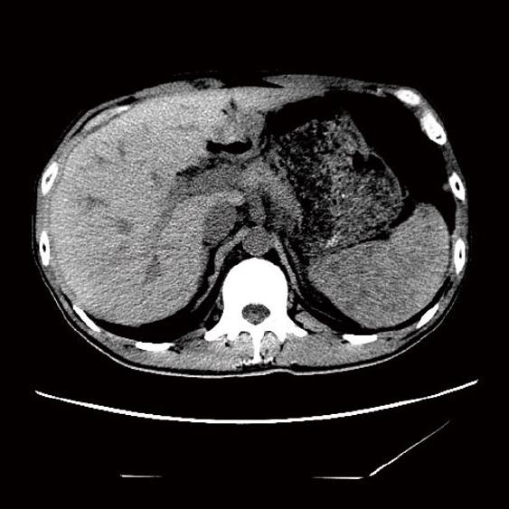
Figure 2 Abdominal computed tomography demonstrated multiple hypodense diffuse lesions in the spleen.
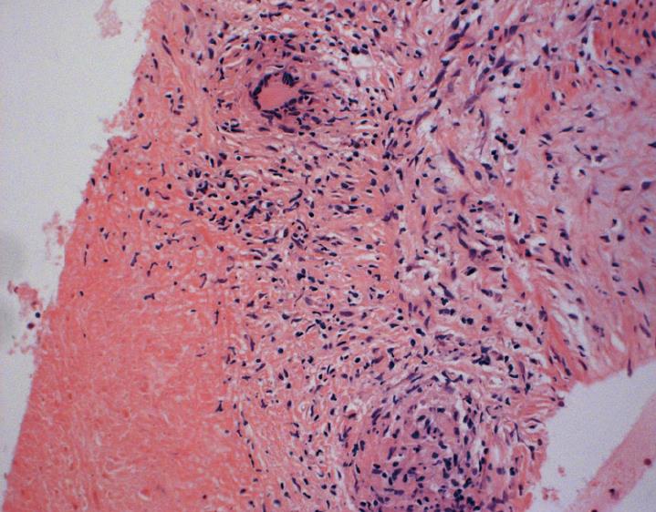
Figure 3 A section of the biopsy specimen showing a granuloma nodule with central areas of caseation surrounded by Langerhan’s giant cells and epithelioid cells (Hematoxylin and eosin stained technique, middle-multiplications).
- Citation: Zhan F, Wang CJ, Lin JZ, Zhong PJ, Qiu WZ, Lin HH, Liu YH, Zhao ZJ. Isolated splenic tuberculosis: A case report. World J Gastrointest Pathophysiol 2010; 1(3): 109-111
- URL: https://www.wjgnet.com/2150-5330/full/v1/i3/109.htm
- DOI: https://dx.doi.org/10.4291/wjgp.v1.i3.109


